OCULUS Keratograph 5M - Topographer
←
→
Page content transcription
If your browser does not render page correctly, please read the page content below
OCULUS Keratograph 5M
              Topographer

   We focus on progress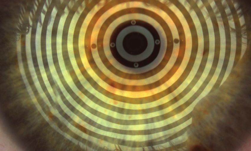
OCULUS Keratograph 5M
More than just a topographer!
The new Keratograph 5M technology is a revolution in corneal topography and Dry Eye analysis.
The high-resolution color camera and the integrated magnification changer offer a new perspective to the tear film
assessment procedure.
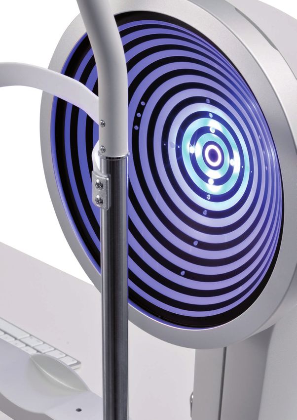
Measurements with Placido disc illumination
The white annular illumination is used to precisely measure
thousands of points on the surface of the cornea. The
infrared annular illumination is available during the tear
film analysis to prevent light reflection and glare.
Measurements with light emitting diodes
The Keratograph 5M features the ideal illumination for
each application: white diodes for the assessment of
tear film particles movement, blue diodes for fluorescein
imaging, infrared diodes for meibography.
In addition to these unique features, the Keratograph 5M precisely measures corneal topography. The built-in real
Keratometer and the automatic measurement activation guarantee perfect reproducibility of the Ks. The data from the
non-contact measurement is automatically analyzed and shown in comprehensive presentation formats.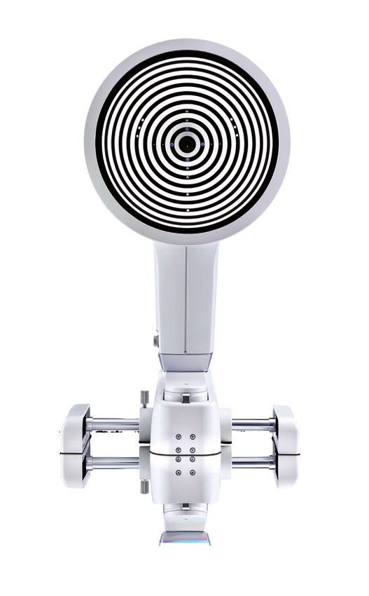
Clear presentation
For more transparency of your services

Actively integrate the Keratograph 5M into your practice as a patient consultation tool. With many visual maps and easy
to understand diagrams, the Keratograph 5M enhances the communication with your patients. Use your Keratograph 5M
as a marketing tool and gain your patients’ confidence.

Build trust
With the Keratograph 5M, you are able to show images that your patients have never seen before. Build trust by providing
professional consultation during examinations and follow-ups.

                                                                   Images speak louder than words
                                                                   With the TF-Scan software, you can analyze and
                                                                   document the tear film break up time and share the
                                                                   results with your patients.
                                                                   Let images and videos speak for themselves during
                                                                   your consultation — this creates a strong
                                                                   physician/patient relationship.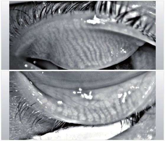
Meibo-Scan
Meibography of the upper and lower eyelid

The new Keratograph 5M is a multi-purpose diagnostic
device that easily and efficiently integrates complex
examination, such as meibography into the
ophthalmological and optometric practices.

Dry Eye is most commonly caused by the Meibomian
Gland Dysfunction (MGD). The Meibo-Scan shows the
morphological changes in the glandular tissue.

                         The new Keratograph 5M is patient-friendly and takes the measurements at a greater distance to
           100 mm
                         the eye. An image section of 26 mm enables optimal examination of the lower and upper eyelids.

3-D display of the meibomian glands
There are different viewing options, such as, the 3-D display, various section planes and the marking of the individual area
of examination to easily evaluate the meibomian glands and the morphological changes in the upper and lower eyelids.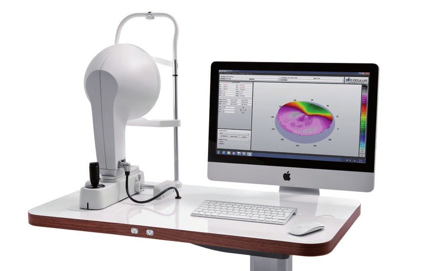
TF-Scan
Visualizing the quality and quantity of the tear film
The OCULUS Keratograph 5M evaluates the tear film with the aid of white or infrared illumination. The new high-definition
color camera makes the finest structures visible. The NIKBUT (non-invasive Keratograph break-up time), the tear meniscus
height, the lipid layer and the tear film particles movement are examined carefully and documented easily. The exams are
non-invasive, user-friendly and reproducible.
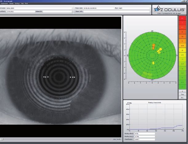
NIKBUT
Evaluation of the tear film break-up-time
The tear film break-up-time measurement with the
Keratograph 5M is touch-free and fully automatic. The
new infrared illumination is invisible to the human eye
and produces no glare during the examination and no
reflex tearing.
The TF-Scan maps the tear film break up time and is a
great tool for follow ups and patient consultation.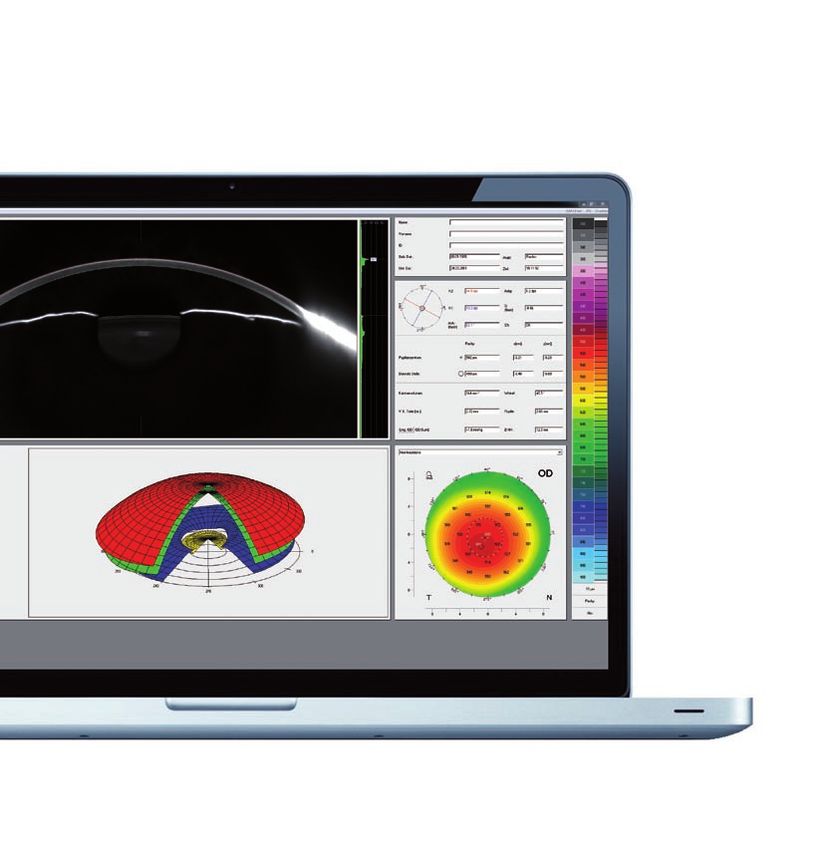
Tear meniscus-height
Evaluation of the tear film quantity
With an integrated measuring guide and various
magnification tools, you can measure the tear meniscus
height and evaluate its characteristics along the lower lid
margin. The result is saved in the patient file.

Lipid layer
Evaluation of the lipid layer thickness
The color and structure of the lipid layer is visible and
can be recorded. This shows the lipid layer thickness,
which correlates with tear film evaporation and dry eye
symptoms. If the lipid layer is too thin or absent, the tear
evaporation rate and the tear film instability increase.

TF dynamics
Evaluating the particle flow characteristics
A video recording with up to 32 images per second
enables the observation of the tear film particle flow
characteristics and shows the tear film viscosity.
R-Scan
Automatic bulbar redness classification

The R-Scan is the first and only technology that
automatically and objectively measures and classifies the
bulbar and the limbal degree of redness.

The R-Scan detects thin blood vessels in the conjunctiva
and evaluates the degree of redness. With automatic
evaluation, there is no need to manually compare the
classification sheets and you can be more confident in
the user-independent results.

                                                    Choice of display                     Precise classification
                                                                                             of bulbar redness

                                                                                                Precise classification
                                                                                                   of limbal redness

Intelligent recognition of the relevant areas
Display of thin blood vessels in the conjunctiva:               Display in red-free light:
Only the thin blood vessels of the conjunctiva are              Under the green spectrum of visible light, blood vessels
detected and evaluated.                                         are richer in contrast and more visible.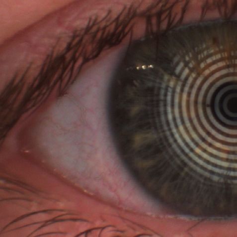
Imaging
High-definition color camera for image and
video documentation
The Keratograph 5M features an optimal imaging function and a new high-definition camera. This opens new doors for
documentation during every day practice. Experience the highest level of multi-functionality, similar to a digital slit lamp.
The selectable illumination and variable magnification serve as the basis for high-definition image and video recordings.
The simple operation easily integrates into a practice routine and allows the efficient documentation of the examinations.

                             For contact lens fitting
                             Easily evaluate the static and dynamic fit of Rigid Gas Permeable and soft
                             contact lenses and the wetting and drying times.

                             For documentation of diagnostic findings
                             The even illumination and high depth of field are especially important for
                             professional documentation of diagnostic findings. The recordings are saved
                             automatically.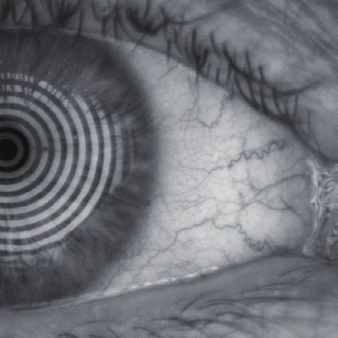
OxiMap® – visualizing the
oxygen transmissibility
Professional patient consultation
The cornea needs oxygen and a good oxygen supply is fundamental for the comfort of a contact lens wearer. New materials
used for soft contact lenses offer excellent oxygen transmissibility. This can be shown with the new OCULUS OxiMap®
display. You can easily show these color maps to your patients and help them choose better contact lenses.

How much oxygen really reaches the cornea ?
Until now, only the oxygen transmissibility values for the center of a contact lens with –3.0 D were available. The
OxiMap® shows the oxygen transmissibility depending on the lens material and the lens thickness. The OxiMap® is
available for the most frequently sold spherical soft contact lenses. This impressive tool assists you in the consultation
with customers to select the most suitable contact lens.

> Oxygen transmissibility for -3.00 D and -10.00 D for identical type of contact lens

Plain and comprehensive visualization assures patient loyalty!
                                                                                                                                                        Dk/t > 125*
Contact lenses act as a potential
barrier to oxygen transport even                                                                            Dk/t > 87*                                  Continuous Wear
when the eyes are open to the
                                                                                                            Extended Wear
atmosphere. Long hours of wearing                                  Dk/t > 35*
comfort can only be guaranteed
with a sufficient oxygen supply. The
                                                                   Daily Wear
color representation of the various
terms of oxygen transmissibility
is based on international
recommendations for daily,
                                                                                                                                                                    cm/sec
extended and continuous wear.                  93 x 10 9
                                                                                                                                                                mL 0²/mL x h Pa
                                               124 x 10 9         cm/sec
                                                                                                                                                                mL 0²/mL x mm Hg

                                             > The OxiMap® color coding of the Dk/T-values and the recommended wearing time
Basic software
Precise analysis. Clear presentation.
The comprehensive basic software contains various analysis for
everyday use; from topography to automatic keratoconus detection
and an extensive contact lens database.
Overview and 3-D displays
The built-in Keratometer guarantees the utmost
measuring precision and reproducibility of the real Ks.
The overview display shows the keratometric values, the
central radii, the corneal astigmatism, the eccentricity
and the corneal diameter. The color topography map
shows the curvature of the anterior corneal surface.
Fourier analysis
The Fourier analysis is an important tool for visualizing
the amount of corneal irregularities. Using the Fourier
analysis, the topography map is divided into individual
components. The first three are standard components
that represent lower order aberration and the fourth map
shows the amount of corneal irregularities or higher
order aberration.Zernike analysis
Zernike analysis provides a means to distinctly describe
irregularities of the cornea. If the indicated aberration
coefficient is elevated, it shows a decline of the optical
quality. The Zernike analysis enables the determination of
the exact position of the apex.
Indices
The Indices display helps with the quick and easy
detection of abnormalities in the topography. The
measured values are compared with a standard database.
If Keratoconus is present, it is detected at an early
stage and categorized according to the topography at
hand. The indices display is helpful during the follow-up
examination and shows whether the irregularities are
changing or staying constant.
Show 2 exams
During the follow-up examinations, it is necessary to
compare the results with the previous exams. With this
feature you can compare the changes in the corneal
topography over time for the contact lens wearers or
patients with progressive conditions, such as
Keratoconus.Compare 3 exams
This display shows the changes in the corneal refraction
power and allows the documentation of refractive changes
caused by refractive surgery or by wearing the Ortho-K
lenses.
Near portion height measurement
This software precisely simulates the near portion height
of rigid bifocal contact lenses and simplifies the complex
fitting process.
Palpebral angle measurement
The measurement of the nasal lower palpebral angle
facilitates the identification of the expected inclination
or stabilization axis when fitting toric contact lenses.
Save time and money by giving this information to the
contact lens manufacturer when you place an order.Pupillometry
The optional pupillometry software measures the pupil
reaction to light with and without glare. The reaction of
both pupils can be compared to detect an anisocoria.
Contact lens fitting
Contact lenses are recommended on an individual basis
and displayed in a list. In order to avoid taking more steps
than necessary when fitting contact lenses, the fluorescein
image can be simulated beforehand. The contact lens
can be rotated and moved around. Fluorescein image
simulation is adjusted automatically. The integrated
and expandable database contains all customary types
of contact lenses and is updated on a regular basis.
The user can determine the order in which contact lens
manufacturers appear.
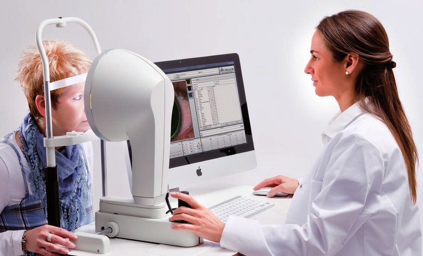
Elegant and functional
OCULUS Keratograph 5M with the
Apple iMac and motorized table
(Optional features)All features at a glance Customize your OCULUS Keratograph 5M Features Keratograph 5M TF-Scan R-Scan Meibo-Scan Imaging Pupillometry OxiMap® Basic software Topography Overview display Color map 3-D presentation Fourier analysis Zernike analysis Indices (automatic keratoconus detection) Compare 3 exams Asphericity Camera image Show 2 exams Contact lens fitting Contact lens database Interfaces to fitting programs of various contact lens manufacturers Palpebral angle measurement Near portion height measurement Data import and export via USB 2.0 Standard Optional Standard and optional features may differ in your country. Contact your local OCULUS representative for details.
Technical data
OCULUS Keratograph 5M
Precision                                                         ± 0.1 dpt
Reproducibility                                                   ± 0.1 dpt
Ring count                                                        22
Working distance                                                  78 - 100 mm
Number of evaluated data points                                   22,000
Camera                                                            Digital CCD camera
Illumination source                                               Placido illumination:          white diodes
                                                                  Placido illumination:          infrared diodes (880 nm)
                                                                  Imaging illumination:          blue diodes (465 nm)
                                                                  Meibography:                   infrared diodes (840 nm))
                                                                  Tear film dynamics:            white diodes
                                                                  Pupillometry illumination:     infrared diodes (880 nm)
Dimensions (W x D x H)                                            275 x 320 - 400 x 485 - 512 mm
Weight                                                            6.8 kg
Minimal PC requirements                                           Processor: Intel Core i3 or better, 4 GB main memory,
                                                                  Hard disk: 500 GB and up, graphic card: Intel HD Graphics 2000 or better,
                                                                  Recommended screen resolution: 1920 x 1080 (full-HD)
   in accordance with Medical Device Directive 93/42/EEC

                                                                                  250 mm
                                                                                 9.84 inches

                                                                                                               19.09 - 20.16 inches
                                                                                                                  485 - 512 mm

                                              320 – 400 mm                         275 mm
                                                                                                                                              Right to design changes reserved.

                                           12.60 – 15.75 inches                  10.83 inches

       WWW.OCULUS.DE                                                     OCULUS Optikgeräte GmbH
                                                                         Postfach • 35549 Wetzlar • GERMANY
                                                                         Tel. +49-641-2005-0 • Fax +49-641-2005-295
                                                                         E-Mail: export@oculus.de • www.oculus.de
                                                                                                                                              18/0412/e/Ha

                                                                         • OCULUS USA, sales@oculususa.com
                                                                         • OCULUS Asia, info@oculus.hk
                  Oculus is certified by TÜV according to                • OCULUS Czechia, oculus@oculus.cz
                  DIN EN ISO 13485/DIN EN ISO 9001                       • OCULUS Iberia, info@oculus.es
You can also read