Pelvic Lymphocele Enlargement after Manual Lymph Drainage: Case Report and Literature Review
←
→
Page content transcription
If your browser does not render page correctly, please read the page content below
MedDocs Publishers

                                                        Annals of Gynecologic Cancer
                                                                                                      Open Access | Case Report

  Pelvic Lymphocele Enlargement after Manual Lymph
      Drainage: Case Report and Literature Review
Jaqueline Munaretto Timm Baiocchi1*; Larissa L Campanholi2; Glauco Baiocchi3
1
 Department of Physical Therapy, Instituto Oncofisio, São Paulo, Brazil.
2
 Department of Physical Therapy, Instituto Sul Paranaense de Oncologia, Ponta Grossa, Brazil.
3
 Department of Gynecologic Oncology, AC Camargo Cancer Center, Sao Paulo, Brazil.

  *Corresponding Author(s): Jaqueline Munaretto                        Abstract
 Timm Baiocchi                                                        Purpose: Lymphocele is a cystic cavity with a fibrous
 Department of Physical Therapy, Oncofisio - Rua Joa-              capsule that contains lymphatic fluid. Our objective is to de-
                                                                   scribe physical therapy approach for leg lymphedema after
 quim Távora, 04015-000, São Paulo, Brazil.
                                                                   endometrial cancer treatment that resulted in an increase
 Tel: +55-11-3255-4727;                                            of a pelvic lymphocele.
 Email: jaqueline@oncofisio.com.br
                                                                       Client description: A 59-year-old woman with endome-
                                                                   trial high-grade adenocarcinoma was referred to our center
                                                                   with a history of pelvic and leg lymphedema followed by a
Received: Oct 30, 2020                                             pelvic lump, subsequently diagnosed as pelvic lymphocele.
Accepted: Jan 21, 2021
                                                                      Intervention: She undertook a physiotherapist-guided
Published Online: Jan 26, 2021                                     complex decongestive therapy program, with manual lymph
Journal: Annals of Gynecologic Cancer                              drainage interventions including intermittent pressure com-
Publisher: MedDocs Publishers LLC                                  pression and use of compression stockings. Limb and pelvic
                                                                   volumes were measured over a 6-month period.
Online edition: http://meddocsonline.org/
Copyright: © Timm Baiocchi JM (2021). This Article is                 Measures and outcome: Within the first month, the pa-
distributed under the terms of Creative Commons                    tient’s excess lower limb volume reduced, and lymphocele
Attribution 4.0 International License                              volume was 300 ml. At 4 months, her lymphocele volume
                                                                   increased to 500ml and at 6-month evaluation the lympho-
                                                                   cele decreased to 17 ml, suggesting spontaneous drainage
                                                                   to the abdominal cavity.
Keywords: lymphedema; lymphocele; lymph drainage.
                                                                      Implications: A woman with pelvic and lower leg lymph-
                                                                   edema caused by endometrial cancer treatment benefited
                                                                   from reduced swelling despite the lymphocele enlarge-
                                                                   ment. This case provides knowledge about lymphocele be-
                                                                   havior after complex decongestive physiotherapy for pelvic
                                                                   and lower leg lymphedema.

                Cite this article: Timm Baiocchi JM, Campanholi LL, Baiocchi G. Pelvic Lymphocele Enlargement after Manual
              Lymph Drainage: Case Report and Literature Review. Ann Gynecol Cancer. 2021; 4(1): 1004.

                                                               1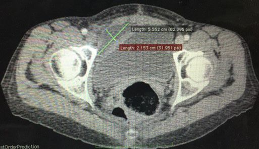
MedDocs Publishers
   Introduction                                                           complete surgical staging on December 19th, 2015, which in-
                                                                          cluded resection of 64 pelvic and retroperitoneal lymph nodes,
   A lymphocele is a cystic mass that can form in the pelvic ret          followed by adjuvant chemotherapy. The patient did not receive
peritoneum or in the paraaortic region after pelvic or pelvic and         adjuvant radiotherapy. Previously she already has undergone a
paraaortic lymphadenectomy. A lymphocele is a collection of               melanoma resection at her left thigh with inguinal lymphad-
lymph bordered by a thick fibrous wall without vascular supply            enectomy.
and epithelial lining, expanding from the retroperitoneum into
the pelvis or the abdominal cavity [1].                                       She was referred to our center after development of lower
                                                                          limb lymphedema. During physical exam, observed a stage II
   The pathophysiological basis for lymphocele development is             lymphedema at the left lower limb, supra pubic and pelvic ede-
an incomplete lymph stasis with post-operative lymph leakage              ma. The complementary exams showed no deep vein throm-
in an amount exceeding the capacity for spontaneous perito-               bosis, left and right saphenous vein insufficiency, and collateral
neal reabsorption and the accumulation of lymph in spaces that            and perforating vein with reflux to the popliteal vein. The lym-
have formed as a result of lymphatic tissue removal [2].                  phoscintigraphy reported a dermic reflux at her left foot and
   Pelvic lymphocele prevalence varies from 0% to 58, 5%. This            preserved lymph drainage at right lower limb. For the lymph-
variation is due to underdiagnosis because the majority is as-            edema treatment it was performed CDT with manual lymph
ymptomatic and are often an incidental finding during postop-             drainage, intermittent pressure compression twice a week and
erative or routine follow up [3]. Moreover, large lymphoceles             the use of 20/30 mmHg compression stocking. The program
may cause symptoms related to compression of adjacent struc-              was delivered under the guidance of a physiotherapist certified
tures such as lower abdominal pain, abdominal fullness, consti-           in lymphedema therapy.
pation, increase of urinary frequency, and edema of the genitals              On January 22th, 2016 she did a computed tomography that
and/or legs [4].                                                          revealed the existence of a lymphocele with 300 milliliters,
   The symptomatic lymphoceles rates are reported from 5%                 asymptomatic. The patient kept her lymphedema treatment,
to 6% of the cases and its treatment consists of drainage, surgi-         twice a week, and started Pilates exercises. In April 2016 she
cal resection, biological glues or alcoholization [5, 6]. Moreover,       had a new tomography that evidenced the growth of the lym-
significate sequelae could develop and include infection of the           phocele into 500 milliliters, evidenced by a symptomatic lower
lymphocele, obstruction and infection of the urinary tract, in-           belly lump. After that the patient developed stage I lymphede-
testinal obstruction, venous thrombosis, pulmonary embolism,              ma at the right lower limb. In her last tomography, on June 22th,
chylous ascites and lymphatic fistula formation [7].                      2016, it was noted a sudden decrease, without clinical interven-
                                                                          tion, of the lymphocele to 17 milliliters, suggesting spontaneous
   On clinical examination, a large lymphocele may present as             drainage to the abdominal cavity.
a palpable pelvic mass and the skin may be reddened and swol-
len. Common signs of all lymphoceles are following: A cyst with              In this article, we present a unique case report of a patient
thick wall with no vascularization, no intraluminal calcifications        that had a benefit from CDT, improving the limb and pelvic vol-
and no signs of solid components. The asymptomatic lympho-                ume despite having an enlargement of a pelvic lymphocele. As
cele is usually a round, unilocular cyst with ground-glass con-           far as we know, any study have addressed the prevalence of
tents and differ from symptomatic which is usually an oval, or            lymphocele in the population with lymphedema, particularly
ovoid, unilocular cyst with low-level or anechoic content and             the relation between lymph drainage and lymphocele enlarge-
presence of debris and septations [1].                                    ment.

    Imaging such as ultrasound or computed tomography estab-                  Considering the pathophysiology of lymphocele, it can be
lishes the diagnosis. Other fluid collections to be considered            argued that CDT, especially MLD may have contributed to the
in the differential diagnosis are urinoma, seroma and hema-               lymphocele enlargement after improvement of the lymph flow.
toma Yet, when lower limb edema is present, venous thrombo-               It is important that oncology health professionals, lymphede-
sis should to be considered [7].                                          ma-trained clinicians and patients be informed about the side
                                                                          effects of the lymphedema treatment and be aware of this com-
    The lymphocele development can be related to the tumor                plications.
aggressiveness, metastatic nodes or by the treatment like num-
ber of lymph nodes harvested and use of radiation [5,6]. In ad-
dition, sentinel lymph node mapping decreases the risk of lym-
phocele formation compared to full lymphadenectomy [8].
    Lymphedema is characterized by the accumulation of pro-
tein rich fluid in the interstitial space because of the lymphatic
damage induced by the inguinal, pelvic or paraaortic lymphad-
enectomy [9]. Lymphedema treatment is made by the Complex
Decongestive Therapy (CDT) which consists of manual lymph
drainage, intermittent pressure compression, skin care, exer-
cises and use of bandaging or compression garments.
   Case report
   A 59 years old woman, diagnosed with endometrial high-                    Figure 1: CT aspect of pelvic lymphocele.
grade adenocarcinoma (Stage IIIA – FIGO) was submitted to

Annals of Gynecologic Cancer                                          2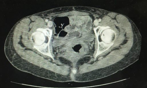
MedDocs Publishers
Conclusion
In this present case, CDP for the lymphedema treatment
contributed to the enlargement of a pelvic lymphocele.
References
1. Weinberger V, Fischerova D, Semeradova I, Slama J, Cibula D, et
al. Ultrasound characteristics of a symptomatic and asymptom-
atic lymphocele after pelvic and/or paraaortic lymphadenecto-
my. Taiwan J Obstet Gynecol. 2019; 58: 266-272.
2. Weinberger V, Cibula D, Zikan M. Lymphocele: Prevalence and
management in gynecological malignancies. Expert Rev Antican-
cer Ther. 2014; 14: 307-317.
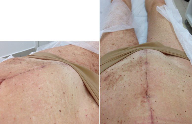
Figure 2: Lymphocele increase after lymph drainage.
3. Achouri A, Huchon C, Bats AS, Bensaïd C, Nos C, et al. Postop-
erative lymphocysts after lymphadenectomy for gynaecological
malignancies: Preventive techniques and prospects. Eur J Obstet
Gynecol Reprod Biol. 2012; 161: 125-129.
4. Metcalf KS, Peel KR. Lymphocele. Ann R Coll Surg Engl. 1993; 75:
387-392.
5. Achouri A, Huchon C, Bats AS, Bensaid C, Nos C, et al. Compli-
cations of lymphadenectomy for gynecologic cancer. Eur J Surg
Oncol. 2013; 39: 81-86.
6. Zikan M, Fischerova D, Pinkavova I, Slama J, Weinberger V, et al.
A prospective study examining the incidence of asymptomatic
and symptomatic lymphoceles following lymphadenectomy in
patients with gynecological cancer. Gynecol Oncol. 2015; 137:
291-298.
7. McCullough CS, Soper NJ, Clayman RV, So SS, Jendrisak MD, et
Figure 3: Pelvic aspect of the lymphocele. al. Laparoscopic drainage of a posttransplant lymphocele. Trans-
plantation. 1991; 51: 725-727.
8. Diniz TP, Drizlionoks E, Faloppa CC, Menezes JN, Mantoan H, et
al. Impact of Sentinel Node Mapping in Decreasing the Risk of
Lymphocele in Endometrial Cancer. Annals of Surgical Oncology.
2020.
9. Hareyama H, Hada K, Goto K, Watanabe S, Hakoyama M, et
al. Prevalence, classification, and risk factors for postoperative
lower extremity lymphedema in women with gynecologic ma-
lignancies: A retrospective study. Int J Gynecol Cancer. 2015; 25:
751-757.
Figure 4: CT aspect after the lymphocele’s regression.
Annals of Gynecologic Cancer 3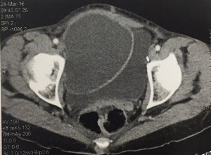


You can also read