Giant Rugae in Helicobacter pylori-Infected Stomachs
←
→
Page content transcription
If your browser does not render page correctly, please read the page content below
COMMENTARY
Clin Endosc 2021;54:1-3
https://doi.org/10.5946/ce.2021.007
Print ISSN 2234-2400 • On-line ISSN 2234-2443

 Open Access

Giant Rugae in Helicobacter pylori-Infected Stomachs
Sun-Young Lee
Department of Internal Medicine, Konkuk University School of Medicine, Seoul, Korea

  See “Endoscopic Characteristics of Rugal Hyperplasia and Related Acid Condition in Helicobacter pylori-Infected Stomach” by Byung
  Chul Kim, Mi Ae Song, Sung Ho Kwon, on page 73-84.

   Giant gastric rugae are often found during upper gastroin-                      levels found in H. pylori-induced corpus gastritis.3
testinal endoscopic examinations. Most of these hyperplastic                          Irrespective of the presence of the gastric rugae, the serum
rugae are induced by the Helicobacter pylori infection in East                     PG I and PG II levels increase with an active H. pylori infec-
Asia, which increases the risk of gastric cancer.1 Nonetheless,                    tion and decrease with the progression to gastric corpus atro-
data on the serum pepsinogen (PG) assay findings of rugal hy-                      phy.4 Furthermore, when there is an active H. pylori infection,
perplastic gastritis (RHG) are scarce. To resolve this issue, Kim                  PG II levels correlate more with the gastric secretory ability
et al. compared data between H. pylori-infected patients with                      than with PG I levels and PG I/II ratios.5 A high PG II level is
and without RHG.2 They found that the serum PG I/II ratio                          a risk factor for diffuse-type gastric cancer,6 whereas low PG I/
was lower in patients with RHG than in H. pylori-infected pa-                      II ratios and PG I levels are risk factors for intestinal-type gas-
tients without RHG. Such differences were significant only in                      tric cancer.7 Giant rugae may progress to Bormann-type IV, a
patients with closed-type chronic atrophic gastritis (CAG) and                     diffuse-type gastric cancer8; hence, high PG II levels are more
not in those with open-type CAG.                                                   reliable than low PG I/II ratios and PG I levels in patients with
   Notable findings of this study were the use of the serum                        RHG.
PG assay and the strict diagnostic criteria for endoscopic di-                        Kim et al. analyzed H. pylori-infected patients with positive
agnosis. As summarized in their figures, Kim et al. followed                       rapid urease test in their study.2 The gastric secretory ability
detailed, endoscopic criteria to diminish interpersonal vari-                      significantly increased in H. pylori-infected patients with
ability.2 By measuring the width of the gastric rugae with biop-                   RHG, as demonstrated by their high serum PG I, PG II, and
sy forceps after air inflation, they found a link between the low                  gastrin levels. Nevertheless, they concluded that most patients
PG I/II ratio and the hypertrophic gastric rugae with a width                      with RHG were in a hypoacidic condition because a hypoac-
≥5 mm in patients with closed-type CAG. The lack of statisti-                      idic stomach was diagnosed when the PG I/II ratio was ≤ 2.7.9
cal significance among patients with open-type CAG might be                        It is important to exercise caution when interpreting these
due to the rarity of this disease and the persistently high PG II                  findings. Gastric acidity cannot be estimated solely from the
                                                                                   PG I/II ratio in H. pylori-infected stomachs because PG levels
                                                                                   increase during an active H. pylori infection. As demonstrated
Received: November 23, 2020 Revised: December 3, 2020
                                                                                   in Figure 3 by Kim et al., high PG II levels were consistently
Accepted: December 4, 2020                                                         found with giant rugae regardless of the degree of CAG.2
Correspondence: Sun-Young Lee                                                     Moreover, most patients with RHG with a low PG I/II ratio
Department of Internal Medicine, Konkuk University School of Medicine, 120-1
Neungdong-ro, Gwangjin-gu, Seoul 05030, Korea                                     ( ≤ 2.7) showed high PG II levels. These patients were different
Tel: +82-2-2030-7747, Fax: +82-2-2030-7748, E-mail: sunyoung@kuh.ac.kr            from those with truly hypoacidic stomachs who showed low
ORCID: https://orcid.org/0000-0003-4146-6686
                                                                                   PG I/II ratios because of the low PG I levels. In other words,
    This is an Open Access article distributed under the terms of the Creative
Commons Attribution Non-Commercial License (http://creativecommons.org/            most patients with RHG included in their study did not have
licenses/by-nc/3.0) which permits unrestricted non-commercial use, distribution,   hypoacidic stomach, but they showed low PG I/II ratios owing
and reproduction in any medium, provided the original work is properly cited.

                                                                                   Copyright © 2021 Korean Society of Gastrointestinal Endoscopy 1
to their high PG II levels. lowed up with gastroscopy and serum PG assays, especially in
Giant rugae findings that suggest the presence of cancerous H. pylori-infected patients after eradication of the infection. If
lesions include poor distention on air inflation and persistent- giant rugae with poor distention and high PG II levels are per-
ly high serum PG II levels; they may indicate scirrhous or sistent even after eradication of H. pylori, abdominal comput-
Borrmann type IV gastric cancer (Fig. 1).10 Other etiologies ed tomography and/or endoscopic ultrasonography should be
for giant rugae include gastric large B cell lymphoma, Mene- performed to exclude gastric cancer lying beneath the rugae.
trier disease, secondary inflammatory changes due to severe In summary, giant rugae show an increased gastric secretory
extragastric inflammation, parietal cell hyperplasia, and the ability in the presence of the H. pylori infection. Nonetheless,
polyposis syndrome (i.e., the Cronkhite-Canada syndrome, PG I/II ratios are low because PG II levels are higher than PG I
the juvenile polyposis syndrome, gastric hyperplastic polyp- levels. Therefore, in H. pylori-infected stomachs with high PG
osis, and familial adenomatous polyposis with fundic gland II levels, a low PG I/II ratio may not indicate hypochlorhydria.
polyps). Regardless of the etiological differences (Table 1), gas- To detect gastric cancer in these patients, the persistence of
tric malignancies are often missed in the giant rugae because giant rugae with poor distention and high serum PG II levels
of the false-negative biopsy findings. Hence, it should be fol- should be checked after H. pylori eradication.
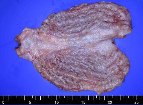
A B
C D
Fig. 1. Endoscopy and serum assay findings of the giant rugae in a 62-year-old man with an Helicobacter pylori infection. (A) Initial endoscopy of the gastric body:
the mucosal surfaces of the gastric rugae showed diffuse redness and poor distention upon air inflation. Biopsies of the gastric rugae revealed an active H. pylori
infection without malignant cells. The serum pepsinogen (PG) I (91.6 ng/mL) and PG II (15.5 ng/mL) levels were elevated, and the PG Ⅰ/Ⅱ ratio was 5.9 on the
day of endoscopy. The serum anti-H. pylori IgG assay revealed seropositivity (61.4 AU/mL) using the Chorus H. pylori IgG assay (DIESSE, Siena, Italy), which has a
sensitivity of 100% and a specificity of 75.4% in Korean.10 (B) Follow-up endoscopy after successful H. pylori eradication was confirmed by the negative urea breath
test: poor distention was still noticed owing to the hard and thick rugae. Compared with the findings before the eradication, a progression to tortuosity was found in the
giant rugae with the regression of diffuse redness. There were no malignant cells or H. pylori in the biopsied specimens. Nevertheless, the serum PG I (100.2 ng/mL)
and PG II (16.8 ng/mL) levels were higher than those before the eradication. The PG Ⅰ/Ⅱ ratio was 6.0, and the serum anti-H. pylori IgG titer was 46.0 AU/mL. (C)
Computed tomography of the abdomen: imaging was performed because of the persistent giant rugae and increased serum PG II levels after H. pylori eradication. A
diffusely enhanced wall thickening of the entire stomach, suggesting Borrmann type IV gastric cancer, was found on a coronal section. (D) Gross image of the resect-
ed stomach: after total gastrectomy, a 19.0×18.0×1.0 cm, diffuse-type, poorly cohesive carcinoma was diagnosed. Cancer cells were invading the visceral peritoneum
(pT4a). Metastases were found in 17 of the 30 regional lymph nodes (pN3b). Resection margins were free of carcinoma.
2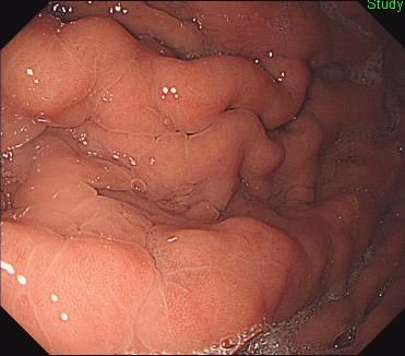
Lee SY. Giant Rugae

Table 1. Endoscopic Findings of Giant Gastric Rugae according to Its Etiology
                                                                                                          Surface of gas-
 Etiology                                    Location              Primary lesion       Consistency                                 Coexisting diseases
                                                                                                           tric mucosa
                                                                                                               Diffuse
                                      Corpus-dominant >                                                     redness and/
 Borrmann type IV gastric                                        Submucosa and/or Hard (poor air
                                      antrum-dominant >                                                      or irregular       Helicobacter pylori infection
  cancer (linitis plastica)                                        proper muscle    inflation)
                                        junctional type                                                     erosions/ul-
                                                                                                              cerations
                                                                                                               Diffuse
                                                                                                            redness and/
                                    All (multifocal, localized     Mucosa and/or      Soft (preserved
 Large B cell lymphoma                                                                                       or irregular       Helicobacter pylori infection
                                         > diffuse type)            submucosa           distention)
                                                                                                            erosions/ul-
                                                                                                              cerations
                                                                                                                   Anemia, edema due to hypoal-
 Menetrier disease (hyper-
                                                                  Mucosa (lamina      Soft (preserved Whitish exu- buminemia, hypergastrinemia,
 trophic protein-losing                    Corpus only
                                                                    propria)a)          distention)      date       low acid secretion owing to
 gastropathy)
                                                                                                                       parietal cell loss, etc.
                                                                                                                               Colon tumors, alopecia, nail
 Cronkhite-Canada syn-                                                                Soft (preserved      Innumerable        atrophy, loss of taste, diarrhea
                                        Corpus > antrum                Mucosa
 drome                                                                                  distention)        hamartomas         due to protein-losing enterop-
                                                                                                                                         athy, etc.
                                                                                                                                Severe inflammation in the
 Secondary inflammatory                                          Outside the gastric Soft (preserved        Edematous
                                                 All                                                                          abdomen (i.e., acute pancreati-
  change                                                               wall            distention)           change
                                                                                                                                            tis)
a)
 In Menetrier disease, foveolar hyperplasia, glandular dilation, eosinophil and/or plasma cell infiltration, and smooth muscle hyperplasia
are often found in the lamina propria.

Conflicts of Interest                                                                   2014;2014:481607.
     The author has no potential conflicts of interest.                             4. Kim JH, Lee SY, Lee SP, et al. The histologic detection of Helicobacter
                                                                                        pylori in seropositive subjects is affected by pathology and secretory
                                                                                        ability of the stomach. Helicobacter 2018;23:e12480.
Funding                                                                             5. Tu H, Sun L, Dong X, et al. Serum anti-Helicobacter pylori immuno-
  This work was supported by the Korean National Research Foundation                    globulin G titer correlates with grade of histological gastritis, mucosal
2016R1D1A1B02008937.                                                                    bacterial density, and levels of serum biomarkers. Scand J Gastroenterol
                                                                                        2014;49:259-266.
ORCID                                                                               6. Baek SM, Kim N, Kwon YJ, et al. Role of serum pepsinogen II and Heli-
                                                                                        cobacter pylori status in the detection of diffuse-type early gastric cancer
     Sun-Young Lee:                      https://orcid.org/0000-0003-4146-6686
                                                                                        in young individuals in South Korea. Gut Liver 2020;14:439-449.
                                                                                    7. Cha JH, Jang JS. Clinical correlation between serum pepsinogen
                                                                                        level and gastric atrophy in gastric neoplasm. Korean J Intern Med
REFERENCES                                                                              2020;35:550-558.
                                                                                    8. Ichinose M, Watanabe M, Kato J. Re: Development of diffuse carcinoma
                                                                                        in the gastric corpus in patients with rugal hyperplastic gastritis. Int J
1. Lee SY. [Helicobacter pylori infection and the Kyoto classification of gas-          Cancer 2013;133:2259.
   tritis]. Korean J Helicobacter Up Gastrointest Res 2019;19:81-87.                9. Iijima K, Koike T, Abe Y, Shimosegawa T. Cutoff serum pepsinogen
2. Kim BC, Song MA, Kwon SH. Endoscopic characteristics of rugal                        values for predicting gastric acid secretion status. Tohoku J Exp Med
   hyperplasia and related acid condition in Helicobacter pylori-infected               2014;232:293-300.
   stomach. Clin Endosc 2021;54:73-84.                                              10. Lee SY, Moon HW, Hur M, Yun YM. Validation of western Helicobacter
3. Massarrat S, Haj-Sheykholeslami A, Mohamadkhani A, et al. Pepsino-                   pylori IgG antibody assays in Korean adults. J Med Microbiol 2015;64(Pt
   gen II can be a potential surrogate marker of morphological changes                  5):513-518.
   in corpus before and after H. pylori eradication. Biomed Res Int

                                                                                                                                                                  3
You can also read