The concordance between endoscopic and histological diagnosis in 114 dogs affected by gastric disease - IZS
←
→
Page content transcription
If your browser does not render page correctly, please read the page content below
The concordance between endoscopic and histological
      diagnosis in 114 dogs affected by gastric disease
                                          Maria Chiara Marchesi*, Cecilia Carlotta Timpano, Sara Busechian,
                                                                      Camillo Pieramati and Fabrizio Rueca
                                      Dipartimento di Medicina Veterinaria, Università degli Studi di Perugia, Perugia, Italy.
        *
            Corresponding author at: Dipartimento di Medicina Veterinaria, Università degli Studi di Perugia, Via San Costanzo 4, Perugia (Italy).
                                                                               Tel.: +39 075 5857604, e-mail: mariachiara.marchesi@unipg.it.

                                                           Veterinaria Italiana 2017, 53 (4), 309-313. doi: 10.12834/VetIt.903.4608.1
                                                                                         Accepted: 04.11.2016 | Available on line: 29.12.2017

Keywords                           Summary
Dog,                               Endoscopy is a common, minimally invasive diagnostic technique that can be used to observe
Gastropathy,                       internal organs, e.g. the stomach, and to obtain mucosal bioptic samples for histo pathological
Gastroscopy,                       examination. The aim of this study was to analyse the concordance between endoscopic
Histological exam.                 and histological evaluation of gastric diseases in dogs. One hundred twenty‑nine medical
                                   records of dogs undergoing gastroscopy have been received and stored by the Veterinary
                                   Hospital of Perugia University (Perugia, Italy) between 2009‑2012. The concordance between
                                   endoscopic and histological reports of acute and chronic gastritis or gastric tumours was
                                   assessed by Cohen’s k coefficient. Considering histological diagnosis as the “gold standard”,
                                   sensitivity, specificity, positive predictive value (PPV), and negative predictive value (NPV) of
                                   the endoscopic report have been calculated. Frequencies of gastritis types differed between
                                   macroscopic and microscopic analyses. The evaluation of histological and endoscopic
                                   agreement was fair (0.35). Endoscopy showed sensitivity of 45%, 88%, and 100% for acute
                                   gastritis, chronic gastritis, and gastric tumours, respectively; and specificity of 84%, 71%, and
                                   100%. The positive predictive value and NPV resulted to be 25% and 93% for acute gastritis,
                                   93% and 60 % for chronic gastritis, 100% and 100% for gastric tumours. The results of this
                                   study show that gastric endoscopy cannot be performed as a screening exam, and that to
                                   optimise diagnosis both endoscopic and histological exam should be conducted.

                                           Concordanza tra la diagnosi endoscopica e istologica
                                                     in 114 cani affetti da patologie gastriche
Parole chiave                      Riassunto
Cane,                              La gastroscopia è una tecnica diagnostica mini‑invasiva che consente di visualizzare il lume
Esame istologico,                  gastrico e prelevare frammenti bioptici di mucosa necessari per le indagini istopatologiche.
Gastropatia,                       Obiettivo del presente studio è stato di analizzare la concordanza tra valutazione endoscopica
Gastroscopia.                      ed istologica di patologie gastriche nel cane. Presso l'Ospedale Universitario Didattico di
                                   Perugia sono state esaminate 129 cartelle cliniche di cani sottoposti a gastroscopia tra il
                                   2009‑2012. I dati ottenuti sono stati analizzati mediante il coefficiente K di Cohen ed è stata
                                   valutata sensibilità, specificità, valore prognostico positivo (PPV) e negativo (NPV) dell’indagine
                                   endoscopica. L'età ha mostrato un effetto significativo riguardo la predisposizione a tumori
                                   gastrici e gastropatia nodulare (P < 0.001). La frequenza dei tipi di gastrite è risultata diversa tra
                                   le interpretazioni macroscopiche e microscopiche. La concordanza tra valutazione istologica
                                   ed endoscopica è stata scarsa (0.35). Usando i tre reperti istologici (gastrite acuta, cronica e
                                   tumori gastrici) come “gold standard”, l'esame endoscopico ha mostrato una sensibilità del
                                   43%, 88% e 100% e una specificità del 84%, 71%, 100% per la gastrite acuta, cronica e tumori
                                   gastrici. PPV e NPV dell'endoscopia sono risultati 25% e 93% per la gastrite acuta, 93% e 60%
                                   per la cronica e 100% per i tumori gastrici. In base ai risultati, la gastroscopia non può essere
                                   effettuata come esame di screening. Considerando le limitazioni legate al campionamento
                                   bioptico e la soggettività nella valutazione endoscopica della mucosa, si raccomanda di
                                   effettuare sia l'esame endoscopico che quello istopatologico.

                                                                                                                                                     309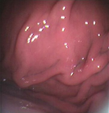
Endoscopic and histological diagnosis in dogs with gastric disease                                                                                            Marchesi et al.

        Introduction                                                              Materials and methods
        Gastric diseases are frequent in dogs (Hall 2000).                        We reviewed 129 medical records of dogs that
        Endoscopy is a common, minimal invasive                                   underwent gastroscopy at the Veterinary Hospital
        diagnostic technique used to evaluate the stomach                         of Perugia University (Perugia, Italy) because of
        and to obtain mucosal bioptic samples from 1 or                           vomiting, lack of appetite, and weight loss, during
        more gastric sites (Mansell and Willard 2003, Evans                       the years 2009‑2012. Cases have been excluded
        et al. 2006). As a result, nowadays the diagnosis                         if no gastric biopsies were obtained or if a mass
        and following treatment of gastric diseases in dogs                       was found inside the stomach. Information gained
        is frequently based on gastroscopy; it also allows                        from medical records consisted of signalment,
        for the collection of mucosal bioptic samples. The                        gastroscopy results, and histological diagnosis.
        histopathological interpretation of these samples                         Gastroscopy was performed on all patients after
        has proved to be the most contentious and                                 general anaesthesia (Medetomidine, Ketamine,
        frustrating step during the diagnostic process for                        and Diazepam) with a 6 mm ø × 1100 mm flexible
        both clinicians and pathologists (Day et al. 2008),                       endoscope (Pentax EG‑1840; Pentax, Japan)
        as it often happens that the number of samples or                         equipped with an instrumental portal to insert
        their quality is inadequate for a safe histological                       biopsy forceps. The animal was positioned in
        evaluation. Different studies have shown (Mansell                         left lateral recumbence and gastroscopy was
        and Willard 2003, Washabau et al. 2010) how quality                       performed according to the technique described
        of tissue samples obtained through a flexible                             by Guilford (Guilford 2005). Three biopsies of each
        endoscope plays a great role in determining                               gastric region were obtained, using 2.4 mm biopsy
        biopsy diagnostic sensitivity. Another important                          forceps. The same, highly experienced endoscopist
        consideration is that often there is no association                       performed all endoscopic investigations. Findings
        among clinical data, endoscopic findings, and                             were first classified according to macroscopic
        histopathological results (Gad 1986, Lidbury at                           view in acute gastritis (Figure 1), chronic gastritis
        al. 2009). To the best of our knowledge, no other                         (Figure 2), or nodular gastropathy (Figure 3).
        study has investigated the concordance between                            Specifically, acute gastritis was diagnosed if
        endoscopic and histopathological findings in a                            mucosa was hyperaemic and had a prominent
        group of dogs with symptoms of gastric diseases.                          light reflectivity, thus suggesting oedema. While
        In order to evaluate the degree of concordance                            a thickened mucosa and a grainy texture were
        between these 2 sets of data, we retrospectively                          indicative of chronic gastritis; moreover, in this
        reviewed the records of 129 dogs undergoing                               condition mucosal folds usually were hypertrophic
        gastroscopy for gastric symptoms.                                         or reduced in number and size. Finally, nodular

        Figure 1. Endoscopy finding of acute gastritis in dogs undergoing         Figure 2. Endoscopy finding of chronic gastritis in dogs undergoing
        gastric endoscopy at Veterinary Teaching Hospital of Perugia University   gastric endoscopy at Veterinary Teaching Hospital of Perugia University
        (Perugia, Italy) between 2009-2012. The mucosa was hyperaemic and         (Perugia, Italy) between 2009-2012. The mucosa was thickened and
        had a prominent light reflectivity, thus suggesting oedema.               has a grainy texture.

310                                                                                                     Veterinaria Italiana 2017, 53 (4), 309-313. doi: 10.12834/VetIt.903.4608.1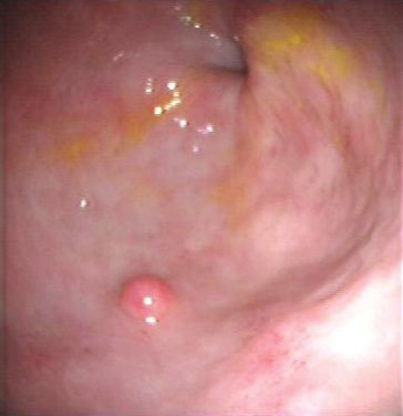
Marchesi et al.                                                                                Endoscopic and histological diagnosis in dogs with gastric disease

                                                                                       The agreement between endoscopic and
                                                                                       histological reports of acute and chronic gastritis
                                                                                       or gastric tumours was assessed by Cohen’s k
                                                                                       coefficient. Considering histological diagnosis as the
                                                                                       “gold standard”, we calculated sensitivity, specificity,
                                                                                       positive predictive value (PPV), and negative
                                                                                       predictive value (NPV) of the endoscopic report.
                                                                                       Statistical analysis was performed using “R” software
                                                                                       (R Core Team 2013).

                                                                                       Results
                                                                                       We examined 129 medical records of dogs referred
                                                                                       for gastric endoscopy because of vomiting, lack of
                                                                                       appetite, and weight loss. Among those animals,
                                                                                       15 dogs were excluded because no gastric biopsies
                                                                                       were collected, or because a mass was found in
                                                                                       the stomach: in the end, 114 dogs were included
           Figure 3. Endoscopy finding of nodular gastropathy in dogs                  in this study.
           undergoing gastric endoscopy at Veterinary Teaching Hospital of             No significant effect of sex was found on endoscopic
           Perugia University (Perugia, Italy) between 2009-2012. The mucosa           or histologic diagnosis, while age showed a
           presented nodules.
                                                                                       significant effect on liability to gastric tumours and
                                                                                       nodular gastropathy (P < 0.001).
           gastropathy was diagnosed if mucosa presented                               Frequencies of gastritis types were different
           disseminated nodules.                                                       between macroscopic and microscopic views: at
                                                                                       endoscopical examination there were 23 diagnoses
           Biopsy specimens were fixed in 10% buffered                                 of acute gastritis (20.1%), 86 of chronic gastritis
           formalin, embedded in paraffin, sectioned at 4 μm                           (75.4%), and 5 of nodular gastropathy (4.5%).
           and stained with Haematoxylin and Eosin. The same                           Frequencies of histological diagnoses were as
           pathologists reviewed all slides, and cases were                            follows: acute inflammation in 11 cases (9.6%),
           classified according to histological presentation in                        chronic inflammation in 91 cases (79.8%), and
           acute gastritis, chronic gastritis, or gastric tumors.                      gastric neoplasia in 5 cases (4.5%). No lesions were
                                                                                       detected in the 7 remaining cases (6.1%). The reports
                                                                                       are summarized in Table I.
           Statistical analysis                                                        Twenty‑eight cases (24.6%) were misdiagnosed
           The role of sex in influencing frequency of                                 at endoscopy. Among the 7 cases with negative
           endoscopical and histological reports has been                              histological report, 3 were diagnosed as acute
           tested using the Fisher’s “exact” test; while the effect                    gastritis during gastroscopy, and the remaining
           of age has been tested by means of a logistic model.                        4 as chronic gastritis. Fifteen cases diagnosed as
                                                                                       acute gastritis during gastroscopy resulted to be
                                                                                       chronic at histological exam, the opposite situation
           Table I. Dogs undergoing gastric endoscopy at Veterinary Teaching           occurred in 6 dogs. The evaluation of histological and
           Hospital of Perugia University (Perugia, Italy) between 2009-2012:          endoscopic agreement by Cohen’s k coefficient (and
           concordance between macroscopical diagnosis (rows) and microscopic          its Confidence Interval, CI) indicated a value of 0.35
           report (columns).                                                           (CI 95%: 0.14‑0.56). If the 7 cases with no histological
                                                       Histology
                                          Acute Chronic Gastric             Total
                                         gastritis gastritis tumor Negative            Table II. Specificity and sensitivity of the endoscopic exams using
                                                                                       the histological findings as the “gold standard” for dogs undergoing
                             Acute          5           15            0      3   23    gastric endoscopy at Veterinary Teaching Hospital of Perugia University
                            gastritis
                                                                                       (Perugia, Italy) between 2009-2012.
             Gastroscopy

                            Chronic         6           76            0      4   86
                            gastritis                                                                                 Specificity            Sensitivity
                             Nodular                                                        Acute gastritis              84%                    45%
                           gastropathy      0            0            5      0    5
                                                                                           Chronic gastritis             71%                    88%
                             Total         11           91            5      7   114        Gastric tumors              100%                   100%

Veterinaria Italiana 2017, 53 (4), 309-313. doi: 10.12834/VetIt.903.4608.1
                                                                                                                                                                 311
Endoscopic and histological diagnosis in dogs with gastric disease Marchesi et al.
lesions were excluded, Cohen’s k coefficient would that, in order to achieve a definitive diagnosis of
have slightly risen to 0.40 (CI 95%: 0.17‑0.63). Using gastric lesions, it is always necessary to perform a
the 3 histological findings (acute gastritis, chronic histological exam on biopsy specimens obtained
gastritis, and gastric tumors) as “gold standard”, by endoscopy. Moreover, the rate of agreement
the endoscopic exam showed a sensitivity of 45%, between these 2 exams may improve if the number
88%, and 100% for acute gastritis, chronic gastritis, of biopsy samples is increased, as pointed out by
and gastric tumours respectively and a specificity of Willard and colleagues (Washabau et al. 2010).
84%, 71%, and 100% (Table II). Using the 3 histological findings (acute gastritis,
Finally, the positive predictive value (PPV) and chronic gastritis, and gastric tumors) as “gold
negative predictive value (NPV) of endoscopy standard”, gastroscopy reached a good negative
resulted to be 25% and 93% for acute gastritis, 93% predictive value (NPV) for acute gastritis and a
and 60 % for chronic gastritis, and 100% and 100% good positive predictive value (PPV) for chronic
for gastric tumours. gastritis. Endoscopy showed a perfect agreement
with histological findings in the diagnosis of
stomach tumours. However, it must be noticed that
Discussion tumours had a limited prevalence in our review
Application of the endoscopic technique was and that histological evaluation is mandatory for
possible in all dogs with gastric symptoms, and it this kind of lesions because of their severity. The
has always allowed for a good visualization of the PPV of endoscopic exam was only 25% in dogs
4 gastric regions. In our study, chronic gastritis was affected by acute gastritis. This could be due to
the most common finding by both endoscopic low ability of the operator to differentiate between
(75.4%) and histological exams (79.8%). As reported a normal mucosa and a fairly inflamed one, as
by other authors (Lidbury et al. 2009, Washabau et al. well as to the high prevalence of acute gastritis.
2010), diagnosis of acute gastritis is rare because it A similar misinterpretation could happen also in
is often a self‑limiting condition or it responds to diagnosing chronic gastritis, due to the difficulty
palliative treatment, therefore biopsy specimens of recognizing a variation of mucosae granularity.
are seldom obtained. In our study, 28 cases Furthermore, assessing the mucosa granularity
were misdiagnosed during endoscopy. We then variations can be another challenge for the
calculated the Cohen’s k coefficient to assess the operator. The subjective aspects in defining the
agreement between endoscopic and histological mucosal texture, colour, and contour described
findings obtaining a k value of 0.35, that indicates above have also been reported by Gurjeet and
a “fair agreement”. The difference in diagnosis Mahendra (Gurjeet and Mahendra 2002).
could be related to the difficulty of revealing some From our results, it can be concluded that gastric
macroscopic lesions by endoscopic exam (Lidbury endoscopy cannot be performed as a screening exam
et al. 2009) or to histological misinterpretations in veterinary medicine. Nonetheless, this technique
due to the inadequate number or quality of biopsy is useful in diagnosing the most common gastric
samples. Different studies have provided ample diseases of the dog. Even if macroscopic evaluation
information about these possibilities (Mansell and of the mucosa during gastroscopy showed a good
Willard 2003, Day et al. 2008, Washabau et al. 2010). ability in diagnosing chronic gastritis and gastric
The actual number of endoscopic specimens could tumours, we do recommend performing biopsies.
also influence the fairness of the concordance, In conclusion, after analysing the concordance
because it is common to obtain inadequate samples between endoscopic and histological diagnosis in
during an endoscopic exam (Washabau et al. 2010). dogs with gastric disease, specifically considering the
However, excluding the 7 cases of macroscopic limitations related to biopsy sampling (Washabau
lesions not histologically confirmed, the Cohen’s k et al. 2010) and subjectivity in endoscopic evaluation
coefficient raises only to 0.40. of the mucosa, it is recommended to conduct both
The Cohen’s k values obtained in this study suggest the endoscopic and histological exam.
312 Veterinaria Italiana 2017, 53 (4), 309-313. doi: 10.12834/VetIt.903.4608.1Marchesi et al. Endoscopic and histological diagnosis in dogs with gastric disease
References
Day M.J., Bilzer T., Mansall J., Wilcock B., Hall E.J., Jergens prevalence of Helicobacter pylori infection. Singapore
A., Minami T., Willard M. & Washabau R. 2008. Med J, 43 (2), 090‑092.
Histopathological standards for the diagnosis of Hall J.A. 2000. Diseases of the stomach. In Textbook of
gastrointestinal inflammation in endoscopic biopsy veterinary internal medicine, 5th ed. Vol. 2. (Ettinger
samples from the dog and cat: a report from the World S.J. and Feldman EC, eds) Philadelphia, WB Saunders,
Small Animal Veterinary Association Gastrointestinal 1162‑1163.
Standardization Group. J Comp Path, 138, S1‑S43.
Lidbury J.A., Suchodolski J.S. & Steiner J.M. 2009.
Evans S.E., Bonczynski J.J., Broussard J.D., Han E. & Baer A retrospective study of 67 dogs with gastric
K.E. 2006. Comparison of endoscopic and full‑thickness histopathological abnormalities (2002‑2007). J Am Vet
biopsy specimens for diagnosis of inflammatory bowel Med Ass, 234 (9), 1147‑1153.
disease and alimentary tract lymphoma in cats. J Am
Mansell J. & Willard M.D. 2003. Biopsy of the gastrointestinal
Vet Med Ass, 229, 1447‑1450.
tract. Vet Clin Small Anim, 33, 1099‑1116.
Gad A. 1986. Erosion: a correlative endoscopic histopathologic
R Core Team, 2013. R: a language and environment for
multicenter study. Endoscopy, 18 (3), 76‑79.
statistical computing. R Foundation for Statistical
Guilford W.G. 2005. Gastrointestinal endoscopy. In Computing, Vienna, Austria. http://www.R‑project.org/.
Veterinary endoscopy for the small animal practitioner
Washabau R.J., Day M.J., Willard M.D., Hall E.J., Jergens
(T.C. McCarthy, ed ), Elsevier, USA.
A.E., Mansell J., Minami T. & Blizer T.W. 2010.
Gurjeet K. & Mahendra Raj S. 2002. A study of the Endoscopic, biopsy, and histopathologic guidelines
concordance between endoscopic gastritis and for the evaluation of gastrointestinal inflammation in
histological gastritis in an area with a low background companion animals. J Vet Intern Med, 24, 10‑26.
Veterinaria Italiana 2017, 53 (4), 309-313. doi: 10.12834/VetIt.903.4608.1
313You can also read