We are IntechOpen, the world's leading publisher of Open Access books Built by scientists, for scientists
←
→
Page content transcription
If your browser does not render page correctly, please read the page content below
We are IntechOpen,
  the world’s leading publisher of
         Open Access books
  Built by scientists, for scientists

 4,200
Open access books available
                                        116,000
                                       International authors and editors
                                                                                        125M
                                                                                         Downloads

                                           Our authors are among the

      154
  Countries delivered to
                                         TOP 1%
                                             most cited scientists
                                                                                   12.2%
                                                                            Contributors from top 500 universities

                            Selection of our books indexed in the Book Citation Index
                                   in Web of Science™ Core Collection (BKCI)

               Interested in publishing with us?
          Contact book.department@intechopen.com
                           Numbers displayed above are based on latest data collected.
                                For more information visit www.intechopen.com
Chapter

Helicobacter pylori: A Pathogen of
Ample Risk to Health
Isidro Favian Bayas-Morejón, Rosa Angélica Tigre-León,
Edison Riveliño Ramón-Curay
and Darwin Alberto Núñez-Torres

Abstract

   Helicobacter pylori is considered a pathogen of global interest because it is a
microorganism of very easy contagion between the hosts or host. Helicobacter pylori
infection is now recognized as a problem that causes chronic gastritis, peptic ulcer
disease, and lymphoproliferative disorders and is a major risk factor for gastric
cancer. The diagnostic methods to detect H. pylori are classified such as direct or
invasive, when the identification is directly, the bacterium obtained from gastric
mucosa biopsy by endoscopy histology with various staining, culture and PCR
techniques, while indirect or noninvasive or serological tests such as the breath test
with urea marked with 13C.

Keywords: H. pylori, pathogen, risk to human

1. Introduction

    Helicobacter pylori is considered a pathogen of global interest because it is a
microorganism of very easy contagion between hosts or susceptible hosts. The first
isolation of H. pylori was in 1982 by Marshall and Warren who ushered us into a
new era of gastric microbiology [1].
    The number of infected by H. pylori has been increased considerably, since
a third of the world population has it, while the rest do not know if they have it
or not; in developing countries there is an infection rate that goes from 60% and
90% of the population, which is not the case in developed countries ranging from
20–40% [2]. Gastroduodenal ulcer diseases are a major factor in the development of
gastric adenocarcinoma and lymphoma [3].
    According to studies conducted, many of the pathogenic species of Helicobacter are
of fecal origin. The transmission to human seems to be associated with the consump-
tion of water and raw or undercooked foods [4, 5]. In Ecuador, according to the statis-
tics, the poverty quintiles reached up to 2015 are 35% according to INEC data, which are
closely related to the lack of basic services such as drinking water and sanitary services,
a common factor in the population being contamination by water and food [6].
    The different methods used for diagnosis range from antigenic screening (Ag)
to molecular techniques. Antigenic screening techniques have been associated with
a high sensitivity of a detection limit of the Helicobacter pylori test with a 95% con-
cordance in specificity compared to the ELISA test [7]. In plate culture it is usually

1
Gastritis - New Approaches and Treatments
considered a difficult and tedious technique; the diagnostic method has the advantage
of typifying the organism and determining its sensitivity to antibacterial agents. The
methods such as endoscopy to obtain a sample through a biopsy are very used nowa-
days; it is a traumatic and invasive procedure that can cause complications such as
infections, perforations, aspiration, bleeding, and incarceration of the endoscope [8].
2. Theoretical framework
2.1 Helicobacter pylori
Helicobacter pylori (H. pylori) is a spiral bacterium that does not form spores and
is Gram-negative, which colonizes the human stomach and is prevalent throughout
the world [9]. It has been associated with peptic ulcer disease, gastric adenocar-
cinoma, and lower grade B-associated lymphoma associated with the mucosa. In
addition, it is thought that the organism is involved in other human diseases such
as hematological and autoimmune disorders, insulin resistance, and metabolic
syndrome [10]. Although almost 50% of the population is infected with H. pylori
worldwide, the prevalence, incidence, age distribution, and sequelae of infection
are significantly different in developed and developing countries.
Helicobacter pylori (previously known as Campylobacter pylori or pyloridis) was
first isolated from humans in 1982 [11]. Since 1994, H. pylori has been considered
carcinogenic to humans, and it has even been associated with other diseases, such as
cerebrovascular accidents, autoimmune thyroiditis, and diabetes mellitus, among
others [12]. This bacterium resides in the stomach of most humans and is usually
found in the deeper portions of the mucus gel that lines the gastric mucosa or
between the mucus layer and the gastric epithelium [13].
The bacterium is one of the most important findings for gastroenterologists,
who for years sought answers to multiple intestinal problems. This is how the
gastroenterologist Walery Jaworski in 1899 after analyzing samples of human
gastric expirations isolated spiral elongated bacteria and called them Vibrio regula,
and the said results were published in the manual of gastric diseases; however, these
findings were not given the importance they deserved to be written in Polish and
not in English [14].
So, it took 79 years for the bacteria to be rediscovered by the Australian doctors
Barry Marshall and Robin Warren, who managed to make the first isolation through a
pure culture in 1979. This rediscovery allowed them to be Nobel Laureates in 2005 [15].
2.2 Microbiological aspects of Helicobacter pylori
Taxonomically, we can describe H. pylori because of its size, shape, color, bio-
chemical function, genus, species, and its relationship with other species. H. pylori
is a slow-growing, spiral-shaped bacterium. It is a small curved bacillus, microaero-
philic, and Gram-negative, and mobile by the presence of flagella. The bacillus has
rounded ends. These microorganisms measure 0.5–1.0 μm wide by 2.5–4.0 μm long,
since they bear a strong resemblance to members of the Campylobacter genus [11].
The multiple genotypic and phenotypic characteristics are different from those
of Helicobacter, so this new genus was established, including H. cinaedi and H.
fennelliae. The two species of Helicobacter that cause diarrheal disease, H. cinaedi
and H. fennelliae, are intestinal microorganisms rather than gastric. As for the
clinical manifestations of the disease they generate, these bacteria are more similar
to Campylobacter than H. pylori [13]. The clinical characteristics of the infections
caused by these Helicobacter are similar to those due to Campylobacter species.
2Helicobacter pylori: A Pathogen of Ample Risk to Health
DOI: http://dx.doi.org/10.5772/intechopen.86789
2.3 Pathways of contagion or infestation of the guests
Although in general there is no difference between the sexes, in some developed
countries, there is a higher prevalence of infection in men than in women [16].
The prevalence of H. pylori infection in adults of any age in developed countries
ranges between 20 and 40% and reaches figures of 60 to 80% in countries consid-
ered third world. The most important difference between countries of high and low
prevalence is the intensity with which the infection is transmitted in childhood and
early adolescence [17].
Epidemiological and microbiological evidences have several transmission routes
that have been proposed in the studies carried out. The gastro-oral, oral-oral, and
fecal-oral routes are the most important routes of transmission [12]. Other routes
of importance are also breastfeeding and iatrogenic transmission which are also
included as alternating forms for the transmission of the pathogen. The possibilities
of spreading the pathogen are of three possible vectors that have been suggested to
maintain the viable form of the bacteria: water, food, and animals.
2.4 Water transmission
The prevalence of H. pylori infection shows a strong correlation with access to water.
Numerous epidemiological studies confirm this, and the World Health Organization
includes it in its list of potential emerging pathogenic microorganisms whose transmis-
sion by water is plausible, although it has not yet been confirmed [18, 19].
Through molecular methods, H. pylori DNA has been detected in wastewater,
drinking water, and other environmental samples throughout the world, and its
survival capacity in water, even chlorinated, has been demonstrated. It has also
been detected in the drinking water distribution network [20]. These findings indi-
cate that contaminated water and food play a vital role in the survival and spread of
H. pylori.
In another study, developed by Moreno et al. [21]; Moreno and Ferrús, [22] H.
pylori was detected in 46% of more than 100 wastewater samples, 40% were of
river water samples and, most strikingly, the 66% were public source.
On the other hand, H. pylori is able to survive in biofilms when it grows under
high C:N conditions [23]. The biofilms formed protect microorganisms from the
action of adverse agents, increase the availability of nutrients for their growth,
and also increase the frequency of transfer of genetic material [24]. Gião et al. [25]
observed that H. pylori formed biofilms after 24 hours of being in an unfavorable
environment. The association of H. pylori with biofilm communities within a water
distribution system could offer the bacterium protection against disinfection and
predation by protozoa, and there are studies that demonstrate the survival of H.
pylori within amoebae of free life[26, 27].
2.5 Foods transmission
Those foods that have a water activity (aw) >0.97 and a pH between 4.9 and 6.0
theoretically provide the ideal conditions for the survival and development of H.
pylori [28, 29].
Vegetables are one of the foods with the highest risk of fecal contamination,
since they are in contact with soil and contaminated irrigation water, which would
mean the spread of H. pylori in the environment and its transmission to humans.
Atapoor et al. [30] and Yahaghi et al. [31] in Iran managed to detect and isolate H.
pylori in percentages higher than 10%, in vegetable samples. Also, Bayas et al. [32]
have detected the pathogen in vegetables by molecular methods.
3Gastritis - New Approaches and Treatments
On the other hand, the ability of H. pylori to survive on lettuce leaves forming
biofilms has been demonstrated [33].
Milk could also act as a vehicle for H. pylori. Several studies have shown that the
bacterium is able to survive in inoculated milk stored in refrigeration for more than
6 days or for 3 days at room temperature [34]. In addition, in an investigation devel-
oped by Fujimura et al. [35], the presence of the H. pylori ureA gene was detected in 13
of 18 samples of raw milk (72.2%) and in 11 of 20 samples of pasteurized milk (55%).
On the other hand, Meng et al. [36] analyzed 11 raw chickens and 18 samples
of tuna meat ready for consumption (sushi). H. pylori was detected by multiple
polymerase chain reaction (m-PCR) in 36% (4/11) of the chickens and 44% (8/18)
of the tuna samples.
Studies have also been conducted on the presence of H. pylori in shellfish.
Fernández et al. [37] detected H. pylori DNA in seawater, plankton, and oysters
from three different regions of Venezuela. They concluded that mollusks could act
as vehicles for H. pylori transmission.
2.6 Detection in human samples
The presence of H. Pylori was focused on a study developed by Samie [38], on
the prevalence of Campylobacter, Helicobacter, and Arcobacter. By molecular meth-
ods, in 322 stool samples from HIV-positive and non-HIV-infected patients in South
Africa, they found that A. butzleri was the third most frequent species (6.2%), after
Helicobacter pylori (50.6%) and Campylobacter jejuni (10.2%).
2.7 Most common pathologies
2.7.1 Gastritis
The term gastritis should be reserved for the histologically demonstrated
inflammation of the gastric mucosa. Gastritis is not the mucosal erythema seen
during endoscopy, nor is it interchangeable with the term “dyspepsia” [13]. On the
other hand, the different etiological factors that cause gastritis are multiple and
heterogeneous; to gastritis it has been classified with a chronological base (acute or
chronic), such as histological typologies, anatomical distribution or its pathogenic
mechanism, clinical correlation, histological data, abdominal pain or dyspepsia,
and endoscopic data in gastric mucosal investigation [13].
The pathogenesis of chronic gastritis by Helicobacter pylori includes two stages:
the first is characterized by the arrival and penetration of the microorganism
into the gastric mucus where it sits and multiplies. In the second stage, there is an
amplification of the inflammatory response, by the interaction of lymphocytes,
neutrophils, macrophages, mastoid cells, and nonimmune cells that, when attracted
to the site of the lesion, release a wide variety of chemical mediators such as cyto-
kines, eicosanoids, reactive oxygen metabolites (oxygen free radicals), and the
complement system, which perpetuate inflammation [39, 40] (Figure 1).
2.7.2 Stomach cancer
It is the uncontrolled growth of stomach cells. Malignant tumors can originate
in each of the three layers: mucosa, muscle, and serosa. This is also known as gastric
cancer that originates in the stomach [41]. The risk factor is considered any caused that
increases the likelihood of having a disease such as cancer, even though several risk com-
ponents do not mean that the person will have the disease; Some scientists connoted that
the risks that take a person to be more prone to suffer stomach cancer are several such as:
4Helicobacter pylori: A Pathogen of Ample Risk to Health
DOI: http://dx.doi.org/10.5772/intechopen.86789
Figure 1.
Second stage of the inflammatory process of the gastric mucosa by H. pylori. Grávalos and González [40].
Incidence according to sex: Stomach cancer is more common in men than in
women [16].
Age: The rate of stomach cancer in people over 50 years increases sharply [42].
Ethnic origin: In the United States, stomach cancer is more common among
Americans of Hispanic origin, black people, and Asians and islanders compared to
white people who are not of Hispanic origin [41].
Geography: On a global scale, stomach cancer is more common in Japan, China,
Eastern and Southern Europe, as well as Central and South America [41].
2.7.3 Risk factors
Several risk factors for gastric cancer have been described, which play a funda-
mental role in their genesis, some of them remain under discussion, and others, on
the contrary, have been confirmed more and more clearly [43].
2.7.4 Genetic
Within the genetic risk factors [41], we have:
• Families of patients with gastric cancer: incidence 2–3 times higher
• Blood group A.
2.7.5 Environmental
Among the environmental risk factors [41], we have:
• Food (variable in each country): dried and salted fish, very spicy foods, and red
meats, among others
• Ingestion of alcohol, hot drinks, and sodium nitrate; chewed tobacco
• Radiation.
5Gastritis - New Approaches and Treatments
2.7.6 Premalignant
Within the premalignant risk factors [41], we have:
• Atrophic gastritis, intestinal metaplasm, and dysplasia.
• Pernicious anemia (20 times more frequent than in normal subjects).
• Gastric polyps: multiple hyperplasia, greater than 2 cm with some degree of
dysplasia 0.4–4% of association with gastric cancer [41].
2.7.7 Stomach lymphoma
People who have suffered from a certain type of stomach lymphoma, known
as lymphoma of lymphatic tissue associated with the mucosa (MALT), have an
increased risk of developing adenocarcinoma of the stomach, probably due to infec-
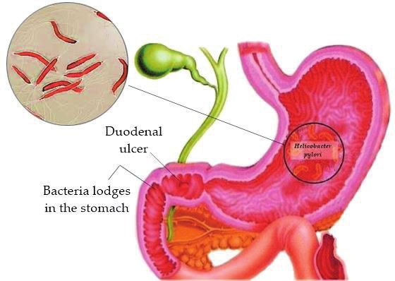
tion with H. pylori [41] (Figure 2).
2.7.8 H. pylori and peptic disorders
The gastric infection produced by H. pylori bacteria in most cases of peptic ulcer
is also important in the appearance of lymphomas that originate in the lymphoid
tissue (MALT) and in gastric adenocarcinoma [13]. The peptic ulcer is an ulcer that
affects the lining of the stomach and is the causes of internal bleeding of the upper
digestive tract with severe complications that lead to an adenocarcinoma [13, 40].
2.7.9 Diagnostic methods of H. pylori infection
The diagnostic methods of H. pylori infection have traditionally been classified
as direct and indirect; the former is based on the “direct” demonstration of the
microorganism by means of the study of samples obtained by gastric biopsy [44].
This technique used is very stressful and uncomfortable for the patient because of
the invasive reason.
Figure 2.
Entrance and lodging of H. pylori in the stomach. Grávalos and González [40].
6Helicobacter pylori: A Pathogen of Ample Risk to Health
DOI: http://dx.doi.org/10.5772/intechopen.86789
The other indirect methods are based on the detection of certain characteristics
of the bacteria, such as the ability to hydrolyze through urea, and based on the
breath test or the response of the immune system through the measurement of
specific antibodies. Its primary advantage is its noninvasive nature [44].
2.7.10 Histological techniques
The presence of the germ can be recognized with the usual hematoxylin and
eosin stain, although it is more easily demonstrated with other stains such as
Giemsa. The histology not only demonstrates the presence of the microorganism
but also informs about the morphological changes of the gastric mucosa [44].
2.7.11 Cultivation of H. pylori
Under optimal conditions H. pylori is extremely difficult to grow, due to its
demanding nutritional requirements and its slow growth. The cultivation of H.
pylori is usually slow, the first colonies usually appear between the fifth and seventh
days, and it may take up to 10 days. Being a microaerophilic microorganism requires
atmospheres with 5–10% of O2, 5–10% of CO2, and 80–90% of N2 at 35–37°C, with a
humidity of 90–95% [45].
The selection and inoculation of the bacteria depend on the number and types
of tests to be carried out as well as on the factors, type of bacteria, clinical impor-
tance of the isolation, availability of the strain, and reliable method of verification
[46]. Plate culture has advantages ranging from typifying the organism to deter-
mining its sensitivity to antibacterial agents, so it is important to study it from the
epidemiological point of view, because it allows knowing the pattern of resistance to
different therapeutic regimens with a specificity of the 100% and a lower sensitivity
than other diagnostic techniques [3]. This microorganism is also urease, oxidase,
and catalase positive, characteristics that are frequently used in the identification of
the microorganism, although its isolation is relatively complex [16].
It is usually considered a difficult and tedious technique. However, adopting a
series of minimal precautions, most laboratories achieve the growth of the microor-
ganism [44].
2.7.12 Serology
Serological techniques only indicate a previous exposure to the microorganism
but do not discriminate between people with active infection and disease in healthy
individuals with prior exposure to infection [44]. Rapid tests are methods for the
detection of antigens and antibodies in serum, plasma, whole blood, and other
fluids, which give results in a few minutes [47]. These serological techniques are
widely used today for rapid diagnosis in laboratories.
The enzyme-linked immunosorbent assay (ELISA) is widely used to perform
epidemiological studies on a considerable number of individuals [48].
In a work done by Siavoshi et al. [49], for the intracellular detection of H. pylori
in yeast identified in oral samples of newborns, the authors detected H. pylori with
immunofluorescence using polyclonal antibodies IgG anti-H. pylori in a rabbit labeled
with FITC, whose concentration was 5000 mg/ml, with a wavelength of 528 nm.
2.7.13 Antigenic screening
This is a chromatographic immunoassay for the qualitative detection of H. pylori
antigen in human stool samples, with a relative sensitivity of 94%, a specificity of
95%, and an accuracy of 97.5%, since it is an in vitro technique ad-bio [50].
7Gastritis - New Approaches and Treatments
Methods Characteristic Advantage Disadvantages
Direct
Histological Habitual staining of Demonstrates the The technique requires
techniques hematoxylin and eosin presence of the samples obtained from a
Giemsa stain microorganism and biopsy
reports on changes in the Proper selection of stain
mucosa fixatives
Direct and indirect
Culture Cultivation of the Isolate the It is difficult to isolate, since
microorganism in microorganism to study H. pylori is very sensitive
specific media under its behavior (in vitro) to drying and to the usual
microaerobic conditions atmospheric conditions
The optimum (it requires the transport
temperature of culture is of samples in the shortest
from 35 to 37°C [53] possible time) [54].
Samples destined for
culture remain viable for
approximately 5 hours and
when stored in saline at 4°C
or for more than 24 hours if
stored at 4°C in a transport
medium specific for H.
pylori [55]
Another disadvantage is
the high contamination
of the environment with
accompanying biota, which
makes it difficult to isolate
H. pylori independently
Indirect
Serological Methods for the Rapid laboratory tests It can induce a false-
techniques detection of antigens negative result
and antibodies (serum,
plasma, and whole blood,
among others)
Enzyme-linked
immunosorbent assay
(ELISA)
Antigenic Chromatographic Rapid laboratory tests Possible false positives due
screening immunoassay for the to cross reactions with other
detection of H. pylori organisms [56]
antigens in stool samples
Molecular DNA amplification of the Great versatility as an Need to have information
Methods pathogen analysis technique, on the target DNA sequence
sequences are amplified Short size of the PCR
from minute amounts of products
target DNA, even from The ease with which DNA
DNA contained in a is amplified requires
single cell avoiding the danger of
contamination inherent to
the multiplier power of the
reaction
Table 1.
Comparison of diagnostic methods for H. pylori.
8Helicobacter pylori: A Pathogen of Ample Risk to Health
DOI: http://dx.doi.org/10.5772/intechopen.86789
2.7.14 Molecular methods
Molecular methods are the names given to all the laboratory techniques used to
isolate DNA or extract it in high purity, visualize it to see its state, cut it and paste it
(Iglesias [51]), or amplify a region in a huge amount of molecules: fragment clon-
ing in bacteria or other vectors such as viruses as well as polymerase chain reaction
(PCR).
Infectious diseases have become the “spearhead” for the development of molecu-
lar diagnostic tests, with more than 50% of the techniques available today. The main
explanation for this development is due to the difficulty of detecting a pathogen
through classical microbiology [52] (Table 1).
3. Conclusion
H. pylori is a microorganism of global interest, given that, in developing coun-
tries, the infection overcomes the 60%. Besides, being microorganisms of difficult
isolation, the used techniques to culture are insufficient, so that molecular methods
and antigen screening are the most recommended for detection, since these tech-
niques are not invasive to patients.
Author details
Isidro Favian Bayas-Morejón*, Rosa Angélica Tigre-León,
Edison Riveliño Ramón-Curay and Darwin Alberto Núñez-Torres
Universidad Estatal de Bolívar, Facultad de Ciencias Agropecuarias Recursos
Naturales y del Ambiente, Centro de Investigación y Desarrollo Biotecnológico
*Address all correspondence to: favian_bm@hotmail.com
© 2019 The Author(s). Licensee IntechOpen. This chapter is distributed under the terms
of the Creative Commons Attribution License (http://creativecommons.org/licenses/
by/3.0), which permits unrestricted use, distribution, and reproduction in any medium,
provided the original work is properly cited.
9Gastritis - New Approaches and Treatments
References
[1] Cava F, Cobas FC. Dos décadas [10] Hasni SA. Role of Helicobacter pylori
de Helicobacter pylori. Scielo. infection in autoimmune diseases.
2003;12(1):1-10 Current Opinion in Rheumatology.
2012;24(4):429-434
[2] Rodríguez Br, González JJ, Carpio L.
Tratamiento para Erradicación [11] Mandell R. Enfermedades
de en una población salvadoreña infecciosas Principios y Practicas.
Terapia Secuencial vs Triple Terapia España: Elsevier España, S.L; 2012
Convencional [tesis doctoral]. 2012
[12] Palomino E. Manuales Venezolanos
[3] Gisbert J, Molina-Infante J. de Nutrición. Obtenido de Helicobacter
Tratamiento actual de la infección por pylori: Rol del agua y los alimentos en su
Helicobacter pylori. Medicina Clínica transmisión. 2016. http://www.scielo.
(Barcelona). 2017;148:20-22 org.ve/scielo.php?script=sci_arttext&pi
d=S079807522012000200005
[4] Bayas Morejón F. Aportaciones
a la epidemiología de arcobacter [13] Harrison. Medicina interna volumen
y helicobaaplicación de métodos 2. España: Mcgraw-hill interamericana
moleculares a su detección e identificación editores, s. A. de C. V. 2012. p. 125
en alimentos [Tesis Doctoral]. Valencia:
Politecnica de Valencia. 2016 [14] Konturek JW. Discovery by
Jaworski of Helicobacter pylori and
[5] Guamán JF, Bayas-Morejón F, Arcos its pathogenetic role inpeptic ulcer,
V, Tigre-León A, Lucio-Quintana A, gastritis and cancer gastric. Journal
Salazar S, et al. Detection of Helicobacter of Physiology and Pharmacology.
pylori from human biological samples 2003;54(3):23-41
(Feces) by antigenic screening and
culture. Jundishapur Journal of [15] Novo Villaverde FJ. GENÉTICA
Microbiology. 2018;11(7):e66721 HUMANA Conceptos, Mecanismos y
Aplicaciones de la Genética en el campo
[6] Dirección Nacional de Vigilancia de la Biomedicina (Vol. 1ra EDICION).
Epidemiológica_MSP—Ecuador. pearson prentice hall. 2007. ISBN:
Anuario. Obtenido de Enfermedades 978-848-322-359-8
Trasmitidas Por Agua Y Alimentos.
2017. https://public.tableau.com/profile/ [16] Martel C, Parsonnet J. Helicobacter
vvicentee80#!/vizhome/ETAS-2014/ pylori infection and gender: A meta-
ANUARIO analysis of population-based prevalence
surveys. Digestive Diseases and
[7] Linear Chemicals S.L. 2017. Website. Sciences. 2006;51(12):2292-2230
Obtenido de: http://www.linear.es/
ficheros/archivos/481_4245125H. [17] Figueroa G, Troncoso M, Toledo MS,
PyloriAgcassette25tcas.pdf Faúndez G, Acuña R. Prevalence of
serum antibodies to Helicobacter pylori
[8] Fundacion Española de Endoscopia VacA and CagA and gastric diseases in
Digestiva. 2009. Complicaciones de la Chile. Journal of Medical Microbiology.
Endoscopia Digestiva Alta. Protocolos y 2002;51(4):300-304
Directrices en Endoscopia
[18] Aziz RK, Khalifa MM, Sharaf RR.
[9] Smolka AJ, Backert SJ. How Contaminated water as a source
Helicobacter pylori infection controls of Helicobacter pylori infection: A
gastric acid secretion. Gastroenterology. review. Journal of Advanced Research.
2012;47(6):609-618 2015;6(4):539-547
10Helicobacter pylori: A Pathogen of Ample Risk to Health
DOI: http://dx.doi.org/10.5772/intechopen.86789
[19] Santiago P, Moreno Y, Ferrús MA. inside Acanthamoeba castellanii.
Identification of viable helicobacter Research in Microbiology.
pylori in drinking water supplies by 2016;167(1):29-34
cultural and molecular techniques.
Helicobacter. 2015;20(4):252-259 [28] Van-Duynhoven YTHP, De-Jonge R.
Transmission of Helicobacter
[20] Eusebi LH, Zagari RM, Bazzoli pylori: A role for food. Bulletin of
F. Epidemiology of Helicobacter pylori the World Health Organization.
infection. Helicobacter. 2014;19 2001;79(5):455-460
(Suppl 1):1-5
[29] Beuchat LR. Ecological factors
[21] Moreno Y, Ferrús MA, Alonso JL, influencing survival and growth of
Jiménez A, y Hernández J. Use of human pathogens on raw fruits and
fluorescent in situ hybridization to vegetables. Microbes and Infection.
evidence the presence of Helicobacter 2002;4(4):413-423
pylori in water. Water Research,
2003;37(9):2251-2256 [30] Atapoor S, Safarpoor Dehkordi F,
Rahimi E. Detection of Helicobacter
[22] Moreno Y, Ferrus MA. Specific pylori in various types of vegetables
detection of cultivable Helicobacter and salads. Jundishapur Journal of
pylori cells from wastewater treatment Microbiology. 2014;7(5):e10013
plants. Helicobacter. 2012;17(5):327-332
[31] Yahaghi E, Khamesipour F,
[23] Percival SL, Suleman L. Biofilms Mashayekhi F, Safarpoor Dehkordi F,
and Helicobacter pylori: Dissemination Hossein SM, Masoudimanesh M, et al.
and persistence within the Helicobacter pylori in vegetables and
environment and host. World Journal salads: Genotyping and antimicrobial
of Gastrointestinal Pathophysiology. resistance properties. BioMed Research
2014;5(3):122-132 International. 2014;1:11. Article ID: 757941
[24] Donlan RM. Biofilms: Microbial [32] Bayas-Morejón IF, González A,
life on surfaces. Emerging Infectious Moreno-Mesonero L, Moreno Y, Ferrús
Diseases. 2002;8(9):881-890 M. Detection of Helicobacter pylori
in vegetables, XXIXth international
[25] Gião MS, Azevedo NF, Wilks SA, workshop on Helicobacter and
Vieira MJ, Keevil CW. Persistence of microbiota in inflammation on
Helicobacter pylori in heterotrophic & cancer, Magdeburg-Germany.
drinking-water biofilms. Applied Helicobacter. 2016;21(Suppl 1):69-177.
and Environmental Microbiology. DOI: 10.1111/hel.12344. [PubMed:
2008;74(19):5898-5904 27531543]
[26] Watson CL, Owen RJ, Said B, Lai S, [33] Ng CG, Hassanbhai AM, Loke MF,
Lee JV, Surman-Lee S, et al. Detection Wong HJ, Goh KL, Vadivelu J,
of Helicobacter pylori by PCR but not et al. Helicobacter pylori biofilm—
culture in water and biofilm samples The probable mode and source
from drinking water distribution of transmission? Helicobacter.
systems in England. Journal of Applied 2014;19(Suppl 1):104
Microbiology. 2004;97(4):690-698
[34] Boehmler G, Gerwert J, Scupin E,
[27] Moreno-Mesonero L, Moreno Y, Sinell HJ. Epidemiology of H. pylori
Alonso JL, Ferrús MA. DVC-FISH in man: Studies on the survival of the
and PMA-qPCR techniques to assess agent in food. Deutsche Medizinische
the survival of Helicobacter pylori Wochenschrift. 1996;103:438-443
11Gastritis - New Approaches and Treatments
[35] Fujimura S, Kawamura T, Kato S, aegastro.es/sites/default/files/archivos/
Tateno H, Wanatabe A. Detection of ayudas-practicas/19_Infeccion_por_
Helicobacter pylori in cow’s milk. Letters in Helicobacter_pylori.pdf
Applied Microbiology. 2002;35:504-507
[45] Ofelia CC, Jorge MQ , Harold BG,
[36] Meng X, Zhang H, Law J, Tsang R, Alfonso CM, Edson GC, Milagros DM,
Tsang T. Detection of Helicobacter pylori et al. Prevalencia de helicobacter pylori
from food sources by a novel multiplex en pacientes sintomáticos de consulta
PCR assay. Journal of Food Safety. externa de la red Rebagliati (EsSalud),
2008;28(4):609-619 Lima, Perú, en el período 2010—2013.
Revista de Gastroenterología del Perú.
[37] Fernández M, Contreras M, Suárez 2016;36(1):49-55
P, Gueneau P, García-Amado MA. Use
of HP selective medium to detect [46] Scott B. Diagnóstico
Helicobacter pylori associated with Microbiológico. Buenos Aires—
other enteric bacteria in seawater and Argentina: Panamericana; 2009
marine molluscs. Letters in Applied
Microbiology. 2007;45:213-218
[47] Secretary of Health Secretaria
de Salud. Guia para la aplicacion de
[38] Samie A. Prevalence of
pruebas rapidas. Mexico: Printed and
campylobacter species, helicobacter
Made in Mexico; 2006
pylori and Arcobacter species in
stool samples from the Venda region,
Limpopo, South Africa: Studies using [48] Hernández Ramírez D, Cabiedes
molecular diagnostic methods. Journal J. Immunological Techniques
of Infection. 2007;54:558-566 that Support the Diagnosis of the
Autoimmune Diseases. México D.F,
[39] Jiménez DF. Mediadores Bacterianos México: Laboratorio de Inmunología,
de la Inflamacion en La Gastritis. Revista Departamento de Inmunología y
Cubana de Medicina. 1999;38:276-283 Reumatología, Instituto Nacional de
Ciencias Médicas y Nutrición Salvador
[40] Grávalos DC, González E. Cancer Zubirán; 2010
gastrico. Sociedad Española de Oncología
Médica. 2017; pp. 1-16. Available from: [49] Siavoshi F, Taghikhani A,
https://seom.org/info-sobre-el-cancer/ Malekzadeh R, Sarrafnejad A,
estomago Kashanian M, Jamal AS, et al. The role
of mother's oral and vaginal yeasts in
[41] American Cancer Society. Obtenido
transmission of Helicobacter pylori to
de Society, American Cancer Atlanta, neonates. Archives of Iranian Medicine.
Ga: American Cancer. 2016, http://www. 2013;16(5):288-294
cancer.org/cancer-de-estomago-pdf
[42] Hinojosa MM. 2017. Obtenido [50] ad-bio H. pylori Ag Prueba Rápida
de: http://www.inen.sld.pe/portal/ en Casete (muestras fecales). 2017;
documentos/pdf/educacion/091115_ Obtenido de: http://www.annardx.
CANCER%20GASTRICO%20-%20 com/productos/images/productos/
JEMH.pdf diagnostica/pruebas-rapidas/ad0192c-
hpylori-ag-rev-cpdf.pdf
[43] Jiménez DF. Cancer gastrico:
Factores de riesgo. Revista Cubana de [51] Iglesias G. Tecnicas de biologia
Oncología. 1998;14:171-179 molecular. Desde Mendel hasta
las moléculas. 2008;1. Obtenido
[44] Gisbert J. Infección por Helicobacter de: https://genmolecular.com/
pylori. 2016. Obtenido de http://www. tecnicas-de-biologia-molecular/
12Helicobacter pylori: A Pathogen of Ample Risk to Health DOI: http://dx.doi.org/10.5772/intechopen.86789 [52] Farfan BM. Biologia Molecular Aplicada Al Diagnostico Clinico. Revista Médica Clínica Las Condes. 2015;26(6):788-793 [53] Ferrús A. Survival and viability of Helicobacter pylori after inoculation into chlorinated drinking water. Water Research. 2007;41(15):3490-3496 [54] Pagola MF. Caracterización de la infección por Helicobacter pylori en pacientes con úlcera gástrica. Scielo/ Medisur. 2009;7(6):3-11 [55] Veenendaal RA, Lichtendahl- Bernards AT, Peña AS, Endtz HP, van Boven CP, Lamers CB. Effect of transport medium and transportation time on culture of Helicobacter pylori from gastric biopsy specimens. Journal of Clinical Pathology. 1993;46(6):561-563 [56] SCREEN. Test Rapido Antigene. Obtenido de: http://www.screenitalia.it/ wp-content/uploads/2017/11/Istruzioni- Screen-H.PyloriSITA-1.pdf 13
You can also read