Premature Ovarian Insufficiency - Medimail October 2019 - SRL ...
←
→
Page content transcription
If your browser does not render page correctly, please read the page content below
Medimail
          October 2019

Premature Ovarian Insufficiency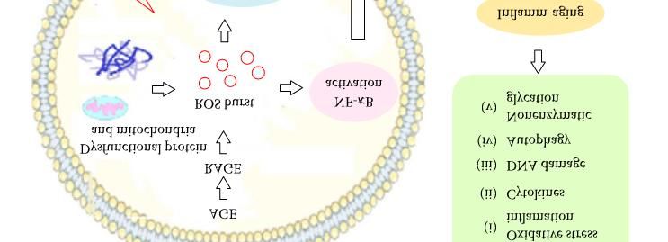
Premature Ovarian Insufficiency (POI)

• POI is a clinical syndrome defined by
  loss of ovarian activity before the age
  of 40 (ESHRE 2015).
• Characterised by menstrual
  disturbance (amenorrhea or
  oligomenorrhea) with raised
  gonadotropins and low estradiol.
• Untreated POI is associated with
  reduced life expectancy, largely due
  to cardiovascular disease.
• Modifiable factors may include:
   – gynaecological surgical practice
                                            Distribution of age at menopause
   – lifestyle: smoking
   – modified treatment regimens for
      malignant and chronic diseases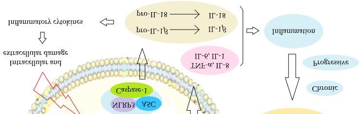
Epidemiology

World
• Incidence of POI before 40 yrs ~1%
• Incidence of POI before 30 yrs - 0.1%
• ~10% to 28% experience primary amenorrhea
• ~4% to 18% exhibit secondary amenorrhea
• Prevalence of menopause varies according to age:
    – 1 : 10,000 at the age of 18-25 years
    – 1 : 1000 in women aged 25-30 years
    – 1 : 100 in the age range 35-40 years
India
• According to the Indian National Family Health Survey (NFHS-3) 2005-2006,
   about 18% married women in the age group of 30-49 yr reached menopause
• 3.1% of women in 30-34 yr and 8% in 35-39 yr were in menopause (NFHS-2)

                    Pallikadavath S. Indian J Med Res. 2016 Sep; 144(3): 366–377
                       Jungari S. Health & Social Work 42(2):1-8 · March 2017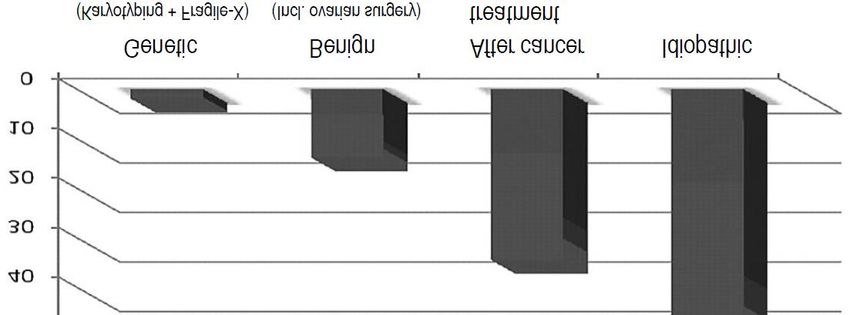
Etiology

•   Chromosomal and genetic defects – X chromosomal abnormalities or
    aneuploidy, Y chromosome abnormalities, fragile-X syndrome, autosomal gene
    defects, gonadal dysgenesis with or without Turner syndrome
•   Autoimmune disorders
•   Infections
•   Iatrogenic cause – surgery, radiotherapy, or chemotherapy
•   Environmental factors
•   Idiopathic – cause remains elusive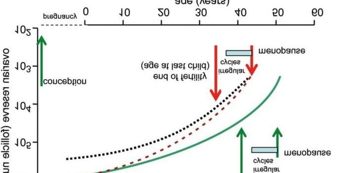
Inflamm-Aging and POI
Inflammatory aging refers to a chronic and low-degree proinflammatory state which occurs with
increasing age and is closely associated with multiple diseases, as excessive inflammation can induce
the inflammatory lesions in certain organs of the body. In recent years, studies have shown that
inflammatory aging plays a significant role in the pathogenesis of POI.

                Journal of Immunology Research. Volume 2019, Article ID 8069898, 7 pages
POI or Diminished Ovarian Reserve?

• POI is distinct from low ovarian
  reserve
• ‘Ovarian Reserve’ encompasses
  both quantity and quality of
  primordial follicles. Low ovarian
  reserve is a condition in which
  ovary loses its normal
  reproductive potential. Low
  ovarian reserve is characterized as
  regular menses and alterations of
  ovarian reserve tests, and can be
  caused by conditions affecting the
  ovaries, but in most cases is a
  consequence of age.
                                                   Decay of ovarian reserve with age

                         Dan Med J. 2016 Oct;63(10). pii: B5292.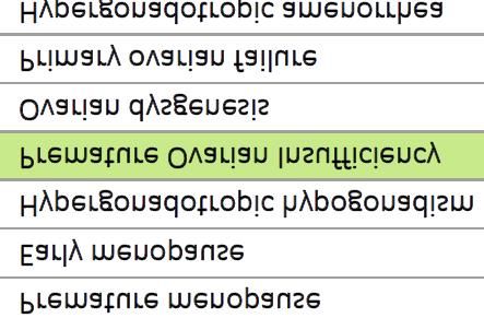
Diagnosis

•   POI is characterised by menstrual
    disturbance, raised gonadotropins,
    and low estradiol.
•   ESHRE 2015 recommends the
    following diagnostic criteria:
     – oligo/amenorrhea for a period of
         4 to 6 months
     – an elevated FSH level > 25 IU/l on
         two occasions > 4 weeks apart
•   ACOG 2014 recommends:
     – Menstrual irregularity for atleast
         3 consecutive months
     – FSH & E2 levels (2 random tests
         atleast 1 month apart)             Different terminologies for POI
     – Prolactin and thyroid function
         test
Clinical States Included in the Spectrum of POI
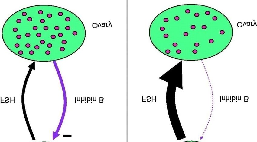
N Engl J Med. 2009 February 5; 360(6): 606–614Follicle stimulating hormone (FSH)
• FSH levels are used as the gold
standard in establishing a
diagnosis of POI.
• Most literature uses FSH>40 IU/l
as criteria for POI diagnosis.
• Since patients with autoimmune
POI should be included in the
diagnosis, ESHRE 2015 use a cut
off level of FSH > 25 IU/l. This is
above the physiological range
for FSH even at the pre-
ovulatory peak.
https://rep.bioscientifica.com/view/journals/rep/140/5/633.xmlConfirmation of Diagnosis of POI International Journal of Women's Health 2014. 6(1):235-243
anti-Müllerian hormone (AMH)

• More direct marker of ovarian reserve and low AMH is more
  prevalent in POI patients.
• Serum AMH levels follow the reduction in follicular number
  over time in healthy women and fall to very low levels prior to
  menopause.
• However, low AMH may also be found in women with regular
  cycles and low ovarian reserve.
• Women attending fertility clinics with low AMH but regular
  menstrual cycles should not be diagnosed with POI.

     Anti-Müllerian hormone (AMH) is not sufficiently
          discriminative for a diagnosis of POI.
Other Investigations in POI
• Chromosomal analysis/ karyotping
• Fragile-X (FMR1) premutation testing
• 21OH-Ab or adrenocortical antibodies (ACA)
• 21-hydroxylase (CYP21)
• Thyroid (TPO-Ab) antibodies Autosomal genetic testing is not at
• Thyroid stimulating hormone (TSH) present indicated in women with POI,
unless there is evidence suggesting a
• Adrenocorticotropic hormone (ACTH) specific mutation (e.g. BPES).
• Plasma renin activity
• ACTH stimulation test
• Vitamin B12
• Ferritin
• FolateKaryotyping (For Diagnosis of Turner Syndrome) • Chromosomal analysis should be performed in all women with non-iatrogenic Premature Ovarian Insufficiency. • For chromosomal analysis for Turner Syndrome, karyotyping is the gold standard; although microarray-based comparative genomic hybridisation (array CGH) and other new technologies exist. • If negative, a second analysis of the karyotype from the gonads (in case of high clinical suspicion).
Test for Y-chromosomal Material

• Every women with a Y chromosome, whether or not
  she has a SRY gene mutation, should be counselled
  about the risk of development of a gonadal tumour.

• Gonadectomy should be recommended for all
  women with detectable Y chromosomal material.
Fragile X Testing

•   Fragile-X syndrome is an X-linked inherited condition caused by a mutation of the
    fragile-X mental-retardation 1 (FMR1) gene.
•   Prevalence of 0.8 to 7.5% in women with sporadic POI (i.e. women without other
    family members with POI) and up to 13% in women with a positive family history
    of POI .
•   Women who carry the premutation (55-200 repeats) do not have an increased risk
    of intellectual disability, but have an increased risk of 13 to 26% to develop POI.
•   The risk of developing POI is not increased in women with the full mutation or
    intermediate sized CGG repeats (45 – 54 repeats).
•   Family members may be carriers and therefore have a risk of developing POI and a
    risk of having (grand)children with fragile-X syndrome. In addition, the patient
    herself may already have daughters, who may be carriers. This requires careful
    counselling before the test is performed, including permission from the patient to
    perform the test. An additional reason to counsel about Fra-X testing is the risk of
    fragile-X-associated tremor/ataxia syndrome (FXTAS), a late onset neurological
    problem predominantly in male carriers of the Fra-X-mutation.
Antibodies
• Autoimmune disorders are more frequent in POI than in the general population,
and POI is more frequent in women with certain autoimmune disorders.
• Addison’s disease and APS type 2 are known to predispose to POI while POI of
adrenal autoimmune origin is the most frequent type observed in 60 to 80% of
patients with autoimmune POI.
– Screening for adrenocortical antibodies (ACA) and/ or 21-hydroxylase autoantibodies
(21OH-Ab) should be considered in every POI patient because of the possibility of
subclinical or latent Addison’s disease in POI patients. Positive 21OH-Ab/ACA test should
be directed to adrenal function testing to rule out Addison’s disease. In the presence of
peripheral 21OH-Ab, SCA on cryostatic sections of ovaries and/or 17α-hydroxylase
antibodies (17α-OH-Ab) and/or P450SCC antibodies can be detected on cryostatic
sections of ovaries in over 90% of cases.
• POI is associated most commonly with thyroid autoimmunity (14–27%) when
adrenal autoimmunity is absent.
– Screening for thyroid antibodies (TPO-Ab) should be performed in women with POI. In
patients with a positive TPO-Ab test, thyroid-stimulating hormone (TSH) should be
measured every year. TSH screening could occur at 5-year intervals if negative.Fertility and Pregnancy • Oocyte donation is an established option for fertility in women with POI. However, women presenting for oocyte donation who are suspected of having POI should be fully investigated prior to oocyte donation, including thyroid and adrenal function as well as karyotype. • Women with POI should have their blood pressure, renal function, and thyroid function assessed prior to pregnancy.
Bone Health

• POI is associated with reduced bone mineral density (BMD).
  Reduced BMD is very likely to indicate that POI is associated with an
  increased risk of fracture later in life.
• Measurement of BMD at initial diagnosis of POI should be
  considered for all women, but especially when there are additional
  risk factors.
• If BMD is normal and adequate systemic estrogen replacement is
  commenced, the value of repeated DEXA scan is low.
• If a diagnosis of osteoporosis is made and estrogen replacement or
  other therapy initiated, BMD measurement should be repeated
  within 5 years. A decrease in BMD should prompt review of
  estrogen replacement therapy and of other potential factors.
Cardiovascular Health

• Women with POI are at increased risk of cardiovascular disease and
  should be advised of risk factors that they can modify through
  behavioural change (e.g. stopping smoking, taking regular weight-
  bearing exercise, healthy weight).
• All women diagnosed with Turner Syndrome should be evaluated by
  a cardiologist.
• Cardiovascular risk should be assessed in women diagnosed with
  POI. At least blood pressure, weight and smoking status should be
  monitored annually with other risk factors being assessed if
  indicated.
• In women with Turner Syndrome, cardiovascular risk factors should
  be assessed at diagnosis and annually monitored (at least blood
  pressure, smoking, weight, lipid profile, fasting plasma glucose,
  HbA1c).
Genetic Counseling

• Relatives of women with non-iatrogenic POI (fragile-
  X premutation) who are concerned about their risk
  for developing POI should be informed that:
   – currently there is no proven predictive test to identify
     women that will develop POI, unless a mutation known to
     be related to POI was detected
   – there are no established POI preventing measures
   – fertility preservation appears as a promising option,
     although studies are lacking
   – their potential risk of earlier menopause should be taken
     into account when planning a family
Tests Done in SRL
TEST METHOD CODE
Fertility Panel, Female, Endocrine
(TSH3G UL, Prolactin, Chemiluminescence 1578
Progesterone, Estradiol, LH, FSH)
Enzyme Immunoassay/
Inhibin B,LH, FSH & Prolactin 1013
Chemiluminescence
Cytogenetics: Blood Lympho
Cell Culture 5814B
Culture
Cytogenetics: Fragile X
Karyotype Cell Culture 5364B
Chromosome Analysis
Fragile X (FMR1) Mutation Triplet Primed PCR Fragment
RD1325
Screen Analysis
Thyroid Antibodies Chemiluminescence 1016
21Thank You
You can also read