Prevalence of Demodex Folliculorum and Demodex Brevis in Patients with Blepharitis and Chalazion
←
→
Page content transcription
If your browser does not render page correctly, please read the page content below
Prevalence of Demodex Folliculorum and Demodex
Brevis in Patients with Blepharitis and Chalazion
Serife AKKUCUK ( serifeakkucuk@hotmail.com )
Mustafa Kemal University: Hatay Mustafa Kemal Universitesi
Ozlem Makbule Kaya
Mustafa Kemal University Faculty of Medicine: Mustafa Kemal Universitesi Tayfur Ata Sokmen Tip
Fakultesi
Lokman Aslan
Private Sevgi Hospital
Talat Ozdemir
Private Sevgi Hospital
Ugur Uslu
Selçuk Üniversitesi Tip Fakültesi: Selcuk Universitesi Tip Fakultesi
Research Article
Keywords: Demodex folliculorum, Demodex brevis, blepharitis, chalazion, eyelash
Posted Date: August 23rd, 2021
DOI: https://doi.org/10.21203/rs.3.rs-241572/v1
License: This work is licensed under a Creative Commons Attribution 4.0 International License.
Read Full License
Page 1/17Abstract Purpose: Demodex folliculorum and Demodex brevis are common ectoparasites on skin that also can lead to blepharitis and chalazion. The aim of our study is to determine the prevalence of Demodex spp. in eyelashes of patients diagnosed with chronic blepharitis and chalazion. Methods: This study included 330 patients diagnosed with chronic blepharitis, 70 patients diagnosed with chalazion and 130 volunteers without any ocular problems. Patient eyelashes were examined under a light microscope at magnifications of × 40, × 100 and × 400. Demodex spp. were determined. Results: Parasite prevalence was significantly higher in blepharitis (75.5%) and chalazion groups (70%) compared to the control group (16.2%) (p
is common in humans, it is known that it has been ignored as the cause of ocular problems for many
years [12,5]. Recent studies indicate that ocular problems such as blepharitis and chalazion may be
associated with mites. It supports the argument that settlement and multiplication of the Demodex mites
in the hair follicles and seborrheic glands may be involved in the etiology of ocular infectious
diseases [13,1].

We aimed to determine the prevalence of Demodex mites in patients with blepharitis and chalazion and
also to determine the relationship between mites and these diseases in this study.

Materials And Methods
Study groups
330 patients with chronic blepharitis, 70 patients with chalazion and 130 volunteers without any eye
disease or ocular symptoms who were admitted to Private Osmaniye Sevgi Hospital Ophthalmology
Outpatient Clinic between 01.11.2018 and 15.11.2019 with various ocular complaints were included in
the study. Those who were pregnant, under the age of 18, who received antiparasitic treatment in the last
1 month, who had eye surgery in the last 6 months, and who could not sign the consent form for any
reason were not included in the study. Study approval numbered 2018/154 was obtained from the
Clinical Research Ethics Committee of Hatay Mustafa Kemal University Tayfur Ata Sökmen Faculty of
Medicine. A short questionnaire about age, gender, general eye complaints (burning, stinging, dryness,
itching, blurred vision, dandruff at the eyelash base), systemic diseases (diabetes, cancer, kidney
disease), pet feeding at home or in their gardens was applied to all participants included in the study and
the consent form was filled out.

Sampling
Samples were taken from the problematic eyes of the patients with unilateral eye disease, the right eyes
of the patients with bilateral eye disease or the volunteers without any eye problems. With the eyelash
epilation method, a total of six eyelashes were taken from each individual's lower and upper eyelids, three
each. The presence of Demodex spp. was examined under a light microscope at x40 and x100
magnifications. The distinction of demodex types was made according to the relevant literature [14,15].

Statistical Analysis
Kolmogorov Smirnov test was used to compare whether the data were normally distributed or not. One-
way Anova, t-test, Chi-Square (χ2), Fisher's Exact and Spearman Correlation tests were used for analysis.
In the evaluation of the results, p
Results
Demodex mite was found in 249 (75.5%) of 330 patients with blepharitis, 49 (70%) of 70 patients with
chalazion, and 21 (16.2%) of 130 volunteers in the control group. The comparison of Demodex spp.
according to the diagnosis groups is presented in Table 1. The parasite prevalence was significantly
higher in both blepharitis and chalazion groups compared to the control group (p
and the presence of Demodex spp. in the groups is presented in Table 4. In all groups, there was no
significant difference between gender and Demodex spp. positivity (p> 0.05).

The presence of symptoms and the relationship between symptoms and mites in patients diagnosed with
blepharitis and chalazion were given in Table 5. In the presence of symptoms, Demodex spp. positivity
was found to be significant in the group with blepharitis (p = 0.0001), while no significant difference was
found in the group with chalazion (p = 0.308).

The most common complaints in patients with blepharitis were 43.3% itching, 36.4% stinging (foreign
body sensation) and 30.3% burning, respectively. However, among these complaints, a significant
difference was found only between stinging and the positivity of Demodex spp. (Χ2 = 9.75; p = 0.002). DF
was found in 116 (96.6%) of 120 mite positive patients with stinging complaints, while 29 (24.1%) were
found to have DB. There was a significant difference between the presence of DF and the complaint of
stinging (p  0.05).

The relationship between education level and mite positivity in the groups is given in Table 6. According
to the correlation coefficient (r) results, a negative correlation was observed between the education level
and the positivity of Demodex spp in the blepharitis and the control group. However, while this negative
correlation was not significant in the control group (p = 0.213), it was significant in the blepharitis group.
It was found that the positivity of Demodex spp. decreased as the education level increased in patients
with blepharitis (r = -0.114; p = 0.039). The positive correlation found in the chalazion group was not
found to be significant (p = 0.562).

There was no significant difference between animal feeding and the positivity of Demodex spp. in all
groups (p> 0.05).

Discussion
It is stated in some studies that Demodex mites, which are frequently detected in humans around the
world, may be pathogenic in chronic eyelid diseases [16,17,1]. While Demodex folliculorum causes
anterior blepharitis, DB positivity shows a strong correlation with the prevalence of posterior blepharitis,
                                                   Page 5/17
meibomian gland dysfunction, recurrent chalazion, and keratoconjunctivitis [12,18]. In the pathogenesis
of blepharitis, it has been reported that mites are vectors in the transport of some bacteria such as
Bacillus, Staphylococcus and Streptococcus, and mites play a role with the bacteria especially in cases
that are resistant to treatment and recurrent after treatment [12,19]. At the final point, mites are
considered as a causative factor for blepharitis [20].
The etiology of chalazion has not been explained and is still unknown. The accumulated secretion
causes an inflammatory reaction in the surrounding tissues. Another factor could be bacterial and / or
Demodex infestation [7,6,13].
Considering the epidemiological studies on blepharitis and chalazion, it is seen that the prevalence of
Demodex spp. ranges from 28.8% to 84% in patients with blepharitis, 63.2% to 91.67% in patients with
chalazion, and 4.6% to 54.9% in the healthy control group. The results of our study are compatible with
the literature [21-24,6,25]. In several studies, no significant difference was found between patients with
blepharitis and the control group in terms of mite positivity [21,22]. However, it is stated in most of the
literature that the presence of parasites is significantly higher in patients with blepharitis and
chalazion [16,24,6,25,13]. In our study, it was found that the presence of Demodex spp. was statistically
significantly higher in patients with blepharitis and chalazion.
It is known that the most common type in almost all studies showing the relationship between blepharitis
and Demodex is DF [16,25-27]. Our study is similar to studies reporting that DF is significantly more
common than DB in eyelash follicles in patients diagnosed with blepharitis.
Studies examining the relationship between chalazion and Demodex show that DF dominance is at the
forefront, as in blepharitis studies [24]. In our study, it was determined that DF was the most common
mite in the chalazion group. In addition, although there was no statistically significant difference between
DF and DB in patients diagnosed with chalazion, the incidence of DB was found to be significantly higher
than blepharitis and control groups. Based on the results, we think that both species may have a role in
the pathogenesis of chalazion.
Although the eyelash hair removal method was commonly used in ocular demodicosis studies, which eye
(right eye, left eye, both eyes, lower or upper eyelids), which eyelashes (cylindrical dandruff eyelashes or
random) and how many eyelashes to be taken could not be made a standard method [28]. Apart from the
eyelash epilation method, resection samples were also evaluated in patients with chalazion [24]. The
average number of mites in the studies is not based on a standard method. In some studies, the average
Demodex number was obtained by dividing the total number of mites by the number of Demodex positive
volunteers [29,28], while in others the total number of mites was divided by the number of all
volunteers [23,27]. The issues such as the different number of eyelashes, giving the average number of
mites according to all patients or positive patients, giving the average per patient or per eyelash vary. This
makes it difficult for researchers to interpret between studies. As a result of our observations and
research, we think that positive individuals should be evaluated while giving the average number of mites.
Because:
Page 6/17a) When non-infested patients are included in the studied patient group, the average "mite density in
infested patients in X group", which is the main focus, is reduced.
b) The main goal in medicine is to develop a solution-treatment for the current problem. "Mite
positive in X group" individuals should be evaluated in order to detect the change in the number of mites
before and after treatment studies.
c) Determining how the number of mites changes when species are found individually or together in
"Mite positive in group X" will help to illuminate the pathogenesis between the mite and the disease.
In addition, we think that the average Demodex numbers should be given per eyelash. Although the
researchers examine different numbers of eyelashes, by giving the results to the eyelash head, we provide
the researchers with ease in interpretation and achieve a standard in the literature. If we list our two
suggestions on this subject:
1) Positive individuals should be taken into consideration while giving average mite numbers.
2) Average mite counts should be given per eyelash.
In our study, we found it appropriate to give results with the average number of mites per eyelash. In the
blepharitis group, the average number of mites when DF + DB coexists were found to be significantly
higher compared to those with only DF or only DB and total mites. It was found that the number of mites
increased in patients with blepharitis if both species were seen together.
Similarly, there is no standardization in the data in studies examining the relationship between chalazion
and Demodex. Schear et al. found 0.804 ± 1.03 DF per eyelash epilled, while the average DF in the control
group was found to be 0.487 ± 0.82 [24]. In our study, the DF number per eyelash was found by Schear et
al. shows similarities with the results of their study. In addition, the average number of mites in DF + DB
coexistence was found to be significantly higher in patients compared to those with DB alone.
It is reported that the increase in the activity of the sebaceous glands that occur with age and the
changes in the sebum composition will facilitate the increase of mites in the elderly [26]. In many studies,
it has been reported that ocular Demodex infestation increases with age, it is seen in 84% of the
population in their 60s and in 100% of the population aged 70 and over [12,16,21,23]. Kasetsuwan et al.
reported the prevalence of ocular demodicosis as 70% over the age of 80 [30]. In other studies, it was
reported that infestation was significantly higher in patients with blepharitis over the age of 50, those over
60 years of age, and those with ocular disease over the age of 70 [31,10,26]. However, in some studies, no
significant difference was found between increasing age and the presence of mites [22,32]. This study
supports the general literature. A statistically significant positive correlation was found between age and
the presence of Demodex spp. in patients with blepharitis. We think that this is caused by the weakening
immune system, increased sebum amount and weakened-deteriorating skin structure in the elderly.
Page 7/17Although there is no statistically significant difference, there are studies reporting that infestation is
higher in women [10], as well as studies reporting that it is common in men [25]. Zeytun and Karakurt
stated that the prevalence of mites is significantly higher in males [28]. Most of the studies argue that
there is no significant difference between gender and Demodex spp. [16,22,23,26,32]. This study supports
the literature. No significant difference was found between gender and mite positivity in patients with
blepharitis.
As a result of the resection samples of patients with chalazion, no significant relationship was found
between gender and the presence of mites. Tarkowski et al. reported that the infestation was in a similar
distribution in men and women with chalazion [24,6]. Our study is the same as the results of the other two
studies. There was no significant relationship between sex and mite positivity in the chalazion group.
It was reported that the symptoms of Demodex spp. positive blepharitis patients did not differ from those
of other blepharitis patients. In some studies, it was reported that most of the patients were
asymptomatic and their complaints generally increased in hot weather [33]. The most common
complaints in patients with infected blepharitis were itching, foreign body sensation (stinging) and
redness. Eyelash sticking, dandruff at the bottom of the eyelashes, mild papillary conjunctivitis,
meibomian gland dysfunction and telangiectasia were seen in patients with chronic blepharitis [28]. In
addition, it was stated that patients had ocular pain, contact lens intolerance, photophobia and
crusting [23]. Symptoms were usually worse in the morning, and several flare-ups and remissions might
ocur [1]. In this study, a significant difference was found between the presence of symptoms and mite
positivity in the blepharitis group. The most common complaints in patients with blepharitis were itching,
stinging (foreign body sensation) and burning, respectively. Although itching ,in our study, was the most
common symptom as in other studies, this symptom was similar in patients with mite negative and
positive blepharitis like the studies conducted by Inceboz et al [33]. In our study, among these symptoms,
only a significant relationship was found between stinging complaint and the presence of DF.
While patients with chalazion have eyelid nodules showing pain, inflammation and sensitivity in the
acute phase, there is a permanent, painless mass in the chronic phase [7]. Cylindrical dandruff was not
common, as most patients with recurrent chalazion had regular eyelid hygiene [13]. In this study, the
complaints seen in patients with chalazion were itching, dandruff at the base of the eyelashes, stinging,
burning and watering, respectively. However, there was no significant difference between mite positivity
and the presence of symptoms.
Studies show that DF is increased in immunocompromised patients such as diabetes, end-stage chronic
renal failure, Behçet's disease, urological cancers and eyelid basal cell carcinomas [11,34]. It has been
reported that DF is an important factor in eye diseases such as blepharitis and that the mite is more
common in immune system disorders. In a study, DF was detected in 27.4% of eyelashes of 42 patients
with Type 2 diabetes and 19% in 42 control group volunteers. It has been reported that infestation is
significantly higher in patients with diabetes [35]. In our study, it was determined that the prevalence of
infestation was higher in patients with systemic disease in both groups. However, no significant
Page 8/17difference was found between the presence of systemic disease and mite positivity in the blepharitis and
chalazion groups. We think that this is due to the low number of patients with systemic diseases in our
target study groups.
There is no information about the relationship between education level and mite positivity in the available
literature. In our study, a negative correlation was observed between education level and the positivity of
Demodex spp. in the blepharitis and control group. However, while this negative correlation was not
significant in the control group, it was significant in the blepharitis group. It has been found that as the
education level increases in patients with blepharitis, the mite positivity decreases. The reason for this
may be the increase in personal hygiene practices and compliance with protection methods due to the
increase in education level. There was no significant difference in the chalazion group.
It was reported that there was no significant relationship between pet feeding and demodicosis in
patients with blepharitis [33]. In our study, no significant difference was found between animal feeding
and mite positivity in blepharitis, chalazion and control groups. We think that this is due to the fact that
mites transmitted from infested animals are host specific and can only cause a temporary dermatitis in
the human body.
As a result, clinicians should warn especially the elderly patients against Demodex mites that infect
eyelashes. Patients with recurrent blepharitis and chalazion who do not respond to the current blepharitis
treatment procedure should be investigated and treated for Demodex spp.
Declarations
ACKNOWLEDGEMENT
Study approval numbered 2018/154 was obtained from the Clinical Research Ethics Committee of Hatay
Mustafa Kemal University Tayfur Ata Sökmen Faculty of Medicine.
The study was supported by Coordination of Scientific Research Projects of Mustafa Kemal University.
We declare that there is no conflict of interest in this study.
References
1. Lindsley K, Matsumura S, Hatef E, Akpek EK (2012) Interventions for chronic blepharitis. Cochrane
Database Syst Rev (5):CD005556. doi:10.1002/14651858.CD005556.pub2
2. Navel V, Mulliez A, Benoist d'Azy C, Baker JS, Malecaze J, Chiambaretta F, Dutheil F (2019) Efficacy
of treatments for Demodex blepharitis: A systematic review and meta-analysis. Ocul Surf 17 (4):655-
669. doi:10.1016/j.jtos.2019.06.004
3. Jackson WB (2008) Blepharitis: current strategies for diagnosis and management. Can J Ophthalmol
43 (2):170-179. doi:10.1139/i08-016
Page 9/174. McCulley JP, Shine WE (2000) Changing concepts in the diagnosis and management of blepharitis.
Cornea 19 (5):650-658. doi:10.1097/00003226-200009000-00010
5. Fromstein SR, Harthan JS, Patel J, Opitz DL (2018) Demodex blepharitis: clinical perspectives. Clin
Optom (Auckl) 10:57-63. doi:10.2147/OPTO.S142708
6. Tarkowski W, Owczynska M, Blaszczyk-Tyszka A, Mlocicki D (2015) Demodex mites as potential
etiological factor in chalazion - a study in Poland. Acta Parasitol 60 (4):777-783. doi:10.1515/ap-
2015-0110
7. Liang L, Ding X, Tseng SC (2014) High prevalence of demodex brevis infestation in chalazia. Am J
Ophthalmol 157 (2):342-348 e341. doi:10.1016/j.ajo.2013.09.031
8. Mansour AM, Chan CC, Crawford MA, Tabbarah ZA, Shen D, Haddad WF, Salti I, Ghazi NG (2006)
Virus-induced chalazion. Eye (Lond) 20 (2):242-246. doi:10.1038/sj.eye.6701816
9. Burkhart CG, Burkhart CN (2008) Similar to acne vulgaris, bacteria may produce the biological glue
that causes plugging of the meibomian gland leading to chalazions. Clin Exp Ophthalmol 36 (3):295;
author reply 295-296. doi:10.1111/j.1442-9071.2008.01714.x
10. Sedzikowska A, Oseka M, Grytner-Ziecina B (2016) Ocular symptoms reported by patients infested
with Demodex mites. Acta Parasitol 61 (4):808-814. doi:10.1515/ap-2016-0112
11. Litwin D, Chen W, Dzika E, Korycinska J (2017) Human Permanent Ectoparasites; Recent Advances
on Biology and Clinical Significance of Demodex Mites: Narrative Review Article. Iran J Parasitol 12
(1):12-21
12. Cheng AM, Sheha H, Tseng SC (2015) Recent advances on ocular Demodex infestation. Curr Opin
Ophthalmol 26 (4):295-300. doi:10.1097/ICU.0000000000000168
13. Yam JC, Tang BS, Chan TM, Cheng AC (2014) Ocular demodicidosis as a risk factor of adult
recurrent chalazion. Eur J Ophthalmol 24 (2):159-163. doi:10.5301/ejo.5000341
14. Desch C, Nutting WB (1972) Demodex folliculorum (Simon) and D. brevis akbulatova of man:
redescription and reevaluation. J Parasitol 58 (1):169-177
15. Nutting WB (1976) Hair follicle mites (Acari: Demodicidae) of man. Int J Dermatol 15 (2):79-98.
doi:10.1111/j.1365-4362.1976.tb00663.x
16. Yula E, Kaya O, Atambay M, Doganay S, Daldal N, Tuzcu E (2013) What is the importance of
Demodex folliculorum and D. brevis in the etiology of blepharitis? Turkiye Klinikleri J Med Sci 33:420-
424
17. Kim JH, Chun YS, Kim JC (2011) Clinical and immunological responses in ocular demodecosis. J
Korean Med Sci 26 (9):1231-1237. doi:10.3346/jkms.2011.26.9.1231
18. Murphy O, O'Dwyer V, Lloyd-McKernan A (2019) Ocular Demodex folliculorum: prevalence and
associated symptoms in an Irish population. Int Ophthalmol 39 (2):405-417. doi:10.1007/s10792-
018-0826-1
19. Liu J, Sheha H, Tseng SC (2010) Pathogenic role of Demodex mites in blepharitis. Curr Opin Allergy
Clin Immunol 10 (5):505-510. doi:10.1097/ACI.0b013e32833df9f4
Page 10/1720. Czepita D, Kuzna-Grygiel W, Czepita M, Grobelny A (2007) Demodex folliculorum and Demodex brevis
as a cause of chronic marginal blepharitis. Ann Acad Med Stetin 53 (1):63-67; discussion 67
21. Kabatas N, Dogan AS, Kabatas EU, Acar M, Bicer T, Gurdal C (2017) The Effect of Demodex
Infestation on Blepharitis and the Ocular Symptoms. Eye Contact Lens 43 (1):64-67.
doi:10.1097/ICL.0000000000000234
22. Kemal M, Sumer Z, Toker MI, Erdogan H, Topalkara A, Akbulut M (2005) The Prevalence of Demodex
folliculorum in blepharitis patients and the normal population. Ophthalmic Epidemiol 12 (4):287-290.
doi:10.1080/092865805910057
23. Koo H, Kim TH, Kim KW, Wee SW, Chun YS, Kim JC (2012) Ocular surface discomfort and Demodex:
effect of tea tree oil eyelid scrub in Demodex blepharitis. J Korean Med Sci 27 (12):1574-1579.
doi:10.3346/jkms.2012.27.12.1574
24. Schear MJ, Milman T, Steiner T, Shih C, Udell IJ, Steiner A (2016) The Association of Demodex with
Chalazia: A Histopathologic Study of the Eyelid. Ophthalmic Plast Reconstr Surg 32 (4):275-278.
doi:10.1097/IOP.0000000000000500
25. Turk M, Ozturk I, Sener AG, Kucukbay S, Afsar I, Maden A (2007) Comparison of incidence of
Demodex folliculorum on the eyelash follicule in normal people and blepharitis patients. Turkiye
Parazitol Derg 31 (4):296-297
26. Wesolowska M, Knysz B, Reich A, Blazejewska D, Czarnecki M, Gladysz A, Pozowski A, Misiuk-Hojlo
M (2014) Prevalence of Demodex spp. in eyelash follicles in different populations. Arch Med Sci 10
(2):319-324. doi:10.5114/aoms.2014.42585
27. Zhu M, Cheng C, Yi H, Lin L, Wu K (2018) Quantitative Analysis of the Bacteria in Blepharitis With
Demodex Infestation. Front Microbiol 9:1719. doi:10.3389/fmicb.2018.01719
28. Zeytun E, Karakurt Y (2019) Prevalence and Load of Demodex folliculorum and Demodex brevis
(Acari: Demodicidae) in Patients With Chronic Blepharitis in the Province of Erzincan, Turkey. J Med
Entomol 56 (1):2-9. doi:10.1093/jme/tjy143
29. Gao YY, Xu DL, Huang l J, Wang R, Tseng SC (2012) Treatment of ocular itching associated with
ocular demodicosis by 5% tea tree oil ointment. Cornea 31 (1):14-17.
doi:10.1097/ICO.0b013e31820ce56c
30. Kasetsuwan N, Kositphipat K, Busayarat M, Threekhan P, Preativatanyou K, Phumee A, Siriyasatien P
(2017) Prevalence of ocular demodicosis among patients at Tertiary Care Center, Bangkok, Thailand.
Int J Ophthalmol 10 (1):122-127. doi:10.18240/ijo.2017.01.20
31. Mongi F, Laconte L, Casero RD (2018) [Demodex genus: colonizing parasites of healthy people or
mites associated with ocular pathology?]. Rev Argent Microbiol 50 (4):369-373.
doi:10.1016/j.ram.2017.09.002
32. Zhang XB, Ding YH, He W (2018) The association between demodex infestation and ocular surface
manifestations in meibomian gland dysfunction. Int J Ophthalmol 11 (4):589-592.
doi:10.18240/ijo.2018.04.08
Page 11/1733. Inceboz T, Yaman A, Over L, Ozturk AT, Akisu C (2009) Diagnosis and treatment of demodectic
blepharitis. Turkiye Parazitol Derg 33 (1):32-36
34. Kaya OA, Akkucuk S, Ilhan G, Guneri CO, Mumcuoglu K (2019) The Importance of Demodex Mites
(Acari: Demodicidae) in Patients With Sickle Cell Anemia. J Med Entomol 56 (3):599-602.
doi:10.1093/jme/tjy225
35. Yamashita LS, Cariello AJ, Geha NM, Yu MC, Hofling-Lima AL (2011) Demodex folliculorum on the
eyelash follicle of diabetic patients. Arq Bras Oftalmol 74 (6):422-424. doi:10.1590/s0004-
27492011000600008
Tables
Table 1. Comparison of the Demodex spp. incidence in groups
Demodex spp. p
Negative Pozitive Total
n (%) n (%) n
Blepharitis 81 (24,5) 249 (75,5) 330Number of Demodex spp.
Group Min- Average Average mite per Total
Max eyelash (n±SD) mite
Species (n/6
(n/6 eyelash) (n)
(n= number of Demodex spp. eyelash) ±SD
detected individual)
Blepharitis DF (n:202) 2-45 7,3±5,7 1,2±0,9 1484
DB (n:5) 2-4 3,6±0,8 0,6±0,1 18
DF+DB (n:42) 4-56 11,6±9,5 1,9±1,6 490
Total+(n:249) 2-56 8±6,6 1,3±1,1 1992
Chalazion DF (n:18) 3-21 7±4,8 1,1±0,8 126
DB (n:10) 2-5 3±1,1 0,5±0,2 30
DF+DB (n:21) 4-19 8±4,7 1,3±0,8 168
Total+ (n:49) 2-21 6,6±4,6 1,1±0,7 324
Control DF (n:20) 1-10 3,4±2,3 0,57±0,38 69
DB (n:1) 2 2 0,3 2
DF+DB (n:0) - - - -
Total+ (n:21) 1-10 3,3±2,2 0,56±0,37 71
Table 3. Relationship between age groups and the positivity of Demodex spp.
Page 13/17Age groups Demodex spp. Demodex spp. r p
Total negative pozitive
n n n
50- Blepharitis
20-29 29 14 15
30- 45 15 30
39
40-49 53 17 36 0,301Table 4. Distribution of the relationship between gender and the presence of Demodex spp by groups.
Sex Demodex spp. Total p
Negative Pozitive
Blepharitis Female n (%) 30 (23,1) 100 (76,9) 130 (100) 0,358
Male n (%) 51 (25,5) 149 (74,5) 200 (100)
Total n (%) 81 (24,5) 249 (75,5) 330 (100)
Chalazion Female n (%) 7 (25,0) 21 (75,0) 28 (100) 0,318
Male n (%) 14 (33,3) 28 (66,7) 42 (100)
Total n (%) 21 (30,0) 49 (70,0) 70 (100)
Control Female n (%) 48 (87,3) 7 (12,7) 55 (100) 0,254
Male n (%) 61 (81,3) 14 (18,7) 75 (100)
Total n (%) 109 (83,8) 21 (16,2) 130 (100)
Table 5. Distribution of the relationship between the presence of Demodex spp and the presence of
symptoms by groups.
Group Demodex spp. Symptoms Total p*
No Yes
Blepharitis Negative n (%) 17 (21,0) 64 (79,0) 81 (100) 0,0001
Pozitive n (%) 13 (5,2) 236 (94,8) 249 (100)
Total n (%) 30 (9,1) 300 (90,9) 330 (100)
Chalazion Negative n (%) 0 (0) 21 (100) 21(100) 0,308
Pozitive n (%) 4 (8,2) 45(91,8) 49 (100)
Total n (%) 4 (5,7) 66 (91,8) 70 (100)
* Fisher's Exact test was used.
Table 6. Distribution of the relationship between education level and Demodex spp.
Page 15/17Demodex spp. Total r p
Level of Education Negative Pozitive
Blepharitis Illiterate n (%) 6 (18,8) 26 (81,3) 32 (100)
Primary Education n (%) 36 (22,2) 126 (77,8) 162 (100)
High school n (%) 17 (21,3) 63 (78,8) 80 (100) -0,114 0,039
University n (%) 21 (38,9) 33 (61,1) 54 (100)
Postgraduate n (%) 1 (50,0) 1 (50,0) 2 (100)
Total n (%) 81 (24,5) 249 (75,5) 330 (100)
Primary Education n (%) 6 (40,0) 9 (60,0) 15 (100)
Chalazion High school n (%) 11 (26,2) 31 (73,8) 42 (100) 0,070 0,562
University n (%) 4 (30,8) 9 (69,2) 13 (100)
Total n (%) 21 (30,0) 49 (70,0) 70 (100)
Primary Education n (%) 14 (63,6) 8 (36,4) 22 (100)
Control High school n (%) 43 (89,6) 5 (10,4) 48 (100)
University n (%) 48 (90,6) 5 (9,4) 53 (100) -0,110 0,213
Postgraduate n (%) 4 (57,1) 3 (42,9) 7 (100)
Total n (%) 109 (83,8) 21 (16,2) 130 (100)
Figures
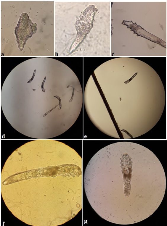
Page 16/17Figure 1
Egg, larva, nymph and adult forms of the parasite; a) D. folliculorum egg (x400), b) Demodex spp., larvae
(x400), c) Demodex spp., nymph (x100), d,e) D. folliculorum egg and adults (x100), f) D. folliculorum
adult (x400), g) D. brevis adult (x400) (Original).
Page 17/17You can also read