Prognostic Factors for Tracheal Restenosis after Stent Removal in Patients with Post-Intubation and Post-Tracheostomy Tracheal Stenosis
←
→
Page content transcription
If your browser does not render page correctly, please read the page content below

Original Article
                       Yonsei Med J 2022 Jun;63(6):545-553
                       https://doi.org/10.3349/ymj.2022.63.6.545                                                      pISSN: 0513-5796 · eISSN: 1976-2437

Prognostic Factors for Tracheal Restenosis after
Stent Removal in Patients with Post-Intubation and
Post-Tracheostomy Tracheal Stenosis
Daegeun Lee, Byeong-Ho Jeong, and Hojoong Kim
Division of Pulmonary and Critical Care Medicine, Department of Medicine, Samsung Medical Center, Sungkyunkwan University
School of Medicine, Seoul, Korea.

Purpose: Long-term tracheal stent placement can increase the risk of stent-related complications; hence, removal of the stent af-
ter stabilization is attempted. However, little evidence has been established regarding the risk factors for tracheal restenosis. We
aimed to identify the risk factors for tracheal restenosis in patients with post-intubation tracheal stenosis (PITS) and post-trache-
ostomy tracheal stenosis (PTTS).
Materials and Methods: We retrospectively analyzed patients with PITS and PTTS between January 2004 and December 2019.
Patients were classified into a success or failure group according to treatment outcomes. Patients with successful stent removal
were defined as patients who did not require additional intervention after stent removal during the follow-up period. Multiple lo-
gistic regression analysis was performed to identify the factors associated with tracheal restenosis.
Results: Among 269 stented patients, 130 patients who had removed the stent were enrolled in this study. During the follow-up
period, 73 (56.2%) patients had a stable clinical course; however, 57 (43.8%) patients had restenosis. The proportion of trauma-in-
duced intubation was higher in the success group than in the failure group (p=0.026), and the median stent length was shorter in
the success group (45 mm) than in the failure group (50 mm, p=0.001). On multivariate analysis, trauma-induced intubation [ad-
justed odds ratio (aOR), 0.329; 95% confidence interval (CI), 0.117–0.927; p=0.036], and stent length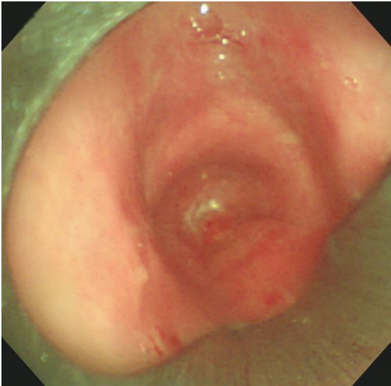
Tracheal Restenosis after Stent Removal

tients, bronchoscopic intervention is considered as an alter-         depending on the characteristics and subtype of tracheal ste-
native to surgical management. Endoscopic techniques for              nosis as well as the patient’s medical condition.
the management of tracheal stenosis include mechanical or                Airway stent insertion was considered when laser treatment
balloon dilatation, laser ablation therapy, and endotracheal          or mechanical dilatation did not satisfactorily maintain airway
stent insertion.9 Tracheal stenting is indicated when airway          patency in the patient. It was implanted using the standard
patency cannot be preserved despite other endoscopic thera-           technique described by Dumon.19 The appropriate stent size
peutic techniques. However, long-term stent placement can in-         was determined by an interventionist by measuring the actual
crease the risk of stent-related late complications, such as mu-      length of stenosis through the scale mark of the flexible bron-
costasis, granulation tissue formation, and stent migration.10,11     choscope. During the study period, three types of silicone stents
Therefore, after maintaining the stent for a certain period, stent    were used for the treatment of tracheal stenosis: Natural stent
removal is attempted in patients who are expected to maintain         (M1S Co. Seoul, South Korea), Dumon stent (Novatech, La Ci-
airway patency. Our previous studies have demonstrated that           otat, France), and Montgomery T-tube (Koken, Tokyo, Japan).
the success rate of tracheal stenting is approximately 40%.12-14      Natural stent was developed at the Samsung Medical Center in
However, approximately 60% of patients require repeated en-           2002 and was used for the treatment of benign tracheobronchi-
doscopic intervention and/or surgical treatment. Furthermore,         al stenosis. Studies in a canine model of tracheal stenosis and
prognostic factors for post-tuberculous bronchial stenosis            in patients with benign tracheobronchial stenosis have dem-
(PTBS) after stent removal have been reported in several stud-        onstrated that the Natural stent is as effective and safe as the
ies15-17; however, to date, prognostic factors for tracheal steno-    Dumon stent.20,21 However, since 2015, the production of Nat-
sis in PITS/PTTS patients after stent removal have not been           ural stent has been suspended due to commercial issues, and
well elucidated. In actual clinical practice, tracheal stenosis is    Dumon stents have been mainly used in clinical practice. A
more likely to cause respiratory failure due to central airway        Montgomery T-tube was considered in patients with a high risk
obstruction. Therefore, it is important to predict which patients     of mucostasis or tracheal stenosis close to the glottis.22,23
are at a high risk of restenosis before considering stent remov-         Following stent placement, patients underwent simple chest
al in patients with tracheal stenosis. Accordingly, we attempt-       radiography and spirometry for evaluation of their condition
ed to identify potential risk factors for tracheal restenosis after   at 1, 3, 6, 9, and 12 months after the intervention. We used bron-
stent removal in patients with PITS and PTTS.                         choscopy and chest CT to reevaluate the stent location and air-
                                                                      way patency if the patients complained of respiratory symptoms
                                                                      and had abnormal findings on chest radiography or before
MATERIALS AND METHODS                                                 planning their stent removal. Stent removal was generally con-
                                                                      sidered when the patients maintained the stent in a stable con-
Patients                                                              dition for more than 6 months and air pockets were detected
We retrospectively reviewed all patients with airway stenosis         on chest CT. To confirm a stable condition after stent removal,
who underwent rigid bronchoscopy between January 2004                 follow-up was performed at least once within 1 month after re-
and December 2019 at Samsung Medical Center, which is a               moval.
1979-bed referral hospital and performs the most rigid bron-             If symptoms and signs of airway stenosis developed after
choscopic interventions in South Korea. The Institutional Re-         removal of the tracheal stent, interventional bronchoscopy
view Board of Samsung Medical Center approved the collec-             and/or stent insertion were repeated in the patient. Surgical
tion, analysis, and publication of the data (IRB No. 2021-07-         management or permanent tracheostomy was performed in
130). The requirement for informed consent was waived due to          patients with ineffective or intolerable tracheal stents.
the retrospective nature of the study.
                                                                      Data collection and clinical outcome
Airway intervention techniques, follow-up and                         We collected data from the study population including demo-
removal of the stent                                                  graphics, etiology, characteristics of tracheal stenosis, stent du-
The airway anatomy was evaluated using chest radiography,             ration, and additional interventional and radiologic data before
computed tomography (CT), and flexible bronchoscopy. Air-             stent removal. We used the American Society of Anesthesiolo-
way interventions were performed according to standard tech-          gists (ASA) physical status classification to evaluate the indi-
niques.9,18 After induction of general anesthesia and intuba-         vidual performance status.24 An ASA physical status ≥3 was
tion with a rigid bronchoscope tube (Bryan Co.,Woburn, MA,            considered a poor performance status. To evaluate the severity
USA), a flexible bronchoscope (BF 1T260 Olympus Corpora-              of tracheal stenosis, we used the Myer–Cotton stenosis grading
tion, Tokyo, Japan) was introduced through the rigid broncho-         system.25
scope tube to examine airway stenosis. Various combinations              We measured the air pocket length, air pocket score, and air
of airway intervention techniques (mechanical dilatation, bal-        pocket density on CT scans before removal of the stent. Based
loon dilatation, laser, and silicone stent insertion) were used       on a previous study,17 an air pocket was defined as a tracheo-

546                                                                                                   https://doi.org/10.3349/ymj.2022.63.6.545
Daegeun Lee, et al.

bronchial air column in the space between the outer surface                    RESULTS
of the stent and the adjacent airway wall. The air pocket score
was calculated as the summation of the number of quadrants                     The overall clinical course of the study population is summa-
containing air pockets in all CT sections extending from the                   rized in Fig. 1. During the study period, 357 patients were di-
proximal to the distal end of the stent. In addition, we intro-                agnosed with PITS (n=215) and PTTS (n=142). In 269 of the
duced the concept of air pocket density, which is defined as the               357 patients who received stents for tracheal stenosis, stent
value obtained after dividing the air pocket score by the stent                removal was performed in 143 patients. After excluding 13 pa-
length.                                                                        tients who were not followed up after stent removal, a total of
   Clinical outcomes were evaluated during the follow-up pe-                   130 patients were enrolled in the study. Of the 130 patients, 73
riod after stent removal. Patients were divided into two groups                (56.2%) had a stable clinical course after removal of the tra-
according to treatment outcomes: success and failure. The                      cheal stent. However, 57 (43.8%) patients underwent re-inter-
success group comprised patients whose airway patency was                      vention for restenosis. Most restenosis cases (88%) developed
maintained without an airway prosthesis during the follow-                     within 3 months of stent removal. Fig. 2 shows representative
up period, and the failure group comprised patients with recur-                cases of success and failure of tracheal stent removal in pa-
rence of symptoms and signs of tracheal stenosis during the                    tients with PITS/PTTS.
follow-up period that required additional endoscopic inter-
vention. Patients with successful stent removal were defined as                Baseline characteristics
those who did not require additional intervention after stent                  The median age was 56 years (IQR, 45–66), and 64 (49.2%) pa-
removal during the follow-up period.                                           tients were male (Table 1). Ninety-eight (75.4%) patients were
                                                                               intubated for medical reasons, while 32 (24.6%) patients were
Statistical analysis                                                           intubated for surgical reasons. No significant difference was
SPSS software (IBM SPSS statistics version 27, IBM Corp., Ar-                  found in baseline characteristics between the two groups ex-
monk, NY, USA) was used for all statistical analyses. Data are                 cept for the proportion of patients who underwent trauma-in-
presented as number (%) or interquartile range (IQR). Cate-                    duced endotracheal intubation, which was more frequent in
gorical variables were compared using Pearson’s chi-square                     the successful group than in the failure group (26.0% vs. 10.5%,
test or Fisher’s exact test. Continuous variables were compared                p=0.026). Due to dyspnea, only 58 (44.6%) of the patients were
using Mann-Whitney U test. Multivariate logistic regression                    able to perform the pulmonary function test. The median forced
analysis with backward stepwise selection was performed to                     expiratory volume in 1 second was 54% of the predicted value
determine the risk factors for tracheal restenosis after stent re-             (IQR, 34–71), and the median forced vital capacity was 82% of
moval. Statistical significance was set at p
Tracheal Restenosis after Stent Removal

 A                                    B                                     C                                    D

 E                                    F                                    G                                     H
Fig. 2. Representative cases of success (A–D) and failure (E–H) of tracheal stent removal. (A–D) A 56-year-old female patient underwent intubation
for 10 days due to subarachnoid hemorrhage. Two months later, she complained of dyspnea. (A) CT scan showing tracheal stenosis at the level of the
thoracic inlet (arrow). (B) Bronchoscopic findings of tracheal stenosis. A 40-mm tracheal stent was inserted and maintained for 19 months. (C) CT
scan before stent removal. (D) Chest radiograph obtained 1 year after stent removal. During the 1-year follow-up period after stent removal, the pa-
tient remained stable without restenosis. (E–F) A 46-year-old female patient underwent intubation for 3 weeks due to epilepsy. She complained of dys-
pnea 2 weeks after extubation. (E) CT scan showing stenosis of the upper trachea (arrow). The patient underwent an emergency tracheostomy for tra-
cheal stenosis (arrow). (F) Bronchoscopy shows a near-complete obstruction of the trachea above the stoma. A 60-mm tracheal stent was inserted and
maintained for 2 years. (G) CT scan before stent removal. Ten days after stent removal, the patient complained of dyspnea again and (H) the CT scan
showed tracheal restenosis (arrow). CT, computed tomography.

The median length of the stenosis was 30 mm (IQR, 25–35                      12 months. Before stent removal, 45 (34.6%) patients under-
mm). The location, severity, and length of stenosis were similar             went additional interventional bronchoscopy. In most of the
between the two groups. Among the stenosis types, granula-                   patients, the stent was removed after confirming the presence
tion was more frequently observed in the failure group than in               of air pockets (106/130, 81.5%). There was no difference be-
the success group (28.1% vs. 12.3%, p=0.024). Forty-one (31.5%)              tween the success (8/57, 14.0%) and failure (16/73, 21.9%)
patients suffered from respiratory failure due to tracheal steno-            groups in the proportion of patients whose air pockets could
sis. Owing to respiratory failure in patients, intubation (n=17),            not be identified due to the lack of available CT scans before
emergency tracheostomy (n=23), and extracorporeal mem-                       stent removal (p=0.267).
brane oxygenation (n=2) were performed before intervention-                     The median air pocket length, air pocket score, and median
al bronchoscopy. The median duration of intubation and tra-                  value of air pocket density were 32.5 mm (IQR, 27.5–40.0), 26
cheostomy was 10 days (IQR, 7–15) and 60 days (IQR, 35–116),                 (IQR 20–36), and 0.57 (IQR 0.38–0.80), respectively. None of
respectively.                                                                these air pocket indices showed a statistically significant dif-
                                                                             ference between the two groups.
Treatment modalities and characteristics of stent
Natural stents were most commonly used in the patients (91/                  Clinical outcomes and prognostic factors for tracheal
130, 70.0%) (Table 3). The median stent length was 45 mm (IQR,               restenosis
40–50 mm). Compared to the failure group, the success group                  Tracheal restenosis occurred in 57 (43.8%) patients after stent
had a shorter stent length (45 mm vs. 50 mm, p=0.001). The                   removal during the follow-up period. The median time-to-re-
proportion of patients with a stent length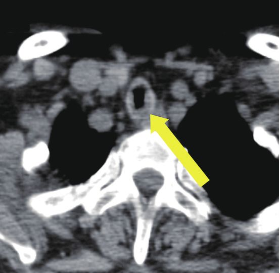
Daegeun Lee, et al.

Table 1. Baseline Characteristics of the Two Groups
                                                          Total (n=130)                 Success (n=73)                 Failure (n=57)             p value
Age, yr                                                   56 (45–66)                     54 (45–70)                     58 (45–64)                  0.877
Sex, male                                                 64 (49.2)                      39 (53.4)                      25 (43.9)                   0.279
BMI, kg/m2                                               22.9 (20.4–25.5)               22.6 (20.4–25.9)              23.1 (20.4–25.3)              0.827
Comorbidities
   DM                                                        39 (30.0)                    20 (27.4)                     19 (33.0)                    0.464
   Neurologic sequelae                                       35 (26.9)                    22 (30.1)                     13 (22.8)                    0.350
   Cardiovascular disease                                    29 (22.3)                    15 (20.5)                     14 (24.6)                    0.585
   Chronic lung disease                                      15 (11.5)                     7 (9.6)                       8 (14.0)                    0.431
Cause of intubation or tracheostomy
   Medical                                                   98 (75.4)                    52 (71.2)                     46 (80.7)                    0.214
     Respiratory failure                                     26 (20.0)                    16 (21.9)                     10 (17.5)                    0.536
     Neurologic disease                                      26 (20.0)                    12 (16.4)                     14 (24.6)                    0.251
     Cardiovascular disease                                  21 (16.2)                    11 (15.1)                     10 (17.5)                    0.704
     Drug intoxication                                       12 (9.2)                      6 (8.2)                       6 (10.5)                    0.652
     Septic shock                                            10 (7.7)                      5 (6.8)                       5 (8.8)                     0.748
     Other*                                                   3 (2.3)                      2 (2.7)                       1 (1.8)                    >0.999
   Surgical                                                  32 (24.6)                    21 (28.8)                     11 (19.3)                    0.214
     Trauma                                                  25 (19.2)                    19 (26.0)                      6 (10.5)                    0.026
     Postoperative                                            7 (5.4)                      2 (2.7)                       5 (8.8)                     0.239
ASA physical status ≥3†                                      51 (39.2)                    30 (41.1)                     21 (36.8)                    0.622
Baseline spirometry (n=58)‡
   FEV1, % predicted                                         54 (34–71)                   51 (30–70)                    61 (43–72)                   0.375
   FVC, % predicted                                          82 (68–92)                   85 (65–96)                    78 (68–90)                   0.332
BMI, body mass index; DM, diabetes mellitus; ASA, American Society of Anesthesiologists; FEV1, forced expiratory volume in 1 s; FVC, forced vital capacity;
COPD, chronic obstructive pulmonary disease.
Data are presented as n (%) or median (interquartile range).
*Diabetic ketoacidosis, hypoglycemia, and obesity; †ASA physical status 3 indicates patients with severe systemic disease (e.g., poorly controlled DM or hyper-
tension, COPD); ‡Spirometry before stent insertion was available for 58 patients.

with a decreased risk of tracheal restenosis (aOR 0.274; 95% CI                      In the present study, the most common cause of trauma (80%)
0.130–0.578; p=0.001) (Table 4).                                                  that required tracheal intubation was brain injury after traumat-
                                                                                  ic events. Therefore, when inferred from the multivariate results,
                                                                                  we can suspect that the prognosis of traumatic brain injury is
DISCUSSION                                                                        better than that of non-traumatic brain injury, such as stroke,
                                                                                  epilepsy, and other medical problems, in patients with tracheal
We compared the differences between the groups that success-                      stents. This result was consistent with previous research sug-
fully maintained and failed to maintain airway patency after                      gesting that patients with traumatic brain injury show greater
stent removal and tried to identify the prognostic factors for re-                functional improvement than those with non-traumatic brain
stenosis in patients with PITS and PTTS. In our study popula-                     injury,29 and successful removal of airway prosthesis is corre-
tion, 56% of the patients had successful stent removal without                    lated with performance status.13
restenosis. We showed that trauma-induced intubation and                             Our study demonstrated that the length of the tracheal stent
stent length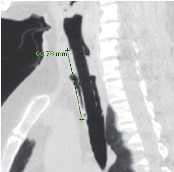
Tracheal Restenosis after Stent Removal

expiration state. Therefore, we believe that the length of the                     restenosis. Furthermore, the tracheal stent is a foreign body,
stent determined during the procedure better reflects the actual                   and a longer stent length results in an increased area of tracheal
length of stenosis and has a meaningful result associated with                     mucosal irritation and inflammation, leading to greater gran-

Table 2. Characteristics of Tracheal Stenosis
                                                           Total (n=130)               Success (n=73)                Failure (n=57)             p value
Etiology of tracheal stenosis                                                                                                                     0.207
   Post-intubation                                          93 (71.5)                    49 (52.7)                    44 (46.7)
   Post-tracheostomy                                        37 (28.5)                    24 (64.9)                    13 (35.1)
Location of stenosis                                                                                                                              0.904
   Subglottis to upper trachea                             101 (77.7)                    57 (78.1)                    44 (77.2)
   Mid to lower trachea                                     29 (22.3)                    16 (21.9)                    13 (22.8)
Severity of stenosis* (myer-cotton grade)                                                                                                         0.570†
   I                                                         6 (4.6)                       4 (5.5)                     2 (3.5)
   II                                                       34 (26.2)                    18 (24.7)                    16 (28.1)
   III                                                      86 (66.2)                    50 (68.5)                    36 (63.2)
   IV                                                        4 (3.1)                       1 (1.4)                     3 (5.3)
Length of stenosis, mm                                      30 (25–35)                   30 (26–35)                   29 (25–35)                  0.886
Stenosis type‡
   Fibrosis                                                112 (86.2)                    64 (87.7)                    48 (84.2)                   0.571
   Granulation                                              25 (19.2)                      9 (12.3)                   16 (28.1)                   0.024
   Malacia                                                  10 (7.7)                       8 (11.0)                    2 (3.5)                    0.184
   Mixed                                                    17 (13.1)                      8 (11.0)                    9 (15.8)                   0.418
Respiratory failure before intervention§                    41 (31.5)                    18 (24.7)                    23 (40.4)                   0.056
Intubation duration, day (n=92)¶                            10 (7–15)                    10 (7–15)                    11 (7–17)                   0.575
Tracheostomy duration, day (n=35)‖                          60 (35–116)                  47 (27–104)                  66 (46–217)                 0.170
Time interval of injury to stenosis, day                    62 (38–107)                  65 (38–115)                  61 (39–96)                  0.620
   PITS                                                     52 (36–87)                   51 (36–85)                   54 (36–89)                  0.936
   PTTS                                                     93 (61–387)                 107 (65–384)                  78 (61–390)                 0.484
PITS, post-intubation tracheal stenosis; PTTS, post-tracheostomy tracheal stenosis; ECMO, extracorporeal membrane oxygenation.
Data are presented as n (%) or median (interquartile range).
*Categorization based on the percentage reduction in cross-sectional area Grade I, ≤50% luminal stenosis; Grade II, 51%–70% luminal stenosis; Grade III,
71%–99% luminal stenosis; and Grade IV, no lumen; †p for trend=0.683; ‡Patients (n=17) had more than one type of stenosis; §Intubation (n=17), tracheostomy
(n=23), ECMO (n=2) state before interventional bronchoscopy; ¶Missing value=38; ‖Missing value=2.

Table 3. Treatment Modalities and Characteristics of Stent
                                                            Total (n=130)                 Success (n=73)                  Failure (n=57)             p value
Stent type
   Natural stent                                             91 (70.0)                      51 (69.9)                      40 (70.2)                    0.969
   Dumon stent                                               35 (26.9)                      21 (28.8)                      14 (24.6)                    0.592
   Montgomery t-tube                                          3 (2.3)                        1 (1.4)                        2 (5.3)                     0.319
Stent length, mm                                             45 (40–50)                     45 (40–50)                     50 (43–50)                   0.001
Stent length 18 months                              54 (41.5)                      31 (42.5)                      23 (40.4)                    0.808
Additional intervention before stent removal*                45 (34.6)                      22 (30.1)                      23 (40.4)                    0.225
Air pocket in CT before stent removal (n=106)†
   Air pocket length, mm                                  32.5 (27.5–40.0)                34.0 (27.8–41.0)               32.5 (26.3–37.5)               0.345
   Air pocket score                                          26 (20–36)                     25 (20–35)                     26 (20–35)                   0.522
   Air pocket density‡                                    0.57 (0.38–0.80)                0.56 (0.39–0.87)               0.58 (0.38–0.73)               0.235
Data are presented as n (%) or median (interquartile range).
*Reasons for additional interventions included stent migration (n=25), granulation tissue overgrowth (n=16), additional stenosis (n=8), mucostasis (n=3), and ma-
lacia (n=2). Nine patients had more than one reason; †24 patients did not have available CT scans before stent removal; ‡Air pocket score/stent length.

550                                                                                                                      https://doi.org/10.3349/ymj.2022.63.6.545
Daegeun Lee, et al.

Table 4. Factors associated with Tracheal Restenosis after Stent Removal
                                                                        Univariate analysis                             Multivariate analysis
                                                                    OR (95% CI)             p value                   aOR (95% CI)            p value
Age                                                              1.000 (0.980–1.020)         0.992                          -                    -
Sex, male                                                        0.680 (0.339–1.367)         0.280                          -                    -
BMI                                                              0.986 (0.907–1.072)         0.747                          -                    -
Comorbidities
   DM                                                         1.325 (0.624–2.815)              0.464                        -                    -
   Neurologic sequelae                                        0.685 (0.309–1.517)              0.351                        -                    -
   Cardiovascular disease                                     1.259 (0.550–2.882)              0.586                        -                    -
   Chronic lung disease                                       1.539 (0.523–4.531)              0.434                        -                    -
Cause of intubation or tracheostomy
   Respiratory failure                                        0.758 (0.315–1.826)              0.537                        -                    -
   Neurologic disease                                         1.655 (0.697–3.928)              0.253                        -                    -
   Cardiovascular disease                                     1.199 (0.470–3.059)              0.704                        -                    -
   Trauma                                                     0.334 (0.124–0.904)              0.031             0.329 (0.117–0.927)           0.036
ASA physical status ≥3                                        0.836 (0.410–1.704)              0.622                        -                    -
Etiology of tracheal stenosis
   Post-intubation                                                 Reference                     -                      Reference                -
   Post-tracheostomy                                          0.665 (0.306–1.444)              0.302                        -                    -
Location of stenosis
   Subglottis to upper trachea                                     Reference                     -                      Reference                -
   Mid to lower trachea                                       1.053 (0.459–2.416)              0.904                        -                    -
Severity of stenosis
   Grade I–II                                                      Reference                     -                      Reference                -
   Grade III–IV                                               0.935 (0.442–1.978)              0.860                        -                    -
Length of stenosis                                            1.015 (0.961–1.072)              0.592                        -                    -
Stenosis type
   Fibrosis                                                   0.750 (0.277–2.032)              0.572                        -                    -
   Granulation                                                2.775 (1.122–6.866)              0.027                        -                    -
   Malacia                                                    0.295 (0.060–1.450)              0.133                        -                    -
   Mixed                                                      1.523 (0.548–4.237)              0.420                        -                    -
Respiratory failure before intervention                       2.067 (0.976–4.378)              0.058                        -                    -
Stent length 30 mm was associated with                  in patients with PITS and PTTS. Additionally, we introduced
a reduced chance of procedural success.31 Eventually, seven of                    the concept of air pocket density, which was defined as the
the patients who had a stent ≥50 mm in our study underwent                        value obtained by dividing the air pocket score with the stent
surgical treatment. Based on these results, if surgical treatment                 length. The air pocket score, length, and density did not differ
is feasible for a patient expected to require a long stent, it may                significantly between the two groups. The previous PTBS study
be an option to consider surgery from the very beginning.                         was conducted without knowledge of the importance of the air
   Verma, et al.17 reported that the extent of air pockets in chest               pocket. However, after knowing the importance of air pockets,
CT was a prognostic factor in patients with PTBS. In this study,                  most patients in this study attempted to remove stents when
the air pocket score was defined as the number of quadrants                       air pockets were present. We believe that these differences in
containing air pockets in section 1 (most proximal section, cra-                  study subjects resulted in a negative result of air pockets for re-

https://doi.org/10.3349/ymj.2022.63.6.545                                                                                                              551
Tracheal Restenosis after Stent Removal
stenosis. 6. Sarper A, Ayten A, Eser I, Ozbudak O, Demircan A. Tracheal ste-
Our study had several limitations. First, since this was a ret- nosis aftertracheostomy or intubation: review with special regard
to cause and management. Tex Heart Inst J 2005;32:154-8.
rospective study based on single-center data, it could not rep-
7. Grillo HC, Donahue DM, Mathisen DJ, Wain JC, Wright CD. Pos-
resent all patients with PITS and PTTS. Furthermore, our cen- tintubation tracheal stenosis. Treatment and results. J Thorac Car-
ter performed the most rigid bronchoscopic interventions in diovasc Surg 1995;109:486-92.
South Korea, and patients with complex airway stenosis and 8. Nouraei SA, Ma E, Patel A, Howard DJ, Sandhu GS. Estimating the
those who failed the procedure were referred to our hospital. population incidence of adult post-intubation laryngotracheal
stenosis. Clin Otolaryngol 2007;32:411-2.
Therefore, a selection bias may have occurred in this study. Sec-
9. Kim H. Stenting therapy for stenosing airway disease. Respirology
ond, since most of the patients had intubation or tracheostomy 1998;3:221-8.
performed in other hospitals, there were incomplete and miss- 10. Ryu YJ, Kim H, Yu CM, Choi JC, Kwon YS, Kwon OJ. Use of silicone
ing data for the exact periods of intubation and tracheostomy stents for the management of post-tuberculosis tracheobronchial
in some patients. Third, we grouped PITS and PTTS together stenosis. Eur Respir J 2006;28:1029-35.
in this study, although our previous study suggested that PITS 11. Shin JH, Song HY, Shim TS. Management of tracheobronchial
strictures. Cardiovasc Intervent Radiol 2004;27:314-24.
and PTTS may have differences in clinical characteristics and 12. Lim SY, Kim H, Jeon K, Um SW, Koh WJ, Suh GY, et al. Prognostic
outcomes.13,32 To overcome these limitations, further prospec- factors for endotracheal silicone stenting in the management of inop-
tive and multicenter studies are required. erable post-intubation tracheal stenosis. Yonsei Med J 2012;53:565-70.
In conclusion, trauma-induced intubation and stent length 13. Shin B, Kim K, Jeong BH, Eom JS, Song WJ, Kang HK, et al. ClinicalDaegeun Lee, et al.
erance and early results in 63 patients. Chest 1996;109:626-9. 31. Freitag L, Darwiche K. Endoscopic treatment of tracheal stenosis.
29. Cullen NK, Park YG, Bayley MT. Functional recovery following Thorac Surg Clin 2014;24:27-40.
traumatic vs non-traumatic brain injury: a case-controlled study. 32. Zias N, Chroneou A, Tabba MK, Gonzalez AV, Gray AW, Lamb CR,
Brain Inj 2008;22:1013-20. et al. Post tracheostomy and post intubation tracheal stenosis: re-
30. Huang S, Xu J, An Z, Yuan P, Xu H, Lv W, et al. Clinical assessment port of 31 cases and review of the literature. BMC Pulm Med 2008;
of airway stent placement in patients with malignant airway le- 8:18.
sions. J Thorac Dis 2018;10:3277-88.
https://doi.org/10.3349/ymj.2022.63.6.545 553You can also read