Prosthetic rehabilitation using a cone morse dental implant and xenogen biomaterial in an aesthetic area: A case report
←
→
Page content transcription
If your browser does not render page correctly, please read the page content below
J Case Rep Images Dent 2020;6:100033Z07JL2020.                                                                    Lopes et al.   1
www.ijcridentistry.com

 CASE REPORT                                                                        PEER REVIEWEDOPEN ACCESS
                                                                                                 | OPEN ACCESS

 Prosthetic rehabilitation using a cone morse dental implant
 and xenogen biomaterial in an aesthetic area: A case report
          João Carlos Amorim Lopes, Saulo Henrique Salviano,
   Marco Antônio Carvalho, Jorge José de Carvalho, Renan Lana Devita,
           Pablo Dallario Ramalho Lucas, Igor da Silva Brum

ABSTRACT                                                            implant, the prosthetic result and the surrounding soft
                                                                    tissues behave more favorably for an aesthetic format
Introduction: With the worldwide growth of aesthetic                more compatible with current standards.
treatments, oral rehabilitation has gained new vestments,
where only the implant placement and their functional               Keywords: Aesthetic, Biomaterial, Cone morse, Dental
activation are not enough. It is necessary to do a whole            implants, Pink aesthetics
three-dimensional planning, taking into account the
remaining bone, possibilities of grafting before, during,                              How to cite this article
or after the implant placement, the final prosthesis and
especially the pink tissues handling and aesthetic. Case             Lopes JCA, Salviano SH, Carvalho MA, de Carvalho
Report: In this case report, we can assess the loss of               JJ, Devita RL, Lucas PDR, Brum IDS. Prosthetic
the dental element 22, which had a broad root lesion                 rehabilitation using a cone morse dental implant and
with almost total loss of the buccal wall. The extraction            xenogen biomaterial in an aesthetic area: A case report. J
of this element was planned and a cone morse dental                  Case Rep Images Dent 2020;6: 100033Z07JL2020.
implant (Systhex® Avantt model, Curitiba, Brazil) was
placed and grafted with a xenogenous biomaterial (Bio
                                                                    Article ID: 100033Z07JL2020
Oss®, Geistlich Pharm, Switzerland). After 9 months, it
was reopened and the provisional prosthesis was made
for tissue management. After this, a porcelain crown                                          *********
was cemented. Conclusion: We can conclude that
when planning correctly and using a cone morse dental               doi: 10.5348/100033Z07JL2020CR

 João Carlos Amorim Lopes1, Saulo Henrique Salviano2,
 Marco Antônio Carvalho3, Jorge José de Carvalho4, Renan
 Lana Devita5, Pablo Dallario Ramalho Lucas2, Igor da Silva
 Brum3                                                              INTRODUCTION
 Affiliations: 1Department of Implantology, Faculty of Den-
                                                                        Oral rehabilitation of toothless spaces with
 tistry, Portuguese Catholic University, Lisboa, Portugal;
 2
   Department of Implantology, Faculty of Dentistry, São
                                                                    osseointegrated dental implants has been a scientifically
 Leopoldo Mandic University, Brasilia, Brazil; 3Department          accepted and well-documented treatment modality for
 of Implantology, Faculty of Dentistry, State University of Rio     years. Since Branemark, in 1908, first discovered the
 de Janeiro, Rio de Janeiro, Brazil; 4Department of Biology         concept of osseointegration, numerous investigations
 (IBRAG), Faculty of Medicine, State University of Rio de           and clinical studies have established titanium as a reliable
 Janeiro, Rio de Janeiro, Brazil; 5Department of Orthodontic,       biomaterial for oral rehabilitation and reconstruction.
 Faculty of Dentistry, State University of Barcelona, Barcelona,    Various modifications to the structure, composition,
 Spain.                                                             and design of titanium dental implants have been made
 Corresponding Author: Igor da Silva Brum, Department of            to improve their physical, mechanical, and optical
 Implantology, Faculty of Dentistry, State University of Rio        properties [1].
 de Janeiro, Rio de Janeiro, Brazil; Email: igor_brum1@hot-             The ultimate goal of a dental implant is to restore
 mail.com
                                                                    missing or extracted teeth, loading anatomical and
                                                                    aesthetical restorations in the long term [2]. Preservation
 Received: 11 May 2020                                              of the alveolar crest and management of the area after
 Accepted: 01 July 2020                                             tooth extraction have a major impact on the volume of
 Published: 20 July 2020                                            hard and soft tissues. The preservation of the socket

                                   Journal of Case Reports and Images in Dentistry, Vol. 6, 2020.
J Case Rep Images Dent 2020;6:100033Z07JL2020. Miguita et al. 2
www.ijcridentistry.com
after extraction is sensitive to the technique, it is not The objective of the present study was to evaluate
100% successful and sometimes unpredictable. Current the gingival, bone, and prosthetic behavior in a case of
techniques can delay the surgical placement of the a extraction of a dental element with periodontal lesion
implant for a few months, and the quality of new bone followed by immediate dental implant placement and
regeneration is questionable [3]. biomaterial grafting. After a nine months healing period,
It is important to follow the clinical protocols for the patient was rehabilitated with a ceramic crown.
placing dental implants in aesthetic areas. Currently, the
principle is the following: each case must be individually
evaluated in order to achieve a satisfactory result: the CASE REPORT
balance of the three-dimensional bone and gingival
architecture with the dental implant and prosthesis. Also Patient
there are other special recommendations for choosing
the best moment for the placement of dental implants in This clinical case study followed the rehabilitation of a
aesthetic areas [4]. The precision in the implant position 31-year-old female patient, leukoderma, who needed oral
in aesthetic areas is more rigorous than in non-aesthetic rehabilitation using biomaterial and dental implants. The
areas. The good management of soft and hard tissues is patient’s main complaint was the pulsating pain she felt
the basis for an aesthetic result. Therefore, the implant in the left lateral incisor in upper arch, which presented
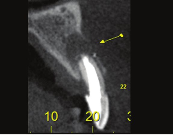
placement in aesthetic areas must follow the exclusive clinically gingival swelling and root exposure (Figure 1)
principles and procedures of the region in question [5]. and radiographically periapical lesion and partial loss of
Although several studies have shown a survival rate the buccal wall (Figure 2).
above 95% of implants in the rehabilitation of the anterior
maxilla in non-compromised patients and 97.9% in single
teeth in the same area after eight years of follow-up, other
authors mention a failure of 10% from the aesthetic point
of view [6]. The dealing with aesthetic complications
in implantology has been very well documented with
scientific evidence, and some protocols have been created,
which raises not only the success rate but the degree of
aesthetic satisfaction [7].
Aesthetic results can be assessed objectively and
subjectively. Subjective assessment can be performed
using the patients’ perception of the aesthetic result that
can be measured using specific questionnaires in which
patients can express their satisfaction or dissatisfaction
[8]. The objective assessment can be performed by a
professional examiner and is based on defined criteria,
aiming at an overall assessment of the harmonic
appearance and the natural integration of the artificial
restoration with the patient’s dentition. Both assessments Figure 1: Initial photo.
must be taken into account to provide a complete overview
of the final aesthetic result. So currently the evaluation
protocols are taken into account when planning, because
the level of satisfaction varies from patient to patient,
which reduces this discrepancy between the perception
of the dentist’s vision and the patient’s desire, obtaining a
more favorable final result for both [9–11].
Data regarding the performance of the implant,
including survival rates, success rates, and peri-implant
bone loss are necessary to understand the advantages and
disadvantages and their aesthetic enhancement [12, 13].
Although immediate implant placement is an attractive
technique because it reduces the number of surgeries
and increases patient satisfaction, it is still premature
to conclude about its long-term results. Brum et al. [14].
claim that the use of biomaterials associated to dental
implants bring excellent aesthetic results in relation to
pink tissues, in addition to having perfect interaction
with the grade IV titanium alloy [15]. Figure 2: Initial radiographic view.
Journal of Case Reports and Images in Dentistry, Vol. 6, 2020.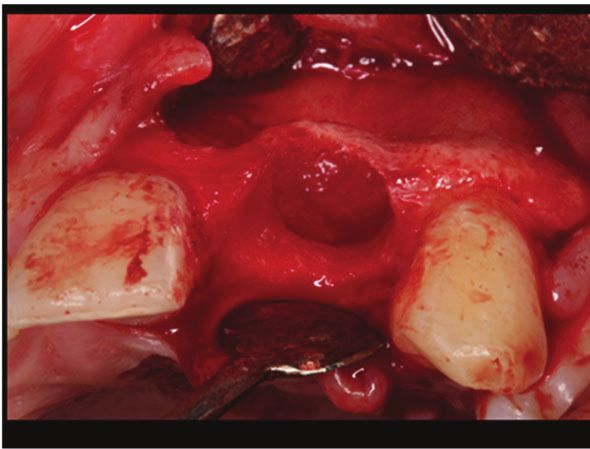
J Case Rep Images Dent 2020;6:100033Z07JL2020. Miguita et al. 3
www.ijcridentistry.com
It was planned to perform the tooth extraction,
implant placement, and biomaterial grafting at the
same surgical moment followed by the placement of a
provisional adhesive prosthesis fixed on adjacent teeth.
Due to an increased mesiodistal space, orthodontic
treatment was suggested in order to correct and adapt it
to normal standards for the region. The follow-up time
from the beginning to the end of the treatment was three
years.
Biomaterial and dental implant
In bone reconstruction surgery, xenogenous
biomaterial Bio Oss® from the company Geistlich Pharm
(Switzerland) was used. The implant used was a 3.5
× 13 mm cone Avantt model from Systhex® company
(Curitiba, Brazil).
Figure 3: Extracted tooth and all inflammatory tissue removed.
Operative, postoperative and prosthetic
management
The patient received antibiotics: 2 g of amoxicillin (4 ×
500 mg capsules) 1 hour after surgery and Clavulin® 785 mg
every 12 hours for 14 days. A rinse of 0.12% chlorhexidine
solution for 1 minute was performed before the operation.
Local anesthesia (2% lidocaine with adrenaline 1:100,000
was administered. Subsequently, an incision was made at
the sulcular level and only one release incision positioned
at the mesial buccal angle of the first upper left premolar.
After this, a full thickness mucoperiosteal displacement
was performed aiming a complete relaxation of the flap
that was elevated to fully expose the alveolar bone, thus,
the upper left lateral incisor was extracted, the endo-
perio lesion was completely removed by curettage and
the dental implant was placed (AvanttSysthex®, 3.5 ×
13mm). All the pre-established concepts for immediate
dental implant placement in alveolus were carried out, Figure 4: Subcrestal placement (buccal wall).
regarding: palatal approach and final positioning about
two to four millimeters subcrestal. The next step was
to carefully fill all “gaps” by the chosen biomaterial
(Bio Oss® from Geistlich Pharm, Switzerland) and then
protect with an absorbable membrane (Bio-Guide® from
Geistlich Pharm, Switzerland) positioned over the entire
length of the wound including the socket entrance. There
was no sign of infection in the entire postoperative course
(Figures 3–7).
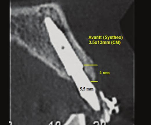
After the reconstruction surgery, the patient
underwent a new tomography where it was possible to
observe the reconstruction of all previously lost alveolar
structure (Figure 8).
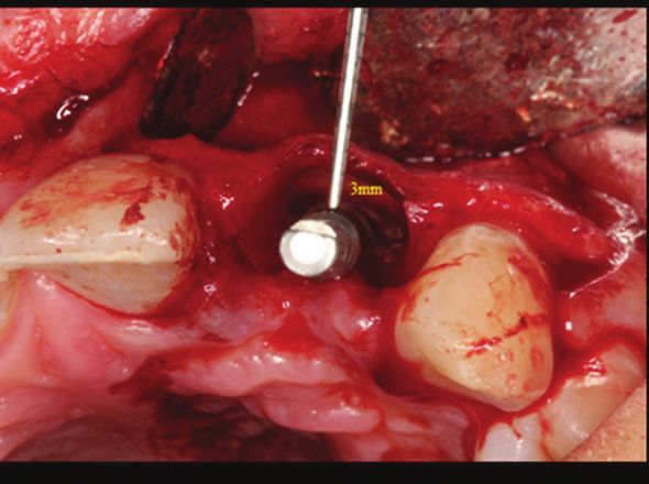
After nine months, the reopening was performed
followed by the provisional implant-supported placement
in order to conditionate the gingival soft tissues (Figure
9). After the transfer and laboratory molding phase, the
final ceramic crown was cemented (Figure 10). The final
photo was obtained after one year of prosthetic function
Figure 5: Palatal positioning favorable for regeneration.
totaling three years of treatment.
Journal of Case Reports and Images in Dentistry, Vol. 6, 2020.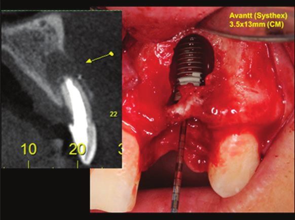
J Case Rep Images Dent 2020;6:100033Z07JL2020.                                                                    Miguita et al.   4
www.ijcridentistry.com

                                                                      Figure 9: Gingival conditioning obtained with the help of the
                                                                      provisional prosthesis.

Figure 6: Filling the gaps with biomaterial.

                                                                      Figure 10: Case completed satisfactorily.

                                                                      DISCUSSION
Figure 7: Closure of the wound.                                          For the placement of an implant in an ideal position,
                                                                      some requirements of soft and hard tissues must be well
                                                                      defined [16]. The authors discuss the best treatment
                                                                      approaches, as well as the limitations associated with
                                                                      aesthetic implant placement.
                                                                         Since anterior maxilla is in greater demand, the
                                                                      authors evaluated data specifically related to this
                                                                      anatomical region and found several parameters and
                                                                      surgical techniques developed to manipulate soft and
                                                                      hard tissue contours to control the aesthetic result in
                                                                      restorations supported by dental implants in this area.
                                                                      These principles were well defined in this case report,
                                                                      in which a more conservative approach was drawn:
                                                                      extraction, immediate dental implant placement,
                                                                      xenogenous biomaterial grafting, reopening followed by
                                                                      gingival conditioning with the provisional prosthesis and
                                                                      definitive prosthesis in porcelain. This provides a more
                                                                      favorable result in the long run [17].
                                                                         A randomized controlled trial with 60 partially
Figure 8: Radiographic view            showing    the   complete      edentulous patients requiring two single crowns supported
reconstruction of the buccal wall.                                    by two dental implants was conducted in six different

                                     Journal of Case Reports and Images in Dentistry, Vol. 6, 2020.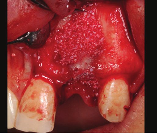
J Case Rep Images Dent 2020;6:100033Z07JL2020.                                                                 Miguita et al.   5
www.ijcridentistry.com

study centers. For three months the dental implants              failures, 3.09%). It is suggested that the placement of
were randomly placed 0.5 mm or 1.5 mm below the bone             dental implants in fresh alveolus affects failure rates [25,
crest in aesthetic and non-aesthetic areas according to          26].
the divided mouth dental region. Two months after the                It is very important to mention that soft tissue
surgery, the provisional acrylic crowns were replaced by         manipulation is an important step in the aesthetic
definitive metal-ceramic crowns. Patients were followed          rehabilitation process, as was well described in [27],
up for three years after completion of treatment.                that found that granulation tissue originating from the
    They concluded that considerable clinical differences        periodontal ligament or connective tissue originally
were not observed when placing implants 0.5 mm or 1.5            covered by keratinized epithelium has the potential to
mm subcrestal, in relation to aesthetic and non-aesthetic        induce keratinization. However, it also appears that the
areas [18]. It corroborates with Gualini et al. [19] who in      deep palatal connective tissue may not have the same
a similar study obtained the same statistical results with       potential to induce keratinization as the palatal connective
the same indications.                                            tissue originating from an immediately subepithelial area.
    In a study [20] with 106 patients who needed a single        Approximately 14 days after surgery the peri-implant
post-extraction dental implant placement, they were              soft connective tissue already resembles a scar tissue in
separated as follows: (immediate group; 54 patients),            the original composition, due to the orientation of the
(delayed group; 52 patients). Four months after the              fibers and vascularization. On the other hand, the peri-
preservation of the alveolus the late implants were              implant supra crestal epithelium can reach a greater final
placed on the delayed group. Dental implants placed              length under certain conditions, such as dental implants
with 35 Ncm torque or more were immediately loaded               placement in fresh sockets, which was well observed in
with provisional non-occlusive unitary crowns and                this case and demonstrated by other authors [28–32].
then replaced after four months by permanent crowns.
As results they obtained: 19 dental implants (35%)
were not loaded immediately in the immediate group,              CONCLUSION
against 39 (75%) in the late positioning group, because
it was not possible to obtain a 35 Ncm torque or more.              We can conclude that by observing the techniques
No patient gave up. Two dental implants (4%) failed in           based on the existing literature, the use of a Cone Morse
the immediate group versus none in the delayed group.            dental implant generates a better prosthetic result, with
A higher number of lesser complications occurred in the          the surrounding soft tissues behaving more favorably
immediate group (8) in comparison with the delayed               for an aesthetic format more compatible with current
group (1). This was statistically significant (p = 0.032).       standards.
Upon delivery of the definitive crowns, four months
after loading, the aesthetics were scored at 12.8 and 12.6
in the immediate and late groups, respectively, with no          REFERENCES
statistically significant difference (p = 0.5). Patients in
both groups were equally satisfied what is in agreement               1.   Tarnow D, Elian N, Fletcher P, et al. Vertical
with other authors [21–23] and with the case reported                      distance from the crest of bone to the height of the
                                                                           interproximal papilla between adjacent implants. J
in this study, where the late loading did not present any
                                                                           Periodontol 2003;74(12):1785–8.
problems after the delivery of the definitive prosthesis,             2.   Benic GI, Mir-Mari J, Hämmerle CH. Loading
and the patient was very satisfied with the aesthetics.                    protocols for single-implant crowns: A systematic
    In a systematic review the authors [24] identified 30                  review and meta-analysis. Int J Oral Maxillofac
eligible studies. A total of 3049 dental implants were                     Implants 2014;29:222–38.
placed in a total of 1435 patients, with a mean of 46.68              3.   Leblebicioglu B, Rawal S, Mariotti A. A review of
years age and a minimum of six months of follow-up. The                    the functional and esthetic requirements for dental
survival rate of delayed loading dental implants (98.38%)                  implants. J Am Dent Assoc 2007;138(3):321–9.
was significantly higher than immediate loading dental                4.   Babu PJ, Alla RK, Alluri VR, Datla SR, Konakanchi A.
                                                                           Dental ceramics: Part I – An overview of composition,
implants (95.21%) (p = 0.001). For marginal bone loss
                                                                           structure and properties. American Journal of
(p = 0.32), dental implant stability coefficients (p =                     Materials Engineeringand Technology 2015;3(1):13–
0.44), and pocket probe depth (p = 0.94), there was no                     8.
significant difference between delayed and immediate                  5.   Slagter KW, Meijer HJA, Bakker NA, Vissink A,
loading.                                                                   Raghoebar GM. Feasibility of immediate placement
    Immediate dental implants placed in newly extracted                    of single-tooth implants in the aesthetic zone: A
areas should be performed with caution, due to                             1-year randomized controlled trial. J Clin Periodontol
significantly lower survival rates than late dental implants               2015;42(8):773–82.
placed in healed cavities. This is in agreement with                  6.   Huynh-Ba G, Meister DJ, Hoders AB, et al. Clinical
                                                                           and patient-centered outcomes of immediately
another author who, in a study with 8241 dental implants
                                                                           placed implants (Type 1) and rarly placed implants
placed in alveolus, obtained (330 failures, 4.00%) in                      (Type 2): Preliminary 3-month results of an ongoing
contrast to 19,410 dental implants in healed sites (599                    randomized controlled clinical trial. Clin Oral

                                Journal of Case Reports and Images in Dentistry, Vol. 6, 2020.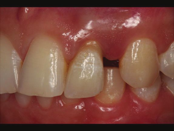
J Case Rep Images Dent 2020;6:100033Z07JL2020.                                                                     Miguita et al.   6
www.ijcridentistry.com

       Implants Res 2016;27(2):241–52.                                        after loading results of a multicentre within-person
   7.  Kolinski ML, Cherry JE, McAllister BS, Parrish                         randomised controlled trial. Int J Oral Implantol
       KD, Pumphrey DW, Schroering RL. Evaluation of a                        (Berl) 2019;12(2):155–67.
       variable-thread tapered implant in extraction sites              21.   Felice P, Pistilli R, Barausse C, Trullenque-Eriksson
       with immediate temporization: A 3-year multicenter                     A, Esposito M. Immediate non-occlusal loading of
       clinical study. J Periodontol 2014;85(3):386–94.                       immediate post-extractive versus delayed placement
   8. Lang NP, Zitzmann NU. Working group 3 of the                            of single implants in preserved sockets of the anterior
       VEWOP. Clinical research in implant dentistry:                         maxilla: 1-year post-loading outcome of a randomised
       Evaluation of implant-supported restorations,                          controlled trial. Eur J Oral Implantol Winter
       aesthetic and patient-reported outcomes. J Clin                        2015;8(4):361–72.
       Periodontol 2012;39(12):133–8.                                   22.   Esposito M, Zucchelli G, Cannizzaro G, et al.
   9. Gupta G, Gupta R, Gupta N, Gupta U. Crown                               Immediate, immediate-delayed (6 Weeks) and
       lengthening procedures-A review article. IOSR Journal                  delayed (4 Months) post-extractive single implants:
       of Dental and Medical Sciences 2015;14(4):27–37.                       1-year post-loading data from a randomised controlled
   10. Chun YP, Raffelt C, Pfeiffer H, et al. Restoring strength              trial. Eur J Oral Implantol 2017;10(1):11–26.
       of incisors with veneers and full ceramic crowns. J              23.   Bavetta G, Bavetta G, Randazzo V, et al. A
       Adhes Dent 2010;12(1):45–54.                                           Retrospective study on insertion torque and implant
   11. Fava J, Lin M, Zahran M, Jokstad A. Single implant-                    stability quotient (ISQ) as stability parameters for
       supported crowns in the aesthetic zone: Patient                        immediate loading of implants in fresh extraction
       satisfaction with aesthetic appearance compared                        sockets. Biomed Res Int 2019;2019:9720419.
       with appraisals by laypeople and dentists. Clin Oral             24.   Felice P, Soardi E, Piattelli M, Pistilli R, Jacotti M,
       Implants Res 2015;26(10):1113–20.                                      Esposito M. Immediate non-occlusal loading of
   12. De Carvalho PFM, Joly JC, Da Silva RC, González-                       immediate post-extractive versus delayed placement
       Martín O. Therapeutic alternatives for addressing                      of single implants in preserved sockets of the
       pink esthetic complications in single-tooth implants:                  anterior maxilla: 4-month post-loading results from
       A proposal for a clinical decision tree. J Esthet Restor               a pragmatic multicentre randomised controlled trial.
       Dent 2019;31(5):403–14.                                                Eur J Oral Implantol 2011;4(4):329–44.
   13. Tischler M. Dental implants in the aesthetic zone.               25.   Mello CC, Lemos CAA, Verri FR, Santos DMD, Goiato
       Dent Today 2016;35(1):104,106.                                         MC, Pellizzer EP. Immediate implant placement into
   14. Brum IS, de Carvalho JJ, Pires JLS, de Carvalho MAA,                   fresh extraction sockets versus delayed implants
       Santos LBFD, Elias CN. Nanosized hydroxyapatite                        into healed sockets: A systematic review and meta-
       and β-tricalcium phosphate composite: Physico-                         analysis. Int J Oral Maxillofac Surg 2017;46(9):1162–
       chemical, cytotoxicity, morphological properties and                   77.
       in vivo trial. Sci Rep 2019;9(1):19602.                          26.   Han CH, Mangano F, Mortellaro C, Park KB.
   15. Brum IS, De Carvalho MAA, Santos PGPD, Devita                          Immediate loading of tapered implants placed in
       RL, Pires JLS, de Carvalho JJ. Ultrastructural                         postextraction sockets and healed sites. J Craniofac
       characterization of the titanium surface degree IV in                  Surg 2016;27(5):1220–7.
       dental implant aluminum free (Acid Attack). Journal              27.   Chrcanovic BR, Albrektsson T, Wennerberg A. Dental
       of Biomaterials and Nanobiotechnology 2020;11:151–                     implants inserted in fresh extraction sockets versus
       160.                                                                   healed sites: A systematic review and meta-analysis.
   16. Kutkut A, Andreana S, Monaco E. Esthetic                               J Dent 2015;43(1):16–41.
       consideration for alveolar socket preservation prior             28.   Sculean A, Gruber R, Bosshardt DD. Soft tissue
       to implant placement: Description of a technique and                   wound healing around teeth and dental implants. J
       80-case series report. Gen Dent 2012;60(6):e398–                       Clin Periodontol 2014;41 Suppl 15:S6–22.
       403.                                                             29.   Tomasi C, Tessarolo F, Caola I, Wennström J, Nollo G,
   17. Canellas JVDS, Medeiros PJD, Figueredo CMS,                            Berglundh T. Morphogenesis of peri-implant mucosa
       Fischer RG, Ritto FG. Which Is the best choice after                   revisited: An experimental study in humans. Clin Oral
       tooth extraction, immediate implant placement or                       Implants Res 2014;25(9):997–1003.
       delayed placement with alveolar ridge preservation?              30.   Zhang C, Tan CK, McFarlane C, Sharma M, Tan
       A systematic review and meta-analysis. J                               NS, Kambadur R. Myostatin-null mice exhibit
       Craniomaxillofac Surg 2019;47(11):1793–1802.                           delayed skin wound healing through the blockade of
   18. Schrott A, Riggi-Heiniger M, Maruo K, Gallucci GO.                     transforming growth factor-β signaling by decorin.
       Implant loading protocols for partially edentulous                     Am J Physiol Cell Physiol 2012;302(8):C1213–25.
       patients with extended edentulous sites--A systematic            31.   Schapher M, Wendler O, Gröschl M. Salivary
       review and meta-analysis. Int J Oral Maxillofac                        cytokines in cell proliferation and cancer. Clin Chim
       Implants 2014;29 Suppl:239–55.                                         Acta 2011;412(19-20):1740–8.
   19. Gualini F, Salina S, Rigotti F, et al. Subcrestal                32.   Mueller CK, Thorwarth M, Chen J, Schultze-Mosgau
       placement of dental implants with an internal conical                  S. A Laboratory study comparing the effect of ridge
       connection of 0.5 mm versus 1.5 mm: Outcome of                         exposure using tissue punch versus mucoperiosteal
       a multicentre randomised controlled trial 1 year after                 flap on the formation of the implant-epithelial
       loading. Eur J Oral Implantol 2017;10(1):73–82.                        junction. Oral Surg Oral Med Oral Pathol Oral Radiol
   20. Salina S, Gualini F, Rigotti F, et al. Subcrestal                      2012;114(5 Suppl):S41–5.
       placement of dental implants with an internal conical
       connection of 0.5 mm versus 1.5 mm: Three-year                                         *********

                                  Journal of Case Reports and Images in Dentistry, Vol. 6, 2020.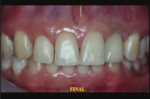
J Case Rep Images Dent 2020;6:100033Z07JL2020.                                                               Miguita et al.   7
www.ijcridentistry.com

Author Contributions                                             critically for important intellectual content, Final approval
João Carlos Amorim Lopes – Conception of the work,               of the version to be published, Agree to be accountable for
Drafting the work, Final approval of the version to be           all aspects of the work in ensuring that questions related
published, Agree to be accountable for all aspects of the        to the accuracy or integrity of any part of the work are
work in ensuring that questions related to the accuracy          appropriately investigated and resolved
or integrity of any part of the work are appropriately           Igor da Silva Brum – Conception of the work, Drafting
investigated and resolved                                        the work, Revising the work critically for important
Saulo Henrique Salviano – Conception of the work,                intellectual content, Final approval of the version to be
Design of the work, Drafting the work, Revising the              published, Agree to be accountable for all aspects of the
work critically for important intellectual content, Final        work in ensuring that questions related to the accuracy
approval of the version to be published, Agree to be             or integrity of any part of the work are appropriately
accountable for all aspects of the work in ensuring that         investigated and resolved
questions related to the accuracy or integrity of any part
of the work are appropriately investigated and resolved          Guarantor of Submission
                                                                 The corresponding author is the guarantor of submission.
Marco Antônio Carvalho – Conception of the work,
Revising the work critically for important intellectual          Source of Support
content, Final approval of the version to be published,          None.
Agree to be accountable for all aspects of the work in
ensuring that questions related to the accuracy or integrity     Consent Statement
of any part of the work are appropriately investigated and       Written informed consent was obtained from the patient
resolved                                                         for publication of this article.
Jorge José de Carvalho – Conception of the work, Design
of the work, Drafting the work, Revising the work critically     Conflict of Interest
for important intellectual content, Final approval of the        Authors declare no conflict of interest.
version to be published, Agree to be accountable for all
aspects of the work in ensuring that questions related           Data Availability
to the accuracy or integrity of any part of the work are         All relevant data are within the paper and its Supporting
appropriately investigated and resolved                          Information files.
Renan Lana Devita – Conception of the work, Analysis
of data, Drafting the work, Revising the work critically         Copyright
for important intellectual content, Final approval of the        © 2020 João Carlos Amorim Lopes et al. This article
version to be published, Agree to be accountable for all         is distributed under the terms of Creative Commons
aspects of the work in ensuring that questions related           Attribution License which permits unrestricted use,
to the accuracy or integrity of any part of the work are         distribution and reproduction in any medium provided
appropriately investigated and resolved                          the original author(s) and original publisher are properly
                                                                 credited. Please see the copyright policy on the journal
Pablo Dallario Ramalho Lucas – Acquisition of data,              website for more information.
Analysis of data, Drafting the work, Revising the work

                                Journal of Case Reports and Images in Dentistry, Vol. 6, 2020.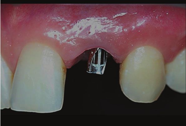
You can also read