Recurrent Syncope as a Presentation of Pulmonary Embolism - Cureus
←
→
Page content transcription
If your browser does not render page correctly, please read the page content below
Open Access Case
Report DOI: 10.7759/cureus.6623
Recurrent Syncope as a Presentation of
Pulmonary Embolism
Kulachanya Suwanwongse 1 , Nehad Shabarek 1
1. Internal Medicine, Lincoln Medical Center, New York City, USA
Corresponding author: Kulachanya Suwanwongse, kulachanya.suwanwongse@gmail.com
Abstract
The diagnosis of pulmonary embolism is challenging particularly when patients present with
vague and/or non-specific symptoms and signs. Misdiagnosis of pulmonary embolism can lead
to death or severe morbidity. We reported a case of a 60-year-old woman presented with
recurrent syncope who later was diagnosed as submassive pulmonary embolism. This case
report highlights the importance of early diagnosis and management of pulmonary embolism
to prevent life-threatening sequels. Pulmonary embolism should be considered as a differential
diagnosis of patients presenting at an emergency department with syncope.
Categories: Pulmonology, Emergency Medicine, Internal Medicine
Keywords: syncope, pulmonary embolism, pe
Introduction
Pulmonary embolism (PE) is accountable for more than 100,000 deaths in the United States
annually [1]. PE presentation varies from asymptomatic with incidentally finding to sudden
cardiac arrest. Early diagnosis and management of PE are important to prevent life-threatening
sequels. Failure to diagnose PE leads to devastating complications, with up to 30% of untreated
patients die [2]. Syncope, as a presentation of PE, has been considered as a
challenging diagnosis [3]. We presented a case of a patient who presented with recurrent
syncope and later was found to have PE, to increase awareness of clinicians in including PE as a
possible cause of recurrent syncope.
Case Presentation
A 60-year-old female presented to the emergency department after an episode of syncope while
walking. She lost consciousness for a minute without convulsion or urinary incontinence. She
reported palpitation, dizziness, mild chest discomfort, and shortness of breath before syncope.
She had a past medical history of hypertension, type 2 diabetes mellitus, and osteoporosis. Her
Received 01/06/2020
medications included sitagliptin, glimepiride, valsartan-hydrochlorothiazide, and raloxifene.
Review began 01/08/2020
Review ended 01/09/2020 She had multiple episodes of syncope (more than 10) accompanying with shortness of breath,
Published 01/10/2020 mild chest discomfort, and palpitation in the past year. She was then admitted to another
hospital and stated that all investigations including electrocardiogram (EKG), 24-hour
© Copyright 2020
Suwanwongse et al. This is an open telemetry, echocardiogram, and cardiac stress test were normal but did not have a chest
access article distributed under the computed tomography (CT).
terms of the Creative Commons
Attribution License CC-BY 3.0., which
permits unrestricted use, distribution,
On initial evaluation, her blood pressure was 148/90 mmHg, heart rate was 93 beats per minute,
and reproduction in any medium, oxygen saturation was 98% on room air and physical exam including heart and lungs and
provided the original author and neurological exam were normal. Blood tests for glucose, electrolytes, creatinine, and complete
source are credited. blood counts were unremarkable except for mild anemia (Hb 10.9 g/dL) and mild elevation of
How to cite this article
Suwanwongse K, Shabarek N (January 10, 2020) Recurrent Syncope as a Presentation of Pulmonary
Embolism. Cureus 12(1): e6623. DOI 10.7759/cureus.6623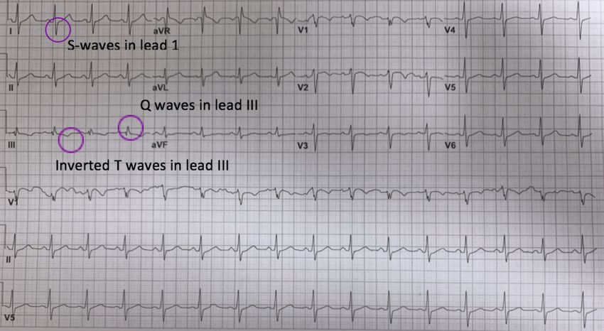
troponin T of 0.015 ng/mL (normal
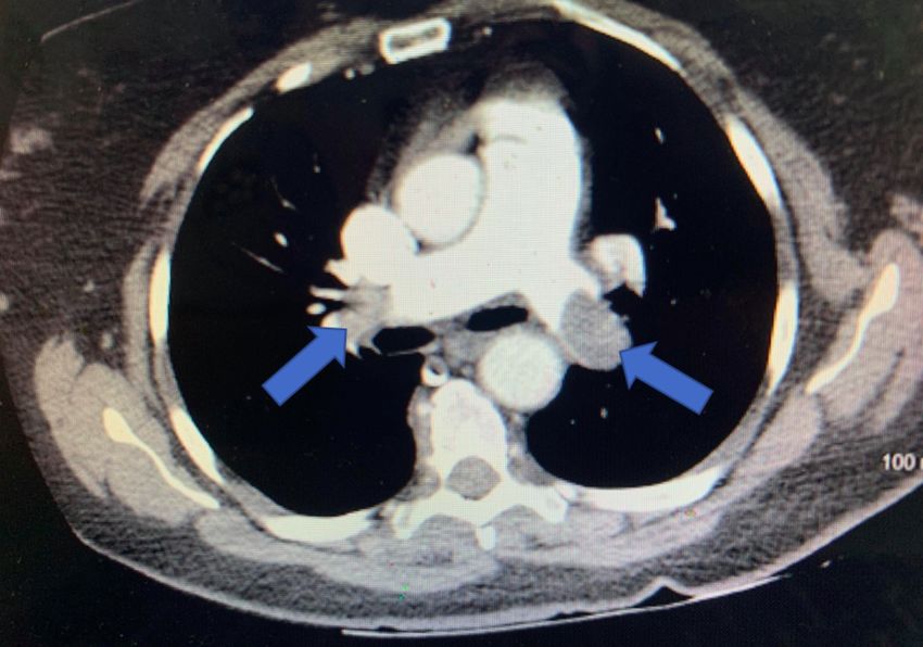
FIGURE 2: CT angiography showed prominent bilateral
                                    pulmonary emboli

                                  Discussion
                                  PE is a differential diagnosis for syncope in most textbooks. However, when patients present
                                  with syncope, PE, a potentially fatal disease requiring urgent attention, is rarely considered
                                  [4]. This case is interesting as our patient had multiple episodes of syncope with extensive
                                  work-up, but without any suspicious of PE until developing severe hypoxemia. PE-induced
                                  syncope can be explained by three possible mechanisms. First, occlusion of more than 50% of
                                  pulmonary vessels leads to right ventricular failure and left ventricular filling impairment,
                                  causing a sudden drop in cardiac output and cerebral blood flow. Second, PE may induce
                                  arrhythmias from right ventricular strain. Third, the embolism itself may provoke a vasovagal
                                  reflex leading to neurogenic syncope [3,5].

                                  Our case highlights the importance of thorough history taking and also illustrates a critical
                                  point in recognition of ‘S1Q3T3’ EKG pattern, which is rare but has high value in helping
                                  diagnosis of PE, especially a new onset ‘S1Q3T3’. The S1Q3T3 sign refers to a prominent S wave
                                  in lead I, Q wave, and inverted T wave in lead III, which reflects right ventricular strain. Any
                                  cause of cor pulmonale can result in an S1Q3T3 pattern on EKG, including PE, pneumothorax,
                                  and bronchospasm [6]. S1Q3T3 pattern has a sensitivity of 54% and a specificity of 62% in the
                                  diagnosis of PE [7]. Although S1Q3T3 is not specific nor sensitive, it is helpful when used with
                                  clinical contexts of patients in guiding the diagnosis of PE.

                                  Conclusions
                                  The diagnosis of PE in patients presenting with syncope is challenging. Physicians should have
                                  PE as a differential diagnosis for patients presented with syncope particularly with
                                  accompanying shortness of breath, respiratory distress, or hypoxemia. Early diagnosis and
                                  treatment of PE are critical to prevent morbidity and mortality from this condition.

2020 Suwanwongse et al. Cureus 12(1): e6623. DOI 10.7759/cureus.6623                                                          3 of 4
Additional Information
Disclosures
Human subjects: Consent was obtained by all participants in this study. Conflicts of interest:
In compliance with the ICMJE uniform disclosure form, all authors declare the following:
Payment/services info: All authors have declared that no financial support was received from
any organization for the submitted work. Financial relationships: All authors have declared
that they have no financial relationships at present or within the previous three years with any
organizations that might have an interest in the submitted work. Other relationships: All
authors have declared that there are no other relationships or activities that could appear to
have influenced the submitted work.
References
1. Tarbox AK, Swaroop M: Pulmonary embolism. Int J Crit Illn Inj Sci. 2013, 3:69-72.
10.4103/2229-5151.109427
2. Meyer NJ, Schmidt GA: Pulmonary embolic disorders: thrombus, air, and fat . Principles of
Critical Care. Hall JB, Schmidt GA, Wood LD (ed): McGraw-Hill, New York; 2014. 4:
3. Thames MD, Alpert JS, Dalen JE: Syncope in patients with pulmonary embolism . JAMA. 1977,
238:2509-2511. 10.1001/jama.1977.03280240055020
4. Paolo P, Anthonie WAL, Martin HP, et al.: Prevalence of pulmonary embolism among patients
hospitalized for syncope. N Engl J Med. 2016, 375:1524-1531. 10.1056/NEJMoa1602172
5. Simpson RJ Jr, Podolak R, Mangano CA Jr, Foster JR, Dalldorf FG: Vagal syncope during
recurrent pulmonary embolism. JAMA. 1983, 249:390-393.
10.1001/jama.1983.03330270054034
6. Chan TC, Vilke GM, Pollack M, Brady WJ: Electrocardiographic manifestations: pulmonary
embolism. J Emerg Med. 2001, 21:263-270. 10.1016/S0736-4679(01)00389-4
7. Ferrari E, Imbert A, Chevalier T, Mihoubi A, Morand P, Baudouy M: The ECG in pulmonary
embolism. Chest. 1997, 111:537-543.
2020 Suwanwongse et al. Cureus 12(1): e6623. DOI 10.7759/cureus.6623 4 of 4You can also read


