The intravesical migration of a lost intrauterine device
←
→
Page content transcription
If your browser does not render page correctly, please read the page content below
J Case Rep Images Urol 2020;5:100011Z15HH2020.                                                                  Hatem et al.   1
www.ijcriurology.com

 CASE REPORT                                                                       PEER REVIEWEDOPEN ACCESS
                                                                                                | OPEN ACCESS

      The intravesical migration of a lost intrauterine device
                                              H Hatem, Jörg Leifeld

ABSTRACT                                                            of their potential complications is intravesical migration,
                                                                    which can present as dysuria, recurrent infections, or
The intravesical migration of intrauterine devices                  dyspareunia.
(IUDs) has rarely been reported. In many cases,                         If there is any suspicion of this condition, it can be
diagnosis is missed or delayed. We present a case of a              investigated simply through ultrasound or plain film.
34-year-old female patient with intravesical migration              The most common approach to removing an IUD is
of an IUD, which was thought to be lost. To check for the           transurethral resection. If large stones are present, open
presence of the IUD, which had dislodged, the patient’s             or laparoscopic approaches are more appropriate. We
gynecologist inserted another one. Later, the patient               report on the case of a patient with intravesical migration
presented with recurrent urinary infections. A diagnosis            of an IUD that was thought to be lost.
was reached through cystoscopy; the migrated device
was visible through the posterior wall. We removed
the device transurethrally. This case is presented to               CASE REPORT
highlight the importance of following up with patients
with IUDs to avoid potential complications.                             We report on a 34-year-old female patient who
                                                                    complained of recurrent cystitis for many years.
Keywords: Intrauterine device, Intravesical, Perforation            To address this issue, the patient went to a urology
                                                                    outpatient clinic. As part of our study, a cystoscopy was
                  How to cite this article                          performed, and the migrated IUD was seen through
                                                                    the posterior wall of the bladder. The IUD had first
Hatem H, Leifeld J. The intravesical migration of                   been inserted seven years earlier, and the patient
a lost intrauterine device. J Case Rep Images Urol                  noted that she had not noticed losing it. Four years
2020;5:100011Z15HH2020.                                             after that, her gynecologist had inserted another one
                                                                    without investigating whether the first one was still
                                                                    intracorporeal. The patient was admitted to our clinic to
Article ID: 100011Z15HH2020
                                                                    remove the IUD.
                                                                        To determine a diagnosis, we performed an abdominal
                           *********                                computed tomography (CT) scan to reveal the position
                                                                    of the migrated device and the status of her anatomical
doi: 10.5348/100011Z15HH2020CR
                                                                    condition.
                                                                        With the patient under general anesthesia,
                                                                    we removed the IUD, performing a superficial,
                                                                    transurethral resection of the bladder mucosa. The
INTRODUCTION                                                        urinary catheter was removed seven days later, after
                                                                    a cystogram showed no evidence for a fistula or
   Intrauterine devices (IUD) are generally considered a
                                                                    extravasation (Figure 1). The recurrent infections have
safe, reliable, and economical contraception method. One
                                                                    since been entirely resolved.
 H Hatem1, Jörg Leifeld2
 Affiliations: 1Senior Doctor, Urology, Borromäus Hospital in
 Leer, Leer, Germany; 2Director, Department for Urology in
 Borromäus Hospital in Leer, Leer, Germany.
 Corresponding Author: H Hatem, Senior Doctor, Urol-
 ogy, Borromäus Hospital in Leer, Leer, Germany; Email:
 dr.h.hatem@hotmail.de

                                                                    Figure 1: A postoperative cystogram (AP and steep oblique
 Received: 21 May 2020                                              position) before removing the catheter showing a regular
 Accepted: 06 August 2020                                           bladder wall and othotop located IUP without any evidence of
 Published: 24 September 2020                                       a fistula or extravasation.

                                       Journal of Case Reports and Images in Urology 5, 2020.
J Case Rep Images Urol 2020;5:100011Z15HH2020. Hatem et al. 2
www.ijcriurology.com
DISCUSSION
The IUD is the most widely used contraceptive
worldwide due to its safety, activity, and affordability.
However, despite its safety, some patients experience
complications, including pelvic inflammatory disease
(PID), uterine perforation, heavy bleeding, dysmenorrhea,
and unplanned pregnancy [1].
Such migration is rare—in 0.003–0.87% [2] or in
1.9–3.6 per 1000 insertions [3]. A dislodged IUD could
be located in different organs, such as the mentum,
rectosigmoid, peritoneum, bladder, appendix, small
bowel, adnexa, or even iliac vein. In about 200 cases of
uterine perforation reviewed from 1991 to 2015, Kart and
colleagues found 90 cases of intravesical migration [4].
Many factors can lead to the migration of the IUD,
such as actions by inexperienced staff or a patient’s Figure 3: A transverse section of the patient’s abdominal CT
atypical anatomical issues, such as an extreme posterior scan reveals the dislocated IUD through the posterior wall of
uterine position [5, 6]. However, it seems that perforation the bladder.
occurs during or after insertion, or as a slow process that
leads to migration, as in our case [2].
Sometimes this migration causes no complaints. In
others, however, it is associated with complications,
including dysuria, dyspareunia, or vesical calculus. In our
case, the patient reported recurrent infections.
A migration diagnosis can be reached through
ultrasound and plain film [7]. In our case, the diagnosis
was confirmed through a cystoscopy performed by an
outpatient urologist. To investigate further, we decided
to perform a CT scan to ensure the extension and location
of the IUD (Figures 2 and 3).
Incomprehensibly, the patient’s gynecologist had not
undergone any examination, such as an ultrasound or
plain film, to check for the presence or position of the first
IUD, which was believed to be lost.
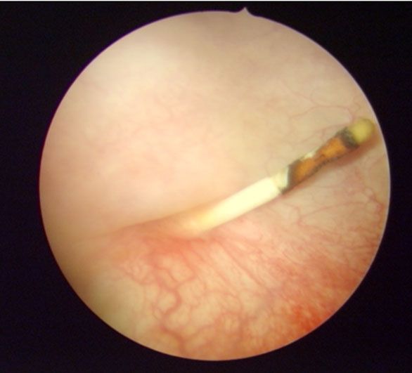
With the patient under anesthesia, we performed a
transurethral resection to remove the IUD (Figure 4).
In standard treatment, a minimally invasive approach
should be chosen [8]. A more invasive approach, such as
a cystotomy, may be required if large stones are present Figure 4: Endoscopic image reveals the IUD through the
or in the case of associated fistula formations [9]. posterior wall of the bladder.
CONCLUSION
Although the intravesical migration of an IUD is
rare, it should be kept in mind, and every female patient
complaining of recurrent infections should be interviewed
regarding her IUD history.
REFERENCES
1. Aggarwal S, Jindal RP, Deep A. Intravesical migration
of intrauterine contraceptive devices with stone
Figure 2: A coronal section of the patient’s abdominal CT scan formation. J Family Med Prim Care 2014;3(4):449–
reveals the dislocated IUD in the bladder and the uterus. 51.
Journal of Case Reports and Images in Urology 5, 2020.
J Case Rep Images Urol 2020;5:100011Z15HH2020.                                                                  Hatem et al.   3
www.ijcriurology.com

    2.   Markovitch O, Klein Z, Gidoni Y, Holzinger M, Beyth      or integrity of any part of the work are appropriately
         Y. Extrauterine mislocated IUD: Is surgical removal      investigated and resolved
         mandatory? Contraception 2002;66(2):105–8.
    3.   Farmer M, Webb A. Intrauterine device insertion-         Jörg Leifeld – Conception of the work, Design of the
         related complications: Can they be predicted? J Fam      work, Acquisition of data, Analysis of data, Interpretation
         Plann Reprod Health Care 2003;29(4):227–31.              of data, Drafting the work, Final approval of the version
    4.   Kart M, Gülecen T, Üstüner M, Çiftçi S, Yavuz U,         to be published, Agree to be accountable for all aspects of
         Özkürkçügil C. Intravesical migration of missed          the work in ensuring that questions related to the accuracy
         intrauterine device associated with stone formation:     or integrity of any part of the work are appropriately
         A case report and review of the literature. Case Rep     investigated and resolved
         Urol 2015;2015:581697.
    5.   Dimitropoulos K, Skriapas K, Karvounis G, Tzortzis
         V. Intrauterine device migration to the urinary
                                                                  Guarantor of Submission
         bladder causing sexual dysfunction: A case report.       The corresponding author is the guarantor of submission.
         Hippokratia 2016;20(1):70–2.
    6.   Vagholkar S, Vagholkar K. Secondary vesical              Source of Support
         calculus resulting from migration of an intrauterine     None.
         contraceptive device. Case Rep Obstet Gynecol
         2012;2012:603193.                                        Consent Statement
    7.   El-Diasty TA, Shokeir AA, el-Gharib MS, Sherif           Written informed consent was obtained from the patient
         LS, Shamaa MA. Bladder stone: A complication of
                                                                  for publication of this article.
         intravesical migration of Lippes loop. Scand J Urol
         Nephrol 1993;27(2):279–80.
    8.   Guner B, Arikan O, Atis G, Canat L, Çaskurlu T.          Conflict of Interest
         Intravesical migration of an intrauterine device. Urol   Authors declare no conflict of interest.
         J 2013;10(1):818–20.
    9.   El-Hefnawy AS, El-Nahas AR, Osman Y, Bazeed              Data Availability
         MA. Urinary complications of migrated intrauterine       All relevant data are within the paper and its Supporting
         contraceptive device. Int Urogynecol J Pelvic Floor      Information files.
         Dysfunct 2008;19(2):241–5.
                                                                  Copyright
                         *********                                © 2020 H Hatem et al. This article is distributed under
                                                                  the terms of Creative Commons Attribution License which
Author Contributions                                              permits unrestricted use, distribution and reproduction in
H Hatem – Conception of the work, Design of the work,             any medium provided the original author(s) and original
Acquisition of data, Analysis of data, Interpretation of          publisher are properly credited. Please see the copyright
data, Drafting the work, Final approval of the version to         policy on the journal website for more information.
be published, Agree to be accountable for all aspects of the
work in ensuring that questions related to the accuracy

               Access full text article on                                           Access PDF of article on
                    other devices                                                         other devices

                                     Journal of Case Reports and Images in Urology 5, 2020.
Submit your manuscripts at
  www.edoriumjournals.com


You can also read