RESEARCH DAY 35TH ANNUAL - ABSTRACTS Poster Presentations - CU Denver
←
→
Page content transcription
If your browser does not render page correctly, please read the page content below
35TH ANNUAL

RESEARCH DAY

             ABSTRACTS
          Poster Presentations

Friday, February 22, 2019 | 12 p.m.-1 p.m.

                  Ed 2 Student Bridge
   University of Colorado Anschutz Medical Campus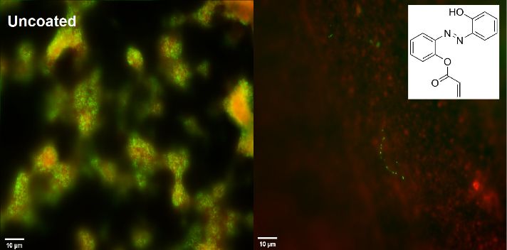
Undergraduates
Designing Opto-mechanically Responsive Molecules as Coatings on Pit and Fissure
Sealants

Kasra Roostan1, Krithika Baskaran2, Dixa Gautam3, Gannon M. Kehe3, Dylan I. Mori3,
Michael J. Schurr4 and Devatha P. Nair3,5
1
 Department of Psychology and Neuroscience, University of Colorado, Boulder, Boulder,
CO, USA; Departments of 2Restorative Dentistry and 3Craniofacial Biology, School of
Dental Medicine, University of Colorado Anschutz Medical Campus, Aurora, CO, USA;
4
 Department of Immunology and Microbiology, School of Medicine, University of Colorado
Anschutz Medical Campus, Aurora, CO, USA, 5Materials Science and Engineering
Program, University of Colorado, Boulder, Boulder, CO, USA

Pits and fissures on the surface of teeth account for 88% of all occlusal caries in children.
Although dental sealants applied on enamel surfaces can restrict bacterial ingress by up to
50%, their efficacy can be enhanced by anti-bacterial strategies that target early cariogenic
colonizers such as Streptococcus mutans (S. mutans). This translational study evaluates
the ability of azopolymer coatings to disrupt biofilms on commercial pit and fissure sealants.

Objectives: This study investigates the mechanical                                Microtensile
properties and biological impact of applying opto-                                bond strength
                                                                                  (µTBS)
mechanically active azopolymer coatings on commercial
pit and fissure sealants.                                                         assessment

Methods: Extracted human molars were prepared with
35% phosphoric acid etch ( NOVO™ , 30s), followed by                                 Azomolecules
the application of sealant and azocoatings (Embrace™                                 used in the
wetbond™).Strenght was assessed via microtensile tests                               study
                                                                           AAZO
(cross-head speed -1 mm/min , n ≥ 3, Mini Bionix II,
MN,USA). Extracted and sealed molars were exposed to             OH-AAZO
S. mutans biofilms grown under sucrose-dependent
conditions and quantified via CFU counts.
                                                                                           Tooth Samples
Results: The mechanical strength of the
sealant was not compromised with the
introduction of the azopolymer (14.0 ± 2
vs 3.2 ± 4 MPa, p = 0.325). The presence              Contr
of azocoatings affected biofilm formation
on the tooth samples (a) and had an
extended impact beyond the substrate as                AAZ
seen via biofilm formation in the wells (b).
No live S. mutans were detected on the
OH-A AZO well.
                                                      OH-AAZO
                                          (a)                                                     (b)
Conclusion: Preliminary results indicate
that azobenzene coatings can be successfully incorporated into commercial pit and fissure
sealants as anti-bacterial coatings.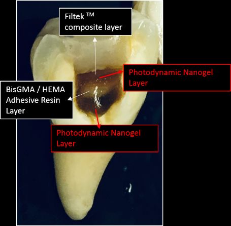
DDS Students
Protective Effect of Theobromine Against Acid Attack on Tooth Enamel

Elizabeth Tremblay and Clifton Carey

Department of Craniofacial Biology, School of Dental Medicine, University of Colorado
Anschutz Medical Campus, Aurora, CO, USA.

Objectives: Theobromine is a potential therapeutic agent and fluoride alternative that
can help protect tooth enamel from the acid produced by bacteria after consumption of
sugars. Our hypothesis is that a 100 ppm solution of Theobromine will protect tooth
enamel from an acid attack. We also want to validate the use of non-destructive µCT
methods for the measurement of enamel erosion compared to transverse
microradiography.

Methods: Both interproximal surfaces of 8 human caries-free teeth were polished with
400 grit paper to make flat surfaces and remove the outer ~50 µm of enamel (i.e., two
surfaces per tooth). A portion of the surfaces were protected with fingernail polish. Four
teeth samples were randomly assigned to the control group of 0 ppm of Theobromine
solution and the other 4 were assigned to the 100 ppm of Theobromine solution. The
samples were soaked in the test solutions for 2 min, briefly rinsed with dH2O then exposed
to a 1% citric acid attack at pH of 3.9 for 10 min. Samples were stored in saliva-like storage
solution for 24 hours before measurement of surface loss by µCT. The measurements
and statistics were blinded.

Results: Enamel loss for the control samples was 13.2±8.0µm; loss for the theobromine
treated samples was 1.5±4.0µm the difference is significant at p=0.0405.

Conclusions: The hypothesis is supported that theobromine protects tooth enamel from
an acid attack at 100ppm concentration. These erosion results are not statistically
different (p>0.05) from results of prior experiments under the same conditions as here
where enamel erosion was determined by transverse microradiography after theobromine
exposure (3.1 ± 1.1 µm, n=16). This confirms the use of the non-destructive µCT for the
measurement of erosive losses.
Intraoral findings in newborns: prevalence and associated factors

Claudia L. Chandler1, Isabelita D. Azevedo2, Manoelito F. Silva, Jr. 3, Johnnatas M.
Lopes2, Antonio Gordon-Nunez4 and Silvana A. Pereira2
1
 ISP Program, School of Dental Medicine, University of Colorado Anschutz Medical
Campus, Aurora, CO, USA; 2Federal University of Rio Grande do Norte – UFRN, Natal-
RN, Brazil; 3State University of Ponta Grossa – UEPG, Ponta Grossa-PR, Brazil;
4
 State University of Paraiba – UEPB, Araruna-PB, Brazil.

Objective: To investigate the prevalence of intraoral characteristics and associated
factors with neonatal and parent variables in a group of Brazilian newborns.

Methods: This cross-sectional study with a descriptive and inferential approach, whose
data was obtained through clinical examination, interview and the collection of medical
record information. The sample was selected from babies up to three days old, of both
sexes, and born between January and December 2013 in the Ana Bezerra University
Hospital, in the city of Santa Cruz-RN, Brazil. The exposure variables included neonatal
(sex, weight, gestational age, type of delivery and Apgar score) and parent (presence of
systemic disease(s), drug use and consanguinity between the parents) variables.
Descriptive analysis and Poisson regression were performed to estimate the ratio of gross
and adjusted prevalence of intraoral findings (Epstein pearls, Bohn’s nodule, dental
lamina cyst and ankyloglossia) with the neonatal and parent variables (p
ISP Students
Detection of White Spot Lesions by PTR-LUM Technology

Minh Trinh and Clifton Carey

Department of Craniofacial Biology, School of Dental Medicine, University of Colorado
Anschutz Medical Campus, Aurora, CO, USA.

Objectives: Early stage WSL are often difficult to detect without some means to indicate
the presence of the demineralization. There is a sensitive caries detection system based
on photothermal radiometry-modulated luminescence (PTR-LUM) technology called the
Canary System. This system uses an infrared laser that penetrates 5mm into the tooth
which creates a fluorescence signal that is related to mineral integrity. The signal is
converted into a “Canary Number” (CN) which is interpreted to indicate the presence of
caries. We wanted to determine if PTR-LUM technology is effective for detecting early
stage WSL. Our hypothesis was that there would be a positive linear relationship between
the CN and visual WSL scores.
Methods: 36 premolars that were extracted for orthodontic therapy were evaluated for
WSL by the Canary System and visually for the presence of WSL under magnification.
CN were determined at the labial gingival, incisal surfaces and left and right interproximal
surfaces. The CN were averaged for each tooth. Visual determination was with the teeth
gently dried and observed under a 10x dissecting microscope for WSL in the same zones
as for the Canary System measurements. The number of zones with WSL for each tooth
were summed to yield the visual WSL score. Digital microphotographs were taken for
reference.
Results: The average CN score was 28.1±5.6, range 19-39, n=36; and the median visual
WSL was 1, range 0-3, n=36. Because the visual WSL score is not parametric, a
Spearman's correlation was run to determine the relationship between the CN and visual
WSL scores. There was a strong, positive monotonic correlation between CN and WSL
scores (Rs=0.743, n = 36, p
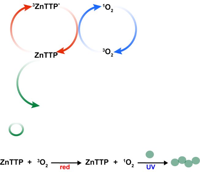
Irradiation of a Dual-Wavelength Photosensitive Molecule to Overcome Oxygen
Inhibition
Kimberly K. Childress,1 Kangmin Kim,2 David J. Glugla,3 Charles B. Musgrave,1,2,4,5
Christopher N. Bowman1,4 and Jeffrey W. Stansbury1,6
Departments of 1Chemical and Biological Engineering 2Chemistry and Biochemistry,
3
 Electrical, Computer, and Energy Engineering and 4Materials Science and Engineering,
University of Colorado, Boulder, Boulder, CO, USA; 5National Renewable Energy
Laboratory, Golden, CO, USA; 6Department of Craniofacial Biology, School of Dental
Medicine, University of Colorado Anschutz Medical Campus, Aurora, CO, USA
Objective: Oxygen inhibition is detrimental to free radical polymerizations and can
result in reduced polymerization kinetics and final mechanical properties.
Photosensitizers have previously been incorporated in monomer formulations to excite
molecular oxygen to its inert singlet state prior to irradiation of a photoinitiator, thereby
avoiding unwanted consumption of free radicals by oxygen. In this work, a single
photoresponsive molecule was shown for the first time to perform the tasks of both
oxygen-scavenging photosensitization and photoinitiation.
Methods: Zinc 2,9,16,23-tetra-tert-butyl-29H,31H-phthalocyanine (ZnTTP) was
intoduced into di(ethylene glycol) ethyl ether acrylate (DEGEEA). Independent
irradiation of ZnTTP’s UV and visible peaks with 365 and 635 nm LEDs, respectively,
was conducted at varying exposure times and intensities. Polymerization conversion
and ZnTTP concentration were measured in real-time via a coupled FT-NIR/UV-Vis
analysis.
Results: Irradiation of ZnTTP’s UV band resulted in polymerization of DEGEEA. At 10
mW/cm2 for 0.2 mM ZnTTP, an induction time of ~290±7 s preceded the onset of
polymerization. Increasing irradiance to 90 mW/cm2 reduced the induction time to 36±3
s, and increasing [ZnTTP]0 from 0.1 to 10 mM reduced the induction time from 479±15 s
to 58±3 s. However, red-light pre-irradiation of the visible band of ZnTTP with a dose of
~6500 mJ/cm2, regardless of the irradiance/expose time combination, resulted in
sufficient singlet oxygen generation to completely eliminate any induction time upon UV-
activated polymerization. Only minor amounts of ZnTTP photodegradation occurred
during the pre-irradiation stage (~4%), leaving sufficient ZnTTP available to promote
efficient photoinitiation.
Conclusions: This work highlights the potential of ZnTTP to perform the polymerization
functions that multiple molecules were previously required to achieve. The utility of
ZnTTP as a photosensitizer and photoinitiator via irradiation of its two absorption bands
resulted in immediate polymerization following adequate singlet oxygen generation.

Funding: IUCRC and ALTANA
Graduate Students
Srsf3-mediated alternative RNA splicing downstream of PDGFRa signaling in the
palatal mesenchyme

Brenna J.C. Dennison1,2, Eric D. Larson3 and Katherine A. Fantauzzo1
1
 Department of Craniofacial Biology; 2Graduate Program in Cell Biology, Stem Cells
and Development; 3Department of Otolaryngology, University of Colorado Anschutz
Medical Campus, Aurora, CO, USA.

Craniofacial development is a critical morphological event during embryogenesis,
defects in which result in highly prevalent human birth defects. In both humans and
mice this process relies on signaling through the platelet-derived growth factor receptor
alpha (PDGFRa). Mutations in human PDGFRA are associated with cleft lip/palate and
mouse models with mutations in this gene similarly display facial clefting phenotypes.
PI3K is the main downstream effector of PDGFRa signaling during mouse
development. We previously performed a mass spectrometry-based phosphoproteomic
screen to identify targets of PI3K/Akt-mediated PDGFRa signaling in primary mouse
embryonic palatal mesenchyme cells (MEPMs), revealing an enrichment for proteins
that regulate RNA splicing.

Objectives: We hypothesize that one of these Akt phosphorylation targets, Srsf3,
mediates tissue-specific alternative RNA splicing downstream of PDGFRa signaling in
the palatal mesenchyme.

Methods and Results: We have biochemically confirmed using immunoprecipitation
assays the PI3K/Akt-mediated phosphorylation of Srsf3 upon PDGF-AA ligand
treatment of MEPMs and further demonstrated that this phosphorylation drives Srsf3
translocation into the nucleus. We revealed that expression of Srsf3 is enriched in the
maxillary processes and palatal shelves of mid-gestation mouse embryos. Moreover,
RNA-sequencing analysis of palatal shelf mesenchyme derived from wild-type versus
autophosphorylation mutant knock-in embryos in which PDGFRa is unable to bind
PI3K identified differentially alternatively-spliced transcripts containing Srsf3 binding
sites that are associated with craniofacial defects. Finally, we showed that ablation of
Srsf3 in the neural crest lineage results in embryos with midline facial clefting, facial
bone hypoplasia and exencephaly.

Conclusions: Taken together, our results point to a novel role for the PDGFRa-
PI3K/Akt-Srsf3 signaling axis in regulating RNA processing during craniofacial
development.
Tailored Nanogels as Antibacterial Agents
Humberto Escobedo1, Dixa Gautum2, Toan Nguyen 3, Michael J Schurr 4, Devatha P. Nair1,2
1
 Department of Pharmaceutical Science, Skaggs School of Pharmacy and Pharmaceutical Sciences,
University of Colorado Anschutz Medical Campus, Aurora, CO, USA;2Department of Craniofacial
Biology, School of Dental Medicine, University of Colorado Anschutz Medical Campus, Aurora, CO,
USA; 3Biomedical Sciences Program, Regis University, Denver, CO, USA; 4Department of
Immunology and Microbiology, School of Medicine University of Colorado Anschutz Medical Campus,
Aurora, CO, USA

The mechanism by which bacteria have evolved to evade targeted, antibiotics is the formation of
biofilms, which renders them 10-1000 times more resistant to common antibiotic drugs. As non-
traditional antibiotic strategies that target biofilms have continued to garner interests, multiple studies
have shown the potential of nanoparticles as a substitute and/or complement to strategies to combat
antimicrobial resistance. We examine the ability of tailored nanogels from 2- 200 nm synthesized to
induce bacterial membrane disruption via passive, non-lytic mechanisms as a promising route to
inhibit and disrupt biofilm growth.

Objective: Study the bacteriostatic effects of tailored nanogels synthesized via two different
mechanisms on biofilm formation of gram-positive and gram-negative bacteria

Methods: Nanogels were synthesized and characterized via both a solution polymerization (NG1, 2-
10 nm) and an emulsion polymerization (NG2, 50-100 nm ) mechanism from monomers 2-
(Dimethylamino)ethyl methacrylate (DMEAMA)/Tetraethyleneglycol dimethacrylate (TTEGDMA) for
NG1 and DMEAMA /Polyethylene glycol (PEG400DMA) for NG2. The impact of the nanogels on
biofilm formation on uropathogenic E. coli (UPEC) and S. mutans was studied to determine its
bacteriostatic effect and quantified via CFU counts.

Results: Nanogels were synthesized
and characterized (GPC and
Zetasizer, NG1 2 nm, NG 2, 100 nm)
and the bacteriostatic effects
quantified via CFU counts indicate
that both there is both a charge and                  Biofilm
concentration dependence on the                      disruption
outcomes.

Conclusion: Tailored Nanogels                                                                   Biofilm
synthesized via two different                                                                  inhibition
mechanisms to control the size and
charge on nanoparticles can impart
an inhibitory and disruptive growth
effect on both gram-negative and gram-positive bacteria. Future work will focus on optimizing the
synthetic protocol for nanogels and determining the ideal charge, size and concentration to prevent
biofilm formation
New Approaches to Reactive Oligomers and Prepolymers in Photo-cured
Systems

Guangzhe Gao1, Parag K. Shah1, Tao Liu1 and Jeffrey Stansbury1, 2
1
 Chemical and Biological Engineering, University of Colorado Boulder, Boulder, CO,
USA; 2Department of Craniofacial Biology, School of Dental Medicine, University of
Colorado Anschutz Medical Campus, Aurora, CO, USA

Objective: The introduction of functional nanogels prepared by a chain-growth free
radical polymerization process has demonstrated utility in enhanced performance of
dental resins for adhesive and composite applications as well as in hydrogels and
controlled-release coatings. Construction of reactive nanogels by a step-growth
mechanism offers access to different nanogel network structure options and properties
that are examined here.

Methods: A series of nanogels were synthesized by diisocyanate addition to a modest
stoichiometric excess of multi-thiols with ambient reactions completed in one hour.
Residual thiol group concentration within nanogels was assessed by Ellman’s test and
this functionality was then used to attach polymerizable methacrylate groups. Nanogel
characterization involved gel permeation chromatography, dynamic light scattering,
rheology and bulk photopolymerization kinetics. Nanogel formulations as dispersions in
TEGDMA over the entire compositional range provided resins that were analyzed for
reaction kinetics, polymer thermomechanical properties and polymerization stress.

Results: Nanogels with sizes less than 10 nm were designed as room temperature liquids
with narrow glass transition temperatures (Tg’s; tandmax = -21 to -10°C). Bulk nanogel
viscosities varied widely (10,000Pa×s). Residual thiol concentrations, which were
subsequently converted to methacrylate linkages, gave 18-24 functional groups/nanogel
on average. All four bulk nanogels underwent rapid, near-quantitative conversion during
ambient photocure. Depending on nanogel structure, polymeric Tg’s obtained upon bulk
nanogel polymerization varied (23-44°C). Photopolymerization of nanogel+TEGDMA
mixtures showed dramatically enhanced reaction rate and conversion compared with
TEGDMA homopolymer. Flexural strength and modulus were maintained (P
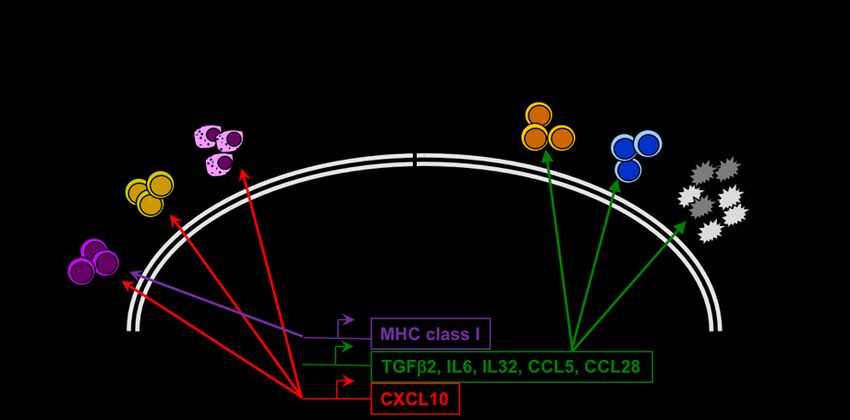
Oncogene Regulation of the HNSCC Immune Microenvironment

Sean Korpela1, Trista Hinz2, Jacob Calhoun2, Raphael Nemenoff3, Sana Karam4,
Ayman Oweida4, Lynn Heasley2,5

Departments of 1Pharmacology, 2Craniofacial Biology, 3Medicine, and 4Radiology,
University of Colorado Anschutz Medical Campus, Aurora, CO, USA; 5Veterans
Administration Eastern Colorado Health Care System (VHAECH), Aurora, CO, USA

Objectives: The Epidermal Growth Factor Receptor (EGFR) is a key component of a
receptor tyrosine kinase (RTK) network that functions as a non-mutated “driver” in head
and neck squamous cell carcinoma (HNSCC) and is the target for the FDA-approved
agent, cetuximab. Individually, HNSCC patients exhibit wide-ranging extent of response
to EGFR inhibitors, even in combination with chemo- or radiotherapy. Herein, we
explored the idea that the EGFR-MEK-ERK pathway acts as a repressor of innate
immune signaling in HNSCC such that targeted drugs de-repress this response to yield
increased paracrine communication with the immune microenvironment which may
directly influence the variability observed in therapeutic responses.

Methods: Human and murine HNSCC cell lines were treated with gefitinib (an EGFR
TKI), AZD8931 (a pan-ERBB inhibitor) or trametinib (a MEK inhibitor) to block
oncogenic signaling through the EGFR-MEK-ERK pathway. The murine HNSCC cell
line, B4B8 was propagated orthotopically in immunocompetent Balb/c mice and treated
for 1 week with AZD8931 (50mg/kg).

Results: Transcriptomic analysis with Affymetrix GeneChips of gefitinib treated human
HNSCC cells revealed marked induction of innate immune response genes. These
findings were validated by qRT-PCR. Induction of CXCL10 protein, was variably
induced by gefitinib, AZD8931 and trametinib, and was IKK-NFkB pathway dependent.
A 1-week treatment of B4B8 tumors in syngeneic mice with AZD8931 elicited tumor
shrinkage and significant increases in immune cell populations.

Conclusions: The data support the overall hypothesis that targeted EGFR-MEK-MAPK
axis inhibitors de-repress an innate immune signaling cascade that induces a variable
spectrum of anti- and pro-tumorigenic chemokines and cytokines that lead to direct
participation of recruited immune cells in the therapeutic response. Identifying
mechanism-based combinations of agents that induce anti-tumorigenic signals or block
pro-tumorigenic signals may ultimately influence the residual disease state and thus
extend progression-free and overall survival in HNSSC patients.
Examining PDGFR dimer-specific dynamics using bimolecular fluorescence
complementation

Madison A. Rogers1,2 and Katherine A. Fantauzzo1
1
 Department of Craniofacial Biology; 2Graduate Program in Cell Biology, Stem Cells and
Development, University of Colorado Anschutz Medical Campus, Aurora, CO, USA

Craniofacial development is a complex morphogenetic process, disruptions in which
result in highly prevalent human birth defects. Signaling through the platelet-derived
growth factor receptors (PDGFRs) plays a critical role in this process in humans and
mice. Pdgfra and Pdgfrb mutant mouse models display varying degrees of midline
clefting as well as subepidermal blebbing. In addition to homodimeric receptor
complexes, we have previously demonstrated that PDGFRa and PDGFRb genetically
and physically interact in the craniofacial mesenchyme to form functional heterodimers.

Objectives: We hypothesize that the various PDGFR dimers have different patterns of
expression and ligand affinities and, further, that each dimer binds to a unique
complement of intracellular signaling molecules to generate distinct cellular outputs
during midface development.

Methods: Here, we highlight ongoing experiments to visualize and purify PDGFR
dimers using bimolecular fluorescence complementation (BiFC), which circumvents
several limitations with antibody-based approaches. We cloned plasmids expressing C-
terminal fusions of each PDGFR with BiFC fragments corresponding to the N-terminal
(V1) or C-terminal (V2) regions of the Venus fluorescent protein.

Results: By transiently transfecting cells with combinations of PDGFR-V1 and -V2
plasmid pairs and stimulating with various PDGF ligands, we detected PDGFRa
homodimer, PDGFRb homodimer and PDGFRa/b heterodimer formation at the cell
membrane and in numerous endosomal compartments, to differing extents. Further, we
employed BiFC coupled with affinity purification following ligand stimulation to
selectively purify and biochemically analyze the various PDGFR dimers, revealing
differences in the timing of receptor autophosphorylation, signal molecule binding and
the amplitude and duration of downstream intracellular signaling.

Conclusions: This approach will likely serve as a powerful tool to quantify the dimer-
specific dynamics of PDGFR activation, signal molecule binding and internalization.
Understanding the role of Prdm1a in zebrafish melanocytes in development and
disease

Brittany Truong1,3, Ritsuko Iwanaga1, David Orlicky2, Craig Ceol4 and Kristin B. Artinger1
1
 Department of Craniofacial Biology; 2Department of Pathology; 3Human Molecular
Genetics and Genomics Graduate Program; University of Colorado Anschutz Medical
Campus, Aurora, CO, USA. 4Program in Molecular Medicine, University of
Massachusetts, USA.

Neural crest cells are highly regulated, transient, multipotent population of cells with the
potential to differentiate into various cell types, including melanocytes among other
derivatives. Mutations in neural crest development genes can lead to misregulation of
neural crest-derived cells, thus promoting defects in development leading to disease.
Prdm1a is a master regulator of neural crest specification during embryonic
development, and loss of prdm1a results in a partial loss of neural crest cell derivatives,
including pigment cells.

Objective: Here, we investigate the role of Prdm1a in melanocyte development and
show that Prdm1a regulates the melanocyte stem cell population through sox10.

Methods/Results: prdm1a zebrafish mutants have increased sox10 expression, but a
decrease in expression of differentiated melanocyte markers, dct, mc1r, mitfa, and tyr.
Pigment production measured by tyrosinase activity is significantly reduced as well.

Conclusion: Our data suggest that Prdm1a is necessary for differentiation of
melanocyte stem cells into mature melanocytes. Understanding the development of
melanocytes and regulatory genes involved will elucidate the mechanisms driving
neural crest derived pigmentation diseases, such as melanoma.
Post-doctoral Fellows
The intersection of biochemistry and developmental biology: Investigating the
disease mechanism of a novel EDNRA mutation linked to severe human
craniofacial birth defects

Stanley M. Kanai1, Amanda Barone Pritchard2, Andre L.P. Tavares1, Nevin A. Lambert3,
Elaine H. Zackai2, and David E. Clouthier1
1
 Department of Craniofacial Biology, University of Colorado Anschutz Medical Campus,
Aurora, CO, USA. 2The Children’s Hospital of Philadelphia, Philadelphia, PA, USA.
3
 Department of Pharmacology and Toxicology, Medical College of Georgia-Augusta
University, Augusta, GA, USA.

Facial morphogenesis requires the establishment of cranial neural crest cell (NCC)
identity in the mandibular and maxillary portions of the first pharyngeal arch. This
process is dependent on G protein-coupled endothelin receptor type A (EDNRA, ETA)
and is highly conserved among gnathostomes, as demonstrated in mouse and zebrafish
gain- and loss-of-function (LOF) models and in the human conditions Auriculocondylar
Syndrome and Mandibulofacial Dysostosis with Alopecia, in which ETA signaling is
altered. Although many ETA dependent gene expression networks have been identified
in mutant models, understanding changes in intracellular signaling that precede gene
expression has proven more difficult.

Objective: To determine whether we can efficiently elucidate how EDNRA disease
alleles impact receptor function and intracellular signaling, we adopted experimental
approaches used in biochemistry and pharmacology.

Method: We used a range of assays based on bioluminescence resonance energy
transfer (BRET) that monitor ETA receptor biology in real-time. We specifically tested
subcellular localization, G protein activation, and G protein recruitment.

Results: To prove the utility of this approach, we examined the consequences of a
novel mutation in EDNRA. An individual with a homozygous missense mutation in exon
seven of EDNRA, p.Glu381Pro, was identified by whole exome sequencing. The
individual died shortly after birth with notable micrognathia/microstomia, an aortic arch
anomaly and a ventricular septal defect. Our BRET assays showed the mutation
prevents G protein-coupling by ETA. Our findings suggest that p.Glu381Pro is a LOF
mutation that renders ETA incapable of recruiting and activating Gaq/Ga11, thus
resulting in severe craniofacial and cardiovascular defects.

Conclusion: These results illustrate that our approach can quickly define the molecular
basis of developmental disorders and thus may impact treatment decisions and options
in the future.
A zebrafish model of frontonasal dysplasia
Jennyfer M. Mitchell, Juliana Sucharov, Elliott Brooks, Kuval Ray and James T. Nichols
Department of Craniofacial Biology, School of Dental Medicine University of Colorado
Anschutz Medical Campus, Aurora, CO, USA
Mutations in the human transcription factor encoding gene ALX3 are associated with
abnormal facial development. ALX3 deficiency is linked to the autosomal recessive
disorder frontonasal dysplasia (FND). FND phenotypes include midline skeletal defects
like palatal clefting and nasal defects. Yet, knock-out of the mouse Alx3 ortholog does
not cause FND phenotypes. The zebrafish palatal skeleton is under similar genetic
control to the amniote palatal skeleton, and therefore is a useful model for
understanding FND.
Objectives: With this study we want to examine the role that alx3 has in palate
development of zebrafish.
Methods and Results: We performed single-cell RNA-sequencing to reveal that the alx
gene family is strongly, and specifically, expressed in zebrafish frontonasal Neural Crest
Cells (NCCs). NCCs give rise to the anterior neurocranium, the zebrafish palate.
CRISPR/Cas9 mutagenesis demonstrates that alx3 function is required for zebrafish
palate formation. We generated two independent alx3 mutant alleles that both cause a
consistent set of robust craniofacial phenotypes. Zebrafish homozygous alx3 mutants
display clefting and shape changes in palatal bones and cartilage. Interestingly,
heterozygous individuals also show palatal defects, contrary to the human inheritance
for this disorder. Both alx3 homozygous mutants and heterozygotes exhibit discrete
patches of reduced chondrogenesis within the palatal skeleton. Because cells are
present in these patches, but do not secrete matrix, we hypothesize that alx3 is required
for skeletal cell differentiation and not NCC migration or proliferation. Since the alx3
paralogs alx1 and alx4a are also expressed in NCC, we mutagenized the entire gene
family. Surprisingly, our current alx1, alx4a and alx4b mutant alleles do not show overt
phenotypes in isolation.
Conclusions: That alx3 is the only gene in the alx family required for craniofacial
development conflicts with previous reports using morpholino knockdown in zebrafish.
Our studies leverage the strengths of the zebrafish system to understand the
developmental, genetic and cellular mechanisms underpinning human FND.
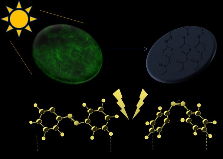
Kill-and-release: Using phenolic azobenzenes to remove Streptoccocus mutans biofilms
from restorative materials
Dylan I. Mori1, Gannon M. Kehe1, Michael J. Schurr2, and Devatha P. Nair1
1
Department of Craniofacial Biology, School of Dental Medicine, University of Colorado
Anschutz Medical Campus, Aurora, CO, USA; 2Department of Microbiology and Immunology,
School of Medicine, University of Colorado Anschutz Medical Campus, Aurora, CO, USA
Secondary caries account for 60-75% of all oral restorative work, as restorations are an ideal
location for bacterial colonization. The Nair lab has demonstrated that while the
photofluidization of azopolymers is effective at removing several types of biofilms,
Streptococcus mutans (S. mutans) biofilms grown under sucrose-rich conditions are
exceptionally adhesive and do not respond to induced photofluidization. This study is aimed at
designing novel antibacterial azopolymers that successfully disrupt S. mutans biofilms (‘kill’)
while maintaining opto-mechanical biofilm removal abilities (‘release’).
Objective: To develop a photoresponsive polymeric coating that can inhibit the growth of S.
mutans biofilms and detach the biofilm from the coating surface.
Methods: Phenolic azobenzene monomer (OH-AAZO) was synthesized, characterized (via
NMR), and polymerized onto a resin to mimic a glassy coating on a dental restoration. The
OH-AAZO’s cytocompatibility was confirmed via ISO9993 using L929 mouse fibroblast cells.
The detachment of an established S. mutans biofilm from the coating surface was initiated via
intermittent light exposures (FlashMaxTM dental lamp, 3 sec flashes) and washes in PBS.
Biofilms were imaged (Zeiss digital microscope) and quantified via serial dilutions and CFU
counts.
Results: In the absence of any external stimuli, the presence of a polymerized OH-AAZO
coating successfully inhibits S. mutans biofilm formation. The effectiveness of OH-AAZO is
greater with higher concentration coatings compared to lower concentration coatings.
Interestingly, a combination of phenolic and azobenzene groups results in the optimal
inhibitory effect. OH-AAZO has also demonstrated long-term S. mutans inhibition in both
sucrose-dependent and -independent conditions.
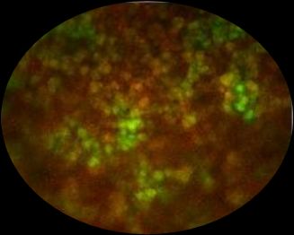
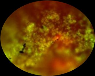
Digital microscope images of an S. mutans biofilm grown on an
uncoated sample (left) and on a substrate coated with OH-
AAZO (right). No live S. mutans was detected on the OH-AAZO
substrate.
Conclusions: Preliminary results indicate that OH-AAZO has potential to prevent S. mutans
biofilm formation on the surface of oral restorations (patent pending) while maintaining the
ability to detach biofilms from the surface. Future work will involve synthesizing new
azobenzene compounds to maximize the detachment forces of dead S. mutans from the
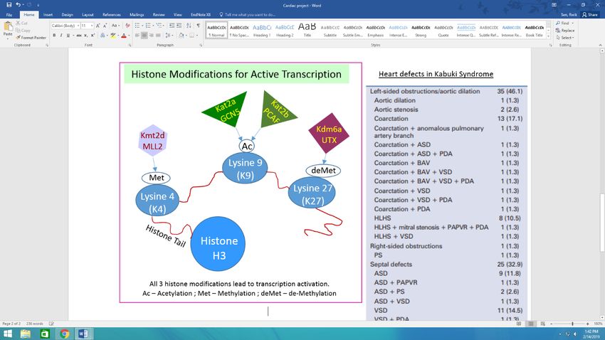
coating surface.Role of chromatin modifiers in craniofacial and cardiac development Rwik Sen1, Sofia A. Pezoa2, Lomeli Carpio Shull1, Laura Hernandez-Lagunas1, Lee A. Niswander2, Tamim H. Shaikh3, and Kristin Bruk Artinger1 1 Department of Craniofacial Biology, University of Colorado Anschutz Medical Campus, Aurora, CO, USA; 2Department of Molecular, Cellular and Developmental Biology, University of Colorado, Boulder, Boulder, CO, USA; 3Division of Pediatrics, Children’s Hospital Colorado, Aurora, CO, USA Objectives: Cranial and cardiac neural crest cells (NCC) undergo cellular growth, patterning, and differentiation in the branchial arches to form cartilage and bone of the face, and the great arteries, cardiac ganglia, and outflow tract myocardium of the heart respectively. Defects in chromatin modifiers result abnormalities in both NCC populations, including in Kabuki syndrome patients, suggesting a role in regulating normal NCC development. Here, we investigated the role of two histone acetyltransferases Kat2a and Kat2b that modify Histone 3 lysine 9 (H3K9) in craniofacial development, a histone H3K4 methyltransferase Kmt2d and two de- methylases Kdm6a/al which target methylated histone H3K27 in cardiac development. Methods: We knocked out these genes in zebrafish using gene editing tools like CRISPR and TALEN, and employed assays to analyze RNA and protein levels, and cartilage formation. These genes are conserved among zebrafish and humans. Results: Single and double kat2a and kat2b zebrafish mutants have an overall shortening and hypoplasia of cartilage elements and disruption of posterior ceratobranchial cartilages, likely due to reduced domains of expression of cartilage and bone-specific markers, sox9a and col2a1, and runx2a and runx2b, respectively. This reduction is likely a result of an overall reduction of H3K9 acetylation in kat2a mutants. Craniofacial patterning genes including tbx15, tgfb3, tgfbrap1 and nf2b are differentially expressed upon RNA sequencing analysis of sox10:GFP positive neural crest cells in kat2a and kat2b mutants. The analysis suggests altered cellular energetics in mutants. Disruption of kmt2d and kdm6a/al result in both cardiac and craniofacial defects, with reduction of heart rates. Ongoing studies will examine the expression of cardiac progenitor markers such as hand2 and mef2ca as well as examination of transcriptome of human Kabuki patient lymphoblastoid cells where kmt2d and kdm6a/al protein levels are reduced. Conclusions: Together, these studies suggest important roles of epigenetic modifiers in craniofacial and cardiac development.
PRDM histone methyltransferases regulate cranial neural crest development

Lomeli C. Shull1, Joseph Gerlach1, Lee Niswander2, Kristin B. Artinger1
1
 Department of Craniofacial Biology, School of Dental Medicine, University of Colorado Denver
| Anschutz Medical Campus, Aurora, CO, USA; 2Department of Molecular, Cellular and
Developmental Biology, University of Colorado, Boulder, CO, USA

Defects in cranial neural crest (cNCC) development are associated with birth defects including
cleft lip/palate and chromatin remodelers have been linked to these craniofacial disorders. The
mechanisms of how chromatin regulators function in normal cNCC development and their
association with congenital diseases remains unknown.

Objective. We investigated the role of lysine methyltransferases, prdm3 and prdm16, during
craniofacial development. prdm3 and prdm16 methylate lysine residue 9 on histone 3 to
repress gene expression.

Methods. CRISPR/Cas9 was used to generate prdm3 and prdm16 zebrafish mutants.
Phenotypes were analyzed by Alcian blue and Alizarin red staining of the craniofacial
skeletons. Transcriptomic (RNA-seq) studies in zebrafish were used to identify mechanisms of
prdm3 and prdm16 in cNCCs during craniofacial development.

Results. Loss of prdm3 or prdm16 in zebrafish causes subtle craniofacial defects including
reduced development of the posterior ceratobrancial cartilages. prdm3-/-;prdm16-/- double
mutants do not survive longer than 3 dpf, however, different combinatorial loss of alleles
causes more severe craniofacial phenotypes at 4 dpf. RNA-seq revealed global changes in
gene expression with loss of prdm3 or prdm16 specifically in pathways associated with cell
junctions, tubulin dynamics and neurogenesis. RNA-seq also revealed an upregulation of
prdm1a in each mutant suggesting genetic compensation between prdm1a, prdm3 and
prdm16. prdm1a knockdown in combinatorial prdm3/prdm16 mutants causes very severe
craniofacial defects at 4 dpf. Characterization of prdm1a-/-;prdm3-/-;prdm16-/- triple zebrafish
mutants is ongoing, as well as defining the mechanism of PRDM orthologs in mediating proper
chondrocyte condensation/adhesion versus activation of cell fate during craniofacial
development.

Conclusion. These studies reveal important roles for Prdm3 and Prdm16 in cNCCs and
formation of the craniofacial skeleton.
ROS modulates ERK signaling after DNA damage

Jordon T. Speidel, Angela M. Ohm, Trisiani Affandi and Mary E. Reyland
Department of Craniofacial Biology, University of Colorado Anschutz Medical Campus,
Aurora, CO, USA

Treatment of head and neck cancers typically involves chemotherapy and/or
radiotherapy. Because of the non-selective nature of these treatments the adjacent
normal, healthy tissue is often also affected. If the salivary gland is injured the secretory
acinar cells can undergo apoptosis, resulting in decreased saliva production leading to
poor health outcomes. Therefore, the modulation of apoptosis in the healthy non-
targeted tissues provides a potential therapeutic target. To this end, our lab has
demonstrated that nuclear localization of PKCδ after DNA damage is necessary for
apoptosis. Additionally, it is well established that DNA damage induces reactive oxygen
species (ROS) generation. However, the interplay between DNA damage induced
PKCδ activation and ROS generation has yet to be elucidated. We hypothesize that
induction of ROS by DNA Damage is regulated by PKCδ and mediates many of the
downstream effects of PKCδ in apoptosis.

Objective: 1) Determine the role of PKCδ in the activation of ERK after DNA damage;
2) Determine the role of ROS in ERK activation after DNA damage; 3) Determine if
PKCδ and ROS work antagonistically or synergistically.

Methods: In vitro studies were performed in rat parotid salivary gland acinar cells
(ParC5) between passages 10-35. Immunoblot analyses were performed using 25ug
total protein lysates. Cellular ROS was detected using CellROX Green (Life Sciences)
by a Gallios flow cytometer (Beckman Coulter). Reverse Phase Protein Array (RPPA)
analysis was performed at MD Anderson’s RPPA Core Facility.

Results: Etoposide induced apoptosis and DNA damage are pERK dependent. PKCδ
overexpression induces ROS dependent DNA damage. We identified the first phase of
ERK activation to be ROS dependent while the second phase is PKCδ dependent and
may be ROS independent.

Conclusions: We have identified that the DNA damage induced biphasic activation of
ERK is both ROS and PKCδ dependent.
PKCδ regulates chromatin conformation and DNA double stranded break repair in
response to irradiation
Trisiani Affandi, Angela M. Ohm, Annie Pham, and Mary E. Reyland
Department of Craniofacial Biology, University of Colorado Anschutz Medical Campus,
Aurora, CO, USA
Treatment for head and neck cancer typically includes irradiation (IR) alone or in
combination with chemotherapy. Although IR kills cancer cells, it also damages normal
tissues in the oral cavity. IR-induced damage to the salivary gland is mediated by
apoptosis, and causes loss of salivary gland function. Our lab has previously shown that
protein kinase C delta (PKCδ) regulates apoptosis in response to DNA-damaging agents,
and that loss of PKCδ function results in radioprotection of normal, but not tumor cells.
Objective: Understanding the mechanism by which nuclear PKCδ regulates DNA
damage-induced apoptosis may allow us to identify new targets for radioprotection of the
salivary gland and oral tissues of HNC patients. We hypothesize that PKCδ regulates
double stranded break repair, and that inhibition of PKCδ activation increases repair of
IR-induced DNA damage through an effect on chromatin accessibility.
Methods: DNA damage was analyzed by γH2AX foci quantification and Comet assay. In
vivo fluorescent reporter assay was used to directly quantify homologous recombination
(HR) and non-homologous end joining (NHEJ). Chromatin remodeling was investigated
by looking at specific histone modifications involved in DNA damage response and by
performing micrococcal nuclease (MNase) assay.
Results: Our data demonstrates that IR-induced γH2AX foci are resolved more rapidly in
PKCδ-depleted cells, which correlates with a more rapid decrease in DNA damage
assayed by Comet. We show that depletion of PKCδ increases DNA repair through both
NHEJ and HR. Furthermore, depletion of PKCδ results in increased activation of DNA-
PK, and the addition of a DNA-PK inhibitor reverses this increase. We show that in PKCδ-
depleted cells, H4K16ac is increased, while expression of nuclear targeted PKCδ
dramatically decreases H4K16ac. Furthermore, depletion of PKCδ results in an increase
in MNase sensitivity, suggesting a more open DNA conformation.
Conclusions: Our data suggest a novel mechanism for control of apoptosis by PKCδ
mediated through regulation of chromatin conformation and DNA repair.
Professional Research Assistants
TKI induced innate immune signaling and response to therapy in EGFR mutant
lung cancer

Ahmed Al Salman1, Natalia J Gurule1, Caroline McCoach3, Trista K Hinz1, Karen Ryall3,
Aik-Choon Tan3, Robert C Doebele3, and Lynn E Heasley1, 3
1
 Department of Craniofacial Biology, School of Dental Medicine, Anschutz Medical
Campus, Aurora, CO, USA; 2MDivision of Medical Oncology, School of Medicine,
Anschutz Medical Campus, Aurora, CO, USA; 3Veterans Administration Eastern
Colorado Health Care System (VHAECH), Aurora, CO, USA

Oncogenic mutations that occur in the receptor tyrosine kinases (RTKs) EGFR, ALK and
ROS1 are present in a significant subset of lung cancer patients, most which present with
advanced stage disease. Tyrosine kinase inhibitors (TKIs) that target tumor cells
dependent on these oncogenic RTKs yield tumor shrinkage but fail to eliminate 100% of
the tumor. Instead, drug tolerant persisters yield residual disease that eventually
contributes to the wide range of therapeutic responses observed in patients.

Objectives: We seek to understand molecular mechanisms that generate residual
disease in response to oncogene specific TKIs. We hypothesize that oncogene targeted
TKIs induce rapid transcriptional reprogramming in the lung cancer cell compartment that
contributes to a varying degree of immune surveillance by the TME through release of
multiple pro- and anti- tumorigenic chemokines and cytokines.

Methods/Results: Preliminary data from RNAseq performed on matched pairs (baseline
and 2 weeks of TKI treatment) of biopsies from two EGFR mutant lung tumors reveals
evidence of marked and variable induction of interferon (IFN) a and g pathways and
effector immune cell signatures. Studies with EGFR mutant cell lines treated with a 3rd
generation EGFR TKI unveils rapid and dynamic transcriptional reprogramming.
Bioinformatic analysis demonstrates induction of the IFNα and IFNγ pathways in
response to TKI, representing increases in pro- and anti-tumorigenic chemokines and
cytokines that instruct adaptive and innate immune cell types. Pro- tumorigenic
chemokines such as IL6 and TGFB2 as well as anti-tumorigenic chemokine CXCL10 are
induced to variable magnitudes and with distinct kinetics across our panel of EGFR
mutant cell lines. Their expression is sensitive to IKK inhibition, suggesting that the NFkB
pathway may be an important transcriptional regulator of EGFR TKI mediated chemokine
and cytokine induction.

Conclusions: Our data demonstrate that inhibitors of oncogenic EGFR induce an innate
immune signaling response in cell line models as well as matched patient biopsies.
Studies are ongoing to test the function of TKI mediated changes to the innate immune
system on response to therapy.
CU Faces of Hope – A craniofacial and dental biorepository

Cristan Carter, David E. Clouthier, Trevor Williams and Kristin B. Artinger
Department of Craniofacial Biology, School of Dental Medicine, University of Colorado
Anschutz Medical Campus, Aurora, CO USA

The craniofacial complex is the most complex region of the human body and thus prone
to genetic insult. As a result, there are wide ranges of syndromes that include
craniofacial abnormalities. The range of these syndromes encompass over one third of
all congenital malformations. Although there have been numerous advances in research
focusing on the causes and treatment of these conditions, there is a critical need to
determine the genetic and developmental underpinnings of these abnormalities. This
lack of knowledge makes it a challenge to translate potential therapeutic and
preventative approaches into the clinic.

Objective: We aim to develop a large biorepository to support a transformational
research program, named CU Faces of Hope, which involves a multidisciplinary team
of biomedical researchers, clinicians and data scientists located across multiple
departments, divisions, institutes and centers at the University of Colorado who will
work together toward a single goal: to compare genetic information from patients
with defects in craniofacial formation and unaffected siblings/parents to identify
mutations that may be causative for the syndromic phenotype.

Methods/Results: The Craniofacial and Dental Biorepository has a goal to enroll up to
2500 individuals with various syndromes associated with craniofacial conditions
between the ages of 6 months (inclusive) and 99 years (inclusive), and 1000 controls.
Clinical data will be obtained from medical health records, radiographs, family history,
and cognitive evaluations that are part of standard clinical encounters. We will collect
both DNA or RNA from blood (or cheek swab) and primary teeth, and whole genome
sequence will be generated for each participant in our project.

Conclusion: The Craniofacial and Dental Biorepository will provide the samples
necessary to significantly advance our understanding craniofacial birth defects, enabling
the development of novel diagnostics and therapeutic tools for patients affected by
craniofacial conditions.
Smart Material Strategies to Combat Contact Lens Microbial Keratitis
Gannon M. Kehe1, Christopher J. Carpenter2, Rooban Nahomi3, Ram Nagaraj3, Michael J. Schurr4,
Devatha P. Nair1,5
1
 Department of Craniofacial Biology, University of Colorado-School of Dental Medicine, Anschutz
Medical Campus, Aurora, CO, USA; 2Department of Biology, Metropolitan State University of Denver,
Denver, CO, USA; Departments of 3Ophthalmology and 4Immunology and Microbiology, University of
Colorado School of Medicine, Anschutz Medical Campus, Aurora, CO, USA; 5Materials Science and
Engineering Program, University of Colorado, Boulder, Boulder, CO, USA

Abstract: Microbial keratitis (bacterial infection of the cornea) is a leading cause of global blindness,
with over two million new cases annually reported. As contact lens (CL) wear significantly increases
the risk of MK via Pseudomonas bacterial infections, non –traditional strategies such as ‘smart’ are of
significant importance. Herein, the athermal photofluidization of azobenzenes in response to transient
light exposure as a novel approach to disrupt and detach biofilms is studied.

Objectives: The aim of this research is to formulate and characterize smart azopolymer coatings that
can disrupt and remove biofilms from the surface of contact lenses in response to transient light
exposure.

Methods: Acrylated azomonomers were
synthesized and characterized (NMR).
Azomonomers were polymerized (double-bond
conversion - FTIR) on the surface of a CL as a
coating (concentrations 100, 50. 0.5 mgs/ml
coatings). Corneal epithelial cell cytocompatibility of
the coated CLs were evaluated and P. aeruginosa
biofilms were grown on CLs and exposed to LED
Light source (3M EliparTM / FlashmaxTM) initiating
the photofluidization of the azo-coatings.
Detachment of biofilms was imaged and quantified
(CFU counts).

Results: The acrylated azomonomers were characterized as CL coatings and concentration
dependent ability to detach biofilms characterized. The initial (before light exposure) and final biofilms
were quantified. Corneal Epithelial cell compatibility indicates that the coatings are non-toxic.

                        PAO1 Biofilm Reduction on Contact Lenses
                        1.0E+09                                               Different concentrations of the azocoating on CLs are studied
                                                                              and their ability to detach biofilms on exposure to light
    CFUs on Substrate

                        1.0E+08                                               (FlashmaxTM, 1 W, 1 min exposure) quantified.
                        1.0E+07                                Initial CFUs
                        1.0E+06                                Final CFUs
                        1.0E+05
                                  AZO 100   AZO 50   AZO 0.5
                                   Light     Light    Light

Conclussion: Our results indicate the azocoatings on CLs can inhibit the growth and attachment of
Pseudomonas aeruginosa biofilms. Future work will focus on optimizing the efficacy of the
azocoatings by tailoring the concentration and structure of the azomolecules
Tuning genetic networks allows development to overcome genetic perturbation Kuval Ray, Juliana Sucharov, Elliott P. Brooks and James T. Nichols Department of Craniofacial Biology, School of Dental Medicine, University Colorado Anschutz Medical Campus, Aurora, CO, USA In humans, loss-of-function mutations in the transcription factor encoding gene MEF2C are associated with many craniofacial dysmorphologies. But, at least one human has been found that is resilient to the mutation, displaying no craniofacial abnormalities. To understand how the same mutation only sometimes causes a phenotype, known as incomplete penetrance, we compare two zebrafish strains that we selectively bred to have either low- or high-penetrance of mef2ca-associated phenotypes. We pushed penetrance to extreme ends of the spectrum by selective breeding. Objectives: We hypothesized that differences in Notch signaling might underlie differences in dlx5a expression and thus differences in penetrance between our low- and high-penetrance strains. Methods and Results: We created a low-penetrance strain in which homozygous mutants survive and develop into fertile adults. We also created a high-penetrance strain in which the mutation is fully lethal and craniofacial phenotypes even manifest in heterozygotes. These strains allow us to probe the mechanisms of genetic resilience. To understand how genetic networks might differ between strains, we examined the mef2ca transcriptional target dlx5a in low- and high-penetrance lines. We find that low- penetrance fish have relatively high dlx5a expression compared to the high-penetrance strain. To test if dlx5a function affects penetrance, we introduced a dlx5a loss-of- function allele into the low-penetrance strain. We find that removing functional copies of dlx5a dose-dependently increases penetrance of mef2ca-associated phenotypes. Notch signaling is known to act oppositely to mef2ca on dlx5a and repress its expression. To test this, we pharmacologically disabled Notch signaling in a high-penetrance family. This treatment reduces the penetrance of mef2ca-associated phenotypes, rescuing mef2ca mutants and phenocopying our low-penetrance strain. Conclusions: It is possible that human MEF2C mutants might use a similar mechanism to overcome deleterious mutations. Thus, we propose that tuning opposing pathways might be a widespread, natural mechanism leading to genetic resilience to many mutations.
Faculty
Remineralization of White Spot Lesions with Apatite-forming Salt

Clifton Carey

Department of Craniofacial Biology, School of Dental Medicine, University of Colorado
Anschutz Medical Campus, Aurora, CO, USA.

Category: Faculty

Objectives: White spot lesions (WSL) are the earliest signs if enamel demineralization
which can develop into cavitations and tooth loss. Currently, no commercial product has
been identified that can reliably reverse WSL. One new product for that contains apatite-
forming calcium-phosphate salts (Ca/P) may induce WSL remineralization. This study
evaluated the remineralization of artificial WSL by use of the SalivaMax™ product that
contains apatite-forming calcium-phosphate salts.
Methods: Fifteen caries-free human premolars were sliced to remove the cusps and the
root to yield tooth discs 5 mm thick. Three groups of 5 discs were joined together with x-
ray transparent epoxy to form columns of teeth samples. One side of the column was
sanded with 600 grit paper to expose flat fresh enamel surfaces. Each set of samples
was attached to a plastic rod for handling and soaked in 1% lactic acid at pH 3.6 for 48 h
to form WSL. Two of the sets were suspended in stirred solutions of SalivaMax™ (1
packet in 35 mL dH2O), and one set was suspended in dH2O (control). SalilvaMax™ and
H2O were replaced daily. All samples were photographed and scanned with microCT,
voxel size 4.4µm, before demineralization, after WSL formation and at 2.5d, 21d and 35d.
Mineral density was determined for sound enamel and the lesion body.
Results: The table shows that there is a significant increase in remineralization at 21
and 35 days exposure to Ca/P-containing SalivaMax™ compared to controls.
                     Time 0 (%)       2.5 days (%)      21 days (%)       35 days (%)
 Control               0.0±0.0           9.5±9.8         10.7±11.0         18.2±10.8
 (dH2O)
 Ca/P treated          0.0±0.0           7.2±6.4          56.7±3.7          73.7±5.4
 t-test p                 -              0.5897            0.0000            0.0000
 Results are percent remineralization average ± standard deviation, n=10 Ca/P treated
 and n=5 controls.
Results are percent remineralization average ± standard deviation, n=10 Ca/P treated
and n=5 controls.
Conclusions: The Ca/P-containing SalivaMax™ is able to cause significant
remineralization of artificial WSLs. This method for remineralizing WSL should be tested
on naturally occurring WSL.
Photodynamic Nanogels as Hybrid layers in Adhesive Dentistry
Rinku .K. Trivedi1,Gannon M. Kehe1, Dylan.l.Mori1, Dixa Gautam1 and Devatha P. Nair1,2
1
Department of Craniofacial Biology, School of Dental Medicine, University of Colorado Anschutz
Medical Campus, Aurora, CO, USA; 2Materials Science and Engineering Program, University of
Colorado, Boulder, Boulder, CO, USA
Achieving optimal resin-dentin bonding is key to long-lasting dental restorations. The 30 ± 11 nm
interfibrillar spaces between the type 1-collagen fibrils serve as diffusion channels for resins to
infiltrate around the collagen to form the hybrid layer that anchors the resin to the tooth. We
hypothesize that by applying a reactive photodynamic azobenzene nanogels (≥ 5 nm) as primer to
form the hybrid layer and the composite-adhesive interface, the mechanical properties of the
adhesive resin can be tailored for optimal adhesion and function.
Objective: The aim of my research is to synthesize photodynamic nanogels and assess the viscosity,
mechanical strength and anti-bacterial properties of the nanogel layers in adhesives
Methods: Nanogels were synthesized using Solvent Yellow7, 2-
Isocyanatoethyl methacrylate, Glycerol and Hexamethylene
Disisocyanate(characterized via triple detector GPC). The viscosity of the
solvated nanogels was evaluated using (CAP2000+;Brookfield,USA) and
while the mechanical strength for two different nanogel concentrations was
assessed via microtensile bond tests (n ≥ 3, Mini Bionix II, MN,USA). S.
mutans biofilms grown on the nanogel-resin samples imaged (Zeiss digital
microscope) and quantified (CFU counts).
Results: Nanogels were successfully synthesized (GPC Mw = 12 kDa, Rh =2
nm). The dry strength of the adhesive was enhanced by the nanogels (41 ± 9 MPa and 27 ± 7 MPa
for the 5wt% and 40 wt% samples vs 20.4 ± 1MPa for the control) while the presence of the nanogels
up to 40 wt% did not influence the anti-bacterial behavior of the BisGMA/HEMA control.
The viscosity of the ethanol solvated
photodynamic nanogels can be tailored via
exposure to light (3M Elipar TM, 1200
mW/cm2 60 s exposure)
Conclusions
Preliminary results indicate that photodynamic nanogels applied as hybrid layers can be tailored for
optimal viscosity and mechanical properties at the adhesive-dentin and adhesive-composite interface.
Future work will include Microtensile dentin bond strength tests and optimizing the viscosity of the
hybrid layer.You can also read