RETINOBLASTOMA BEST PRACTICE GUIDELINES 2019 - KENYA NATIONAL RETINOBLASTOMA STRATEGY - Ministry ...
←
→
Page content transcription
If your browser does not render page correctly, please read the page content below
MINISTRY OF HEALTH

RETINOBLASTOMA
BEST PRACTICE GUIDELINES 2019

         KENYA NATIONAL
    RETINOBLASTOMA STRATEGY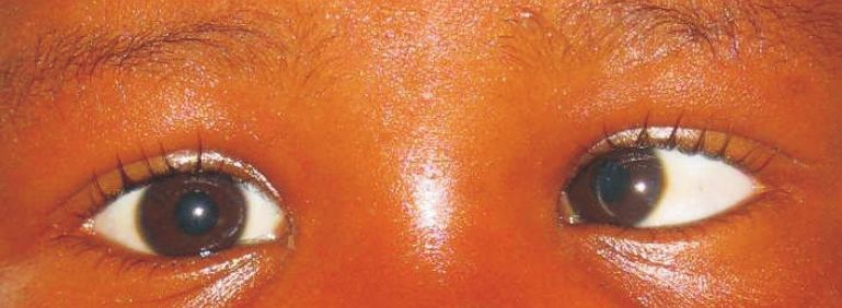
Kenya National Retinoblastoma Strategy Best Practice Guidelines 2019
Kenya National Retinoblastoma Strategy Best Practice Guidelines 2019

                                                    TABLE OF CONTENTS
ACKNOWLEDGEMENTS..................................................................................................................ii

FOREWORD ...................................................................................................................................iii

INTRODUCTION.............................................................................................................................iv

DEFINITIONS & ACRONYMS ...........................................................................................................v

SCREENING - RECOMMENDATIONS ...............................................................................................1

FEATURES AND CLASSIFICATION OF RETINOBLASTOMA CENTRES

RECOMMENDATION ......................................................................................................................2

REFERRAL AND DIAGNOSIS - RECOMMENDATIONS ......................................................................3

GENETIC COUNSELING - RECOMMENDATIONS .............................................................................4

HISTOPATHLOGY - RECOMMENDATIONS .......................................................................................5

TREATMENT - RECOMMENDATIONS..............................................................................................6

        Ocular treatments ................................................................................................................6

        Radiotherapy........................................................................................................................7

        Extraocular disease ..............................................................................................................7

FOLLOW-UP - RECOMMENDATIONS ..............................................................................................8

        Ophthalmology follow-up ....................................................................................................8

        Oncology follow-up ..............................................................................................................8

PSYCHO SOCIAL CARE AND ACCESS TO SERVICES - RECOMMENDATIONS.....................................9

REFERENCES ................................................................................................................................10

ANNEXES

        Annex 1 Management for Retinoblastoma ........................................................................11

        Annex 2 Retinoblastoma Chemo Protocols........................................................................11

        Annex 3 Retinoblastoma Pathology Request Proforma .....................................................14

        Annex 4 Retinoblastoma Pathology Report Proforma .......................................................15

LIST OF TABLES

Table 1: Vision screening guidelines from the CPS ........................................................................2
Kenya National Retinoblastoma Strategy Best Practice Guidelines 2019
ACKNOWLEDGMENT
This is the first revision of the Kenya National Retinoblastoma Strategy Best Practice
Guidelines and has drawn from the expertise of many individuals from medical and social
sciences.
As the guidelines continue to assure good quality of care for children with
retinoblastoma, they embrace and emphasize the multidisciplinary approach in
retinoblastoma care.
The continued learning over the last 5 years from the different disciplines involved has
been instrumental in this revision.
The Ministry of Health wishes to extend appreciation to all who contributed and
supported the revision and production of this document for quality improvement in
retinoblastoma care.
We are sincerely grateful to our heroes; the survivors of retinoblastoma and their families
who inspire for best possible care.
The value of experience and insight from individuals with clinical, laboratory,
pharmaceutical and non-technical backgrounds cannot be underestimated and indeed
greatly enriched the revision of these guidelines.
The process of revising these guidelines was guided by the Ministry of Health;
Ophthalmic Services Unit with close collaboration of the University of Nairobi;
Department of Ophthalmology and with support from the College of Ophthalmology East
Central and Southern Africa (COECSA).
We thank all team members who participated in the revision of these guidelines (see
annex 5). It is with deep commitment to the cause that culminated in these revised
guidelines and it is our sincere hope that the same commitment will remain to see
through the continued improvement of the survival of children with retinoblastoma and
the realization of Universal Health Coverage.
DR MICHAEL M. GICHANGI
HEAD: OPHTHALMIC SERVICES UNIT
iiKenya National Retinoblastoma Strategy Best Practice Guidelines 2019
FOREWORD
Best Practice Guidelines are key tools in the realization of Universal Health Coverage.
Retinoblastoma, a curable cancer of the eye mainly affects children under the age of five
years. The survival of children afflicted by this cancer remains low in Kenya while we know
that it is completely curable if diagnosed and treated early. This has been demonstrated
by the high cure and survival rate in developed where survival is over 95%.
These Best Practice Guidelines have been revised in response to the felt needs,
improvement in the health system, changing patterns in the practice of retinoblastoma
care, and overall National development. The revision process involved gathering relevant
evidence and building consensus, agreeing on what is feasible in Kenya.
The document outlines cardinal characteristics of the disease and best approach to the
management at different levels of care. The management herein referred to includes
timely referral and communication within the health system. It stipulates clearly what
should be done in different circumstances and is recommended for use at all levels of
care.
Over the last five years, following the implementation of the Best Practice Guidelines, we
have observed significant improvement in the outcome of children managed for
retinoblastoma with an estimated doubling of survival from 26% to over 50%.
The revision of these Best Practice Guidelines took in to account the already existing
umbrella National cancer policy and strategy, with emphasis on early detection of cancer.
They are incorporated in the National Cancer strategy 2018-2022 and also clearly relates
to the “Mother and Child Health (MCH) booklet” in early identification of eye problems in
the infant, including retinoblastoma.
We encourage that all children accessing immunization also receive a spot eye check by
the Primary Health care worker as required by the MCH booklet.
We recommend that all health workers continue to familiarize themselves with these
revised guidelines so that all children may live and see to adulthood.
We also recommend that relevant training institutions give more attention to clinical
practice guidelines like this one, at these professional formative stage as we journey
towards sustained Universal Health Coverage in Kenya.
DR PACIFICA K. ONYANCHA
HEAD: DEPARTMENT OF MEDICAL,
PREVENTIVE AND PROMOTIVE SERVICES
iiiKenya National Retinoblastoma Strategy Best Practice Guidelines 2019
INTRODUCTION
Retinoblastoma is the most important primary cancer of the retina and affects young
children mostly under the age of 5 years with over 90% of cases being diagnosed by the
third birthday. The retinoblastoma story provides not only a model for what can be
achieved in the conquest of cancer through research and education but also provides a
model of oncogenesis with exciting insights into specific genetic alterations required to
transform normal cells into cancerous cells.
The incidence of retinoblastoma is 1:15,000-20,000 live births and has a fairly uniform
worldwide distribution with no sexual or racial predilection; Nyamori et al found the
incidence in Kenya to be 1:17200 live births1. Retinoblastoma is curable if detected and
treated early. In the developed world most children treated for retinoblastoma survive,
but in Africa most of them die from advanced disease. In Kenya Nyawira et al
demonstrated the mortality of children diagnosed with retinoblastoma to be greater
than 60% and found this to be due to late presentation2.
The Kenya National Retinoblastoma Strategy was established in 2008 with aim of
improving the survival of children with retinoblastoma in Kenya through:
1. Creating awareness about retinoblastoma among the health care workers
as well as the general population to ensure early diagnosis;
2. Improving the quality of medical treatment of children with
retinoblastoma through access to standard treatments and quality
histopathology reports;
3. Family psychosocial support; and
4. Resource mobilization.
These Best Practice Guidelines are a milestone in the clinical care of children with
retinoblastoma in Kenya. It is the culmination of three years of deliberations and
consultations with different professionals involved in the care of children with
retinoblastoma and included ophthalmologists, paediatric oncologists,
histopathologists, ophthalmic clinical officers, nurses, parents and child life specialist
drawn from all over Kenya. A team from Canada assisted in the process and these
Guidelines have borrowed heavily from the Canadian Guidelines but with modifications
to suit the situation in Kenya. The purpose of these guidelines is to ensure common
standards of evidence-based care to improve the quality of care for children with
retinoblastoma Kenya, in a bid to improve their survival.
ivKenya National Retinoblastoma Strategy Best Practice Guidelines 2019
DEFINITIONS
Level of Evidence
Level 1 Randomized controlled trials (RCTs) (or meta-analyses) without
important limitations
Level 2 RCTs (or meta-analyses) with important limitations; Observational
studies (non-RCTs or cohort studies) with overwhelming evidence
Level 3 Other observational studies (prospective cohort studies, case-
control studies, case series)ConsensusInadequate or no data in
population of interest Anecdotal evidence or clinical experience;
100% agreement of KNRbS members
ACRONYMS
CPS Canadian Paediatric Society
CT Computerised Tomography
EBR External Beam Radiation
EUA Examination Under Anaesthesia
IIRC International Intraocular Retinoblastoma Classsification
KNRbS Kenya National Retinoblastoma Strategy
MCH Maternal and Child Health
MRI Magnetic Resonance Imaging
Rb Retinoblastoma
RbCoLab Retinoblastoma Collaborative Laboratory
RCT Randomized Clinical Trial
vKenya National Retinoblastoma Strategy Best Practice Guidelines 2019
SCREENING-RECOMMENDATIONS
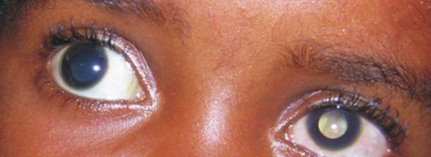
1. We recommend that all infants and children in whom someone has observed a
white pupil (either in person or in a photograph) have a full dilated-eye examination
including red reflex test within 72 hours by an ophthalmologist, or medical
practitioner who is fully aware of the importance of leukocoria as a sign of Rb.
[Consensus]
2. We recommend that any child with strabismus or suspected strabismus be seen by
their primary doctor:
a. We recommend that the red reflex test be applied to any child with strabismus or
suspected strabismus. [Consensus]
b. We recommend urgent referral (within 72 hours) to an ophthalmologist of any child
with strabismus / suspected strabismus and an abnormal red reflex. [Consensus]
c. We recommend that the child in (b) above be seen by secondary or tertiary Rb
centres within 72 hours for the above signs of abnormality, which constitutes an
emergency. [Consensus]
3. We adapt the recommendations of the Canadian Paediatric Society3 with respect
to the suggested timing of vision screening for the general population. [Consensus]
4. We recommend consulting with the Kenyan Mother and Child Health booklet for
information related to retinoblastoma screening. [Consensus]
1Kenya National Retinoblastoma Strategy Best Practice Guidelines 2019
Table 1. Vision screening guidelines from the CPS3.
Age Screening Guideline
n A complete examination of the skin and external eye structures including
the conjunctiva, cornea, iris, and pupils.
n An inspection of the red reflex to rule out lenticular opacities or major
posterior eye disease.
Newborn to
n Failure of visualization or abnormalities of the reflex are indications for
3 months
an urgent referral to an ophthalmologist.
n High-risk newborns (at risk of retinopathy of prematurity and family
histories of hereditary ocular diseases) should be examined by an
ophthalmologist.
n Conduct examination as above.
n Ocular alignment should again be observed to detect strabismus. The
6 to 12 months corneal light reflex should be central and the cover-uncover test for
strabismus normal.
n Fixation and following a target are observed.
n Conduct examination as above.
3 to 5 years n Visual acuity testing should be completed with an age- appropriate tool.
n Screen as above whenever routine health examinations are conducted.
6 to 18 years n Examine whenever complaints occur.
All children should be screened in their preschool years for amblyopia or its risk factors, as
well as for ocular diseases that may have serious consequences, such as retinoblastoma
and cataracts. It remains the responsibility of the child's pediatrician to ensure that these
tests are performed by the most qualified personnel4.
FEATURES AND CLASSIFICATION OF RETINOBLASTOMA CENTRES - RECOMMENDATION
We recommend that Rb treatment centres in Kenya be classified as follows:
Primary able to make a preliminary diagnosis of retinoblastoma.
able to make a clinical diagnosis and treat by enucleation & send eye for
Secondary pathological exam by a qualified pathologist (preferably an ocular
pathologist)
able to confirm diagnosis of retinoblastoma with ultrasound and/or
Tertiary CT/MRI, treat retinoblastoma by enucleation and more complex cases
by chemotherapy, focal therapy and radiation.
[Consensus]
2Kenya National Retinoblastoma
Kenya National
StrategyRetinoblastoma
Best Practice Guidelines
Strategy Best
2019Practice Guidelines 2019
REFERRAL AND DIAGNOSIS - RECOMMENDATIONS
1. We recommend that any child with signs consistent with Rb be referred to an
ophthalmologist to receive a full retinal examination with dilated pupil. A
detailed history must be taken to confirm a diagnosis of Rb. [Consensus]
2. We recommend that secondary and tertiary Rb centres accept direct referrals of
patients with suspected Rb from primary healthcare providers, such as clinical
officers, nurses and general practitioners. [Consensus]
3. We recommend that primary healthcare providers obtain and record complete
contact details including telephone contacts, and immediately refer all Rb cases
to a secondary or tertiary Rb centre. [Consensus]
4. We recommend that all children referred with any possibility of Rb be seen
within 72 hours, or as soon as possible, at the secondary or tertiary Rb centre for
thorough ocular and systemic examination. It is the responsibility of the referring
clinician to make person-to-person contact, to emphasize the urgency to the
parents. [Consensus]
5. We recommend that difficult unilateral cases (e.g. very young child; potential to
save the eye; unilateral multifocal and/or germline RB1 mutation), positive family
history, or risk for extraocular disease and bilateral cases be referred from a
secondary centre to a tertiary centre. [Consensus]
6. We recommend that any child with high-risk pathological features (see “Follow-
up” chapter) be referred to tertiary centre. [Consensus]
7. We recommend that the receiving Rb centre promptly inform the referring
physician of the diagnosis, management and outcome of the referral, and invite
the referring physician to remain involved with the care and follow-up of the
child, as appropriate. [Consensus]
8. We recommend that in order to reduce risks associated with radiation exposure,
if possible all children with Rb requiring imaging have an MRI of the head and
orbits at diagnosis, rather than a CT scan to check for evidence of intracranial
cancer and the extent of the disease, but this should not delay treatment (see
Treatment chapter). [Consensus]
3Kenya National Retinoblastoma Strategy Best Practice Guidelines 2019
GENETIC COUNSELLING – RECOMMENDATIONS
1. We recommend antenatal history include a detailed family history of eye disease,
and referral to an ophthalmologist when this history is positive. [Consensus]
For all individuals with Rb and/or family history of Rb, except those excluded by genetic
testing (see recommendation #9):
2. We recommend that expectant couples undergo early prenatal counselling and
their infants undergo perinatal management to facilitate the earliest possible
treatment of tumours. [Level 2]5,6
3. We recommend counselling for patients, parents and other relatives to discuss Rb,
the risk and hereditary pattern of Rb, pregnancy options, post-delivery surveillance
screening protocols to diagnose tumors early in infants at risk, and treatment
options. [Level 3]7,8
4. We recommend that each at-risk family member be screened as soon as possible
after birth frequently until age 7 years, according to the empiric risk of developing
Rb [Level 2]5,6,8
5. We recommend awareness counselling about the risk of other cancers in adult
survivors and relatives. [Level 2]6,9,10
6. We recommend that children with Rb be offered repeated genetic counselling as
they grow up, so that they completely understand their options and appropriate
care for themselves and their children. [Consensus]
When RB1 gene mutation identification is available:
7. We recommend RB1 gene mutation identification testing for the first affected
person (proband) in each Rb family. [Level 2]
8. When the RB1 gene mutation in a proband/family becomes known, we recommend
genetic testing for all at-risk relatives. [Level 2]
9. We recommend that surveillance be discontinued for relatives determined to be
NOT at risk by genetic testing. [Level 2]
4Kenya National Retinoblastoma Strategy Best Practice Guidelines 2019
HISTOPATHLOGY – RECOMMENDATIONS
1. We recommend that the ophthalmologist who enucleates the eye of a child with
retinoblastoma communicates with the pathologist who will report the
histopathology of the enucleated eye at the time the eye is submitted to the
laboratory.[Consensus]
2. We recommend that the ophthalmologist who enucleates an eye of a child with
retinoblastoma uses the standard request form recommended by the Kenya
National Retinoblastoma Strategy Working Group when submitting the specimen
for histopathology (Annex 3).[Consensus]
3. We recommend that the enucleated eye be fixed in at least 100mls of buffered
formalin for at least 24 hours before grossing.[Consensus]
4. We recommend that the grossing of the enucleated eye be done by a trained
pathologist and the processing should be done by a trained histotechnologist under
the supervision of a pathologist experienced in ocular pathology. [Consensus]
5. We recommend that the sections include the pupil-optic nerve section as well as
superior and inferior calottes.[Consensus]
6. We recommend that pathologist reporting the pathology of an eye enucleated for
retinoblastoma uses the standard reporting format recommended by the Kenya
National Retinoblastoma Strategy Working Group (Annex 4).[Consensus]
7. We recommend that the ophthalmologist involved in the child's case review the
pathology of every enucleated eye together with the involved
pathologist.[Consensus]
8. We recommend that the turnaround time for eyes enucleated for retinoblastoma
be not more than two weeks.[Consensus]
5Kenya National Retinoblastoma Strategy Best Practice Guidelines 2019
TREATMENT– RECOMMENDATIONS
1. We recommend that children with Rb be cared for by a multidisciplinary team, that
provides coordinated and collaborative care in and shared between specialized
centres, with expertise and up-to-date protocols and equipment for optimal
management of Rb. [Consensus]
2. We recommend that tertiary Rb centres work together to assure optimal care for
each child. This might include cross-referrals and cross-consultations to access
specific technical or human resources. [Consensus]
Ocular treatments
3. We recommend that enucleation be performed for IIRC Groups D and E eyes when
the other eye is normal or Group A. [Consensus]
4. We recommend that unilaterally affected eyes be enucleated unless that unilateral
eye is Group A. [Consensus]
5. We recommend considering enucleation of any affected eye when optimal salvage
treatment and follow up are not possible.
6. We recommend that upfront enucleation WITHOUT pre-enucleation
chemotherapy be performed for any IIRC Group E eyes, which pose a risk for
systemic metastases. Pre-enucleation chemotherapy is dangerous, since it may
mask features of extraocular extension causing understaging and undertreating of
systemic disease. [Level 2]12,13
6. We recommend that pathologist reporting the pathology of an eye enucleated for
retinoblastoma uses the standard reporting format recommended by the Kenya
National Retinoblastoma Strategy Working Group (Annex 4).[Consensus]
7. We recommend that the ophthalmologist involved in the child's case review the
pathology of every enucleated eye together with the involved
pathologist.[Consensus]
8. We recommend that the turnaround time for eyes enucleated for retinoblastoma
be not more than two weeks.[Consensus]
9. We recommend chemo reduction for tumors in group B, C and D eyes in bilateral
disease, or for recurrences after other therapy prior to focal therapy. [Consensus]
10. We recommend that cryotherapy through a conjunctival incision may be used for
posterior Rb refractory to laser focal therapy. [Consensus]
6Kenya National Retinoblastoma
Kenya National
StrategyRetinoblastoma
Best Practice Guidelines
Strategy Best
2019Practice Guidelines 2019
11. We recommend the use of pre-chemotherapy cryotherapy 24–72 hours before
chemotherapy to increase drug penetration into the eye, particularly for vitreous
seeding, but not in the presence of retinal detachment.[Consensus]
12. We recommend systemic chemotherapy with focal therapy for the primary
treatment of bilateral IIRC Group B, C or D eyes, but not Group A. If the better eye is
group A, B or C, we recommend enucleation of group D eyes. If both eyes are group
D we recommend chemoreduction and focal therapy for both eyes in an attempt to
save vision as much as possible. [Consensus]
13. We recommend the use of Intravitreal chemotherapy for eyes that have vitreous
seeds refractory to systemic chemotherapy [Consensus]
Radiotherapy
14. We recommend that ocular radiotherapy be used only as salvage therapy for the
only remaining eye after chemotherapy and focal therapy have failed to control the
tumour. [Consensus]
Extraocular disease
For the purpose of decision making for treatment:
15. We recommend that patients be categorized to intention to cure, or intention to
palliate. Patients with high risk features on histology, or patients who present with
proptosis without evidence of systemic metastasis, should be categorized as
intention to cure. Those with evidence of metastasis clinically or on investigations
should be categorized as palliation.[Consensus]
16. We do not recommend exenteration of the orbit for Rb, since chemotherapy will
provide more effective palliation, even for massive proptosis. [Level 2]16
17. We recommend that the ophthalmologist involved in the child's case review the
pathology of every enucleated eye for high-risk features, including invasion of optic
nerve, sclera, choroid or anterior segment, that could predispose to extraocular
disease or metastasis. [Level 2]
18. When high-risk features are observed, including invasion of optic nerve, sclera,
choroid or anterior segment, we recommend treatment with prophylactic
chemotherapy. [Level 2]
19. We recommend that for extraocular Rb (in absence of systemic metastasis)
treatment protocols generally include, but not be limited to: orbital radiation for
orbital recurrence postenucleation and systemic chemotherapy. [Level 2]
7Kenya National Retinoblastoma Strategy Best Practice Guidelines 2019
20. We recommend that metastatic disease be treated palliatively, with 2 to 3 courses
of chemotherapy as per national guidelines or spot radiotherapy for symptoms
relief. We recommend abandoning chemotherapy if there is no response after 3
courses. Where possible, hospice care should be included early in the management
of these patients. These patients should be kept as close to their families as
possible. [Consensus]
FOLLOW-UP - RECOMMENDATIONS
1. We recommend that all survivors of Rb receive individualised, lifelong follow-up
and surveillance, counselling and interventions for late effects of disease and
treatment, delivered by a multidisciplinary team. [Consensus]
Ophthalmology follow-up
2. We recommend that following completion of treatment, EUAs for children at risk of
developing new Rb tumours continue as often as every 3 weeks, or at longer
intervals as tumour activity decreases, until risk of new tumours and recurrences is
low, and the child is able to cooperate in clinic. The frequency of examinations will
be highest when the child has a proven RB1 germline mutation, family history of
disease and/or bilateral Rb. [Level 2] 5,11,17
3. We recommend that EUA or clinic visits for retinal exam continue every 6 months to
age 5 and thereafter annually for life. [Consensus]
4. We recommend that children shown to not carry the RB1 mutant allele of their
family through a blood test do not require EUA or intense surveillance. [Consensus]
5. We recommend the examination of an enucleated socket for infection, fit of
prosthesis and implant exposure or extrusion at every EUA and clinic visit.
[Consensus]
6. We recommend prescribing and monitoring the use of protective eyewear for
children who are functionally uniocular. [Consensus]
7. We recommend that Rb survivors of school age with significantly reduced visual
fields or visual acuity less than 6/12 undergo visual assessment and referral to a low
vision centre for additional assistance when appropriate. [Consensus]
Oncology follow-up
8. We recommend that RB survivors treated with chemotherapy or EBR , undergo
oncology clinic follow up every 6 months or as per national guidelines. [Consensus]
9. We recommend that persons known to be carrying an RB1 germline mutation or
8Kenya National Retinoblastoma Strategy Best Practice Guidelines 2019
have bilateral Rb, or non-germline Rb survivors treated with chemotherapy or EBR,
be seen in oncology clinic for for comprehensive follow-up including counseling for
life. [Consensus]
10. We recommend that MRI replace CT scan if possible, for all patients unless proven
not to have RB1 germline mutations since diagnostic radiation may increase their
already significant risk of secondary non-Rb malignancies. [Level 2]18
PSYCHOSOCIAL CARE AND ACCESS TO SERVICES - RECOMMENDATIONS
1. We recommend that ongoing psychosocial support and timely and equal access to
care is important for all Rb children and their families. [Consensus]
2. We recommend that Rb families have easy and equitable access to [Consensus]:
Ÿ A social worker or clinical psychologist with expertise in Rb or childhood
cancer, from the time of diagnosis onwards.
Ÿ` Structured psychosocial and developmental assessments at diagnosis and key
points during treatment.
Ÿ A child life health worker from diagnosis onwards.
Ÿ Accurate, understandable, as-needed information in a variety of formats.
Ÿ Risk/informed consent information meeting parent language/age/education
level.
Ÿ Advocacy services by professionals or community agencies for parents
requiring additional support to access appropriate services.
Ÿ A centralized referral source providing links to hospital and community
support groups.
Ÿ Long-term psychosocial support from diagnosis through adulthood.
Ÿ Genetic counselling for all family members.
Ÿ Molecular diagnosis when available.
Ÿ Financial support for costs related to treatment.
Ÿ Visual rehabilitation services.
Ÿ Aids for low vision
Ÿ Prosthetic eye service.
Ÿ Paediatric palliative care and bereavement services.
9Kenya National Retinoblastoma Strategy Best Practice Guidelines 2019
REFERENCES
1. Nyamori JM, Kimani K, Njuguna MW, Dimaras H. The incidence and distribution of
retinoblastoma in Kenya. Br J Ophthalmol 2012;96(1):141-3.
2. Nyawira G, Kahaki K, Kariuki-Wanyoike M. Survival among retinoblastoma patients at
the Kenyatta National Hospital, Kenya. J Ophthalmol East Cent & S Afr 2013;17(1):15-9.
3. Community Paediatrics Committee CPS. Vision screening in infants, children and youth.
Paediatrics & Child Health 2009;14:246-8.
4. Community Paediatrics Committee CPS. Vision screening in infants, children and youth.
In: Paediatrics & Child Health; 1998; Reaffirmed February 2007:261-2.
5. Richter S, Vandezande K, Chen N, et al. Sensitive and Efficient Detection of RB1 Gene
Mutations Enhances Care for Families with Retinoblastoma. Am J Hum Genet
2003;72(2):253-69.
6. Kleinerman RA, Tucker MA, Abramson DH, Seddon JM, Tarone RE, Fraumeni JF, Jr. Risk
of soft tissue sarcomas by individual subtype in survivors of hereditary retinoblastoma.
J Natl Cancer Inst 2007;99(1):24-31.
7. Lohmann DR, Gallie BL. Retinoblastoma: Revisiting the model prototype of inherited
cancer. Am J Med Genet 2004;129C(1):23-8.
8. Musarella MA, Gallie BL. A simplified scheme for genetic counseling in retinoblastoma.
J Pediatr Ophthalmol Strabismus 1987;24(3):124-5.
9. Eng C, Li FP, Abramson DH, et al. Mortality from second tumors among long-term
survivors of retinoblastoma. Journal of the National Cancer Institute 1993;85(14):1121-
8.
10. Fletcher O, Easton D, Anderson K, Gilham C, Jay M, Peto J. Lifetime risks of common
cancers among retinoblastoma survivors. J Natl Cancer Inst 2004;96(5):357-63.
11. Houdayer C, Gauthier-Villars M, Lauge A, et al. Comprehensive screening for
constitutional RB1 mutations by DHPLC and QMPSF. Hum Mutat 2004;23(2):193-202.
12. Kingston JE, Hungerford JL, Madreperla SA, Plowman PN. Results of combined
chemotherapy and radiotherapy for advanced intraocular retinoblastoma. Arch
Ophthalmol 1996;114(11):1339-43.
13. Zhao J, Dimaras H, Massey C, et al. Pre-enucleation chemotherapy for eyes severely
affected by retinoblastoma masks risk of tumor extension and increases death from
metastasis. J Clin Oncol 2011;29(7):845-51.
14. Uusitalo MS, Van Quill KR, Scott IU, Matthay KK, Murray TG, O'Brien JM. Evaluation of
chemoprophylaxis in patients with unilateral retinoblastoma with high-risk features on
histopathologic examination. Arch Ophthalmol 2001;119(1):41-8.
15. Kremens B, Wieland R, Reinhard H, et al. High-dose chemotherapy with autologous
stem cell rescue in children with retinoblastoma. Bone Marrow Transplant
2003;31(4):281-4.
16. Bellaton E, Bertozzi AI, Behar C, et al. Neoadjuvant chemotherapy for extensive
unilateral retinoblastoma. Br J Ophthalmol 2003;87(3):327-9.
17. Abramson DH, Du TT, Beaverson KL. (Neonatal) retinoblastoma in the first month of
life. Arch Ophthalmol 2002;120(6):738-42.
18. Brenner DJ, Hall EJ. Computed tomography--an increasing source of radiation
exposure. N Engl J Med 2007;357(22):2277-84.
10Kenya National Retinoblastoma Strategy Best Practice Guidelines 2019
ANNEX 1
MANAGEMENT FOR RETINOBLASTOMA
All E eyes Enucleate
Unilateral RB Group B-D Enucleate
Bilateral RB Group A-D Salvage
Group A Focal Laser or Cryoptherapy
Group B-D Chemoreduction + focal treatment
Pathology pT2a No Chemotherapy
Pathology pT2b-PT3a 4 courses of chemotherapy
Pathology pT3b and worse 6 courses of Chemotherapy + Radiotherapy
Proptosis without metastasis Chemotherapy before enucleation
Metastasis Palliative chemotherapy/radiotherapy
ANNEX 2
RETINOBLASTOMA CHEMO PROTOCOLS
DATE: ______________________ CYCLE: _________________________
Weight: _____________ kg. Height: ________cm. Surface area: _________ m2
Day 1 = ____/_____/_____ (dd/mm/yy)
INDICATIONS:
1. PROPTOSIS:
a. Metastatic workup negative: Use TREAT-TO-CURE CEV PROTOCAL (Pre-
Chemotherapy CT head/orbits; lumbar puncture; bone marrow if abnormal
CBC/Differentiate/Platelet; bone scan optional): May give 1-2 cycles CEV for shrinkage
prior eye enucleation (not orbital exenteration); Give a total 4-cycle CEV; Orbit
radiation as soon as possible (may give concurrent to chemotherapy except on the CEV
day).
b. Metastatic workup positive: Use PALLIATIVE PROTOCOL.
2. UNFAVORABLE HISTOLOGY (sclera involvement/massive choroid disease/post-laminar
tumor in optic nerve/large volume anterior chamber tumor/ciliary body infiltration):
a. Metastatic workup negative: Use TREAT-TO-CURE CEV PROTOCOL (Pre-
Chemotherapy CT head/orbits; lumbar puncture; bone marrow if abnormal
CBC/Differentiate/Platelet; bone scan optional): Give a total 4-cycle CEV; Orbit
radiation if residue tumor after enucleation (tumor at cut margin of optic nerve or long
post-laminar tumor involvement).
b. Metastatic workup positive or optic chiasm involvement: Use PALLIATIVE
PROTOCOL.
11Kenya National Retinoblastoma Strategy Best Practice Guidelines 2019
3. BILATERAL RETINOBLASTOMA:
a. One IIRC Group E or D eye: Enucleation at diagnosis.
b. Both Group E eyes: Bilateral enucleation at diagnosis.
c. One or both IIRC Group A eyes: Give 2-cycle CEV only on failing repeated focal
laser/cryotherapy.
d. One or both IIRC Group B eyes: Give 2-cycle CEV at diagnosis (may repeat 2-cycle CEV
later if needed; if necessary more than once).
e. One or both IIRC Group C eyes: Give 4-cycle CEV at diagnosis (may repeat 2-cycle CEV
later if needed; if necessary more than once).
f. Both IIRC Group D eyes: Give 4-cycle CEV at diagnosis (may repeat 2-cycle CEV later if
needed; if necessary more than once).
4. UNILATERAL RETINOBLASTOMA: Enucleation at diagnosis, except for IIRC Group A eyes that
are treated with focal laser/cryotherapy. NO CHEMOTHERAPY INDICATED TO TRY TO
SAVE UNILATERALLY INVOLVED EYES.
5. METASTATIC DISEASE: Use dosages as in treat to cure but number of courses should be
determined by comfort and pain relieve for the patient. Keep the patient as close to
the family as possible.
RETINOBLASTOMA CEV CHEMO PROTOCOL ORDER SHEETS
DATE: ______________________ CYCLE: _________________________
Weight: _____________ kg. Height: ________cm. Surface area: _________ m2
Day 1 = ____/_____/_____ (dd/mm/yy)
TREAT-TO-CURE CEV PROTOCAL: PRE-CYCLE 1:
☐ CBC, differential count, platelet count
☐ Urea, creatinine, sodium, potassium, chloride, calcium, magnesium, phosphate
☐ ALT, AST, alkaline phosphatase, bilirubin conjugated & unconjugated
☐ Audiogram
☐ If under age 1 years, do 12-hour creatinine clearance (12-hour urine collection with
Foley catheter & serum creatinine taken with the 12 hours):
☐ IF creatinine clearance >100 ml/min/1.73 m2: carboplatin 28 mg/kg per dose
☐ IF creatinine clearance 76-99 ml/min/1.73 m2: carboplatin 600 mg/m2 per dose
☐ IF creatinine clearance 50-75 ml/min/1.73 m2: carboplatin 420 mg/m2 per dose
☐ IF creatinine clearance 25-49 ml/min/1.73 m2: carboplatin 280 mg/m2/dose
☐ IF creatinine clearance ≤24 ml/min/1.73 m2: NO carboplatin
TREAT-TO-CURE CEV PROTOCAL: PRE-EACH CYCLE:
☐ CBC, differential count, platelet count
If possible, EUA day before chemotherapy for 3-spot single freeze/thaw pre-
chemotherapy cryotherapy to increase drug penetration into vitreous (for eyes
without retinal detachment)
12Kenya National Retinoblastoma Strategy Best Practice Guidelines 2019
TREAT-TO-CURE CEV PROTOCAL: PRE-MEDICATIONS & POST-MEDICATIONS:
1. IV Dimenhydrinate (Gravol/Dramamine) ___ mg (2 mg/kg/dose; maximum 50 mg/dose)
2. IV Ondansetron ___ mg (5 mg/m2/dose):
☐ First dose (Dimenhydrinate+Ondansetron) prior to chemotherapy.
☐ Second dose (Dimenhydrinate only) at 3-hours.
☐ Third dose (Dimenhydrinate+Ondansetron) at 6-hours.
TREAT-TO-CURE CEV PROTOCAL DRUGS:
A. 3 years old or YOUNGER:
1. Carboplatin ___ mg (28 mg/kg/dose) in ___ ml (9 ml/kg) 5% dextrose water IV over 30
minutes.
2. Vincristine ___ mg (0.05 mg/kg/dose; maximum of 2.0 mg/dose) IV push
3. Etoposide ___ mg (12 mg/kg/dose), mix well, in ___ ml 0.9% NaCl (30 ml/kg/dose) IV over 30
minutes.
4. Post-chemo, run IV fluid of choice at ___ ml/hr (10 ml/kg/hr) [we use D5W 0.45 saline with
KCl 20 mmol/Liter] for 5 hours.
B. OLDER THAN 3 years:
1. Carboplatin ___ mg (600 mg/m2/dose) in ___ ml (9 ml/kg) 5% dextrose water IV over 30
minutes.
2. Vincristine ___ mg (2 mg/kg/dose; maximum of 2.0 mg/dose) IV push
3. 8Etoposide ___ mg (300 mg/m2/dose), mix well, in ___ ml 0.9% NaCl (30 ml/kg/dose) IV
over 30 minutes.
4. Post-chemo, run IV fluid of choice at ___ ml/hr (10 ml/kg/hr) [we use D5W 0.45 saline with
KCl 20 mmol/Liter] for 5 hours.
C. IF ALLERGIC TO ETOPOSIDE:
1. 3 YEARS OLD OR YOUNGER: Substitute Teniposide ___ mg (12 mg/kg/dose), mix well, in ___
ml 0.9% NaCl (30 ml/kg/dose) IV over not more than 30 minutes.
2. OLDER THAN 3 years: Substitute Teniposide ___ mg (300 mg/m2/dose), mix well, in ___ ml
0.9% NaCl (30 ml/kg/dose) IV over not more than 30 minutes.
13Kenya National Retinoblastoma Strategy Best Practice Guidelines 2019
ANNEX 3
RETINOBLASTOMA PATHOLOGY REQUEST PROFORMA (version 2019)
Patient name: Lab specimen number:
Date of birth: (dd/mmm/yyyy): / / Sex: Female Male
Hospital: Ward: OP/IP number:
Date of procedure: (dd/mmm/yyyy): / / Time of collection: am pm
Surgeon's name:
Telephone: Email:
CLINICAL INFORMATION PROVIDED BY DOCTOR (as per request form)
Laterality: Right Left Bilateral Trilateral
Previous treatment: None Chemotherapy Other (specify):
Clinical assessment: Optic nerve involvement
Extra-orbital involvement
Recurrence (specify): _______________________________________
Metastasis (specify): ________________________________________
Other notes (e.g. nodal involvement, etc) : ___________________________________________
Family history of retinoblastoma? Yes No Unknown
Sign: ________________________________________________________________________
14Kenya National Retinoblastoma
Kenya National
StrategyRetinoblastoma
Best Practice Guidelines
Strategy Best
2019Practice Guidelines 2019
ANNEX 4
RETINOBLASTOMA PATHOLOGY REPORT PROFORMA (version 2019)
Patient name: Lab specimen number:
Date of birth (dd/mmm/yyyy): / / Sex: Female Male
Hospital: Ward: OP/IP number:
Date of procedure: / / Date received: / /
Time of collection: : am pm
Doctor's name:
CLINICAL INFORMATION PROVIDED BY DOCTOR (as per request form)
Laterality: Right Left Bilateral Trilateral
Previous treatment: None Chemotherapy Other (specify):_____________
Clinical assessment: Optic nerve involvement Extra-orbital involvement
Recurrence (specify): _______________________________________
Metastasis (specify): _______________________________________
Other notes (e.g. nodal involvement, etc): _____________________
Family history of retinoblastoma? Yes No Unknown
MACROSCOPIC EXAMINATION
Type of specimen: Eye Orbital biopsy Other (specify): ______________
Side: Left Right
Structures included: Medial rectus Other:
Extra-ocular muscle marked for orientation: Medial rectus Other: None
Specimen dimensions: Anteroposterior: cm Horizontal: cm
Vertical: cm Optic nerve length: cm
Optic nerve thickness/diameter:
Distal end: mm cannot determine (specify):
Proximal end: mm cannot determine (specify):
Tumour dimensions after grossing: Base at cut edge: mm
Height at cut edge: cm
Cannot determine (specify):
Growth pattern: Endophytic Exophytic Diffuse
Cannot determine (specify):
15Kenya National Retinoblastoma Strategy Best Practice Guidelines 2019
RETINOBLASTOMA PATHOLOGY REPORT
MICROSCOPIC EXAMINATION
Percentage of retinal involvement: %
Microscopic involvement of ocular structures.
None Sclera Optic disc
Vitreous Extrascleral extension Vortex veins
Ciliary body Iris Anterior chamber
Angle/Schlemm's canal Cornea Lens
Other (specify):
Choroid; maximum extent of choroidal invasion: mm Notes:
Optic Nerve within lamina cribrosa
prelaminar
retrolaminar ; specify extent of involvement: mm
Status of tumour at resection margin: Present Absent
Surgical margins Cannot be assessed
Tumour at margins.
None
pT STAGING (EYE)
pTX Unknown evidence of intraocular tumor
pT0 No evidence of intraocular tumor
pT1 Intraocular tumor(s) without any local invasion, choroidal invasion or pre- or
intralaminar involvement of the optic nerve head
pT2 intraocular tumor(s) with local invasion
pT2a concomitant focal choroidal invasion pre- or intralaminar involvement of the optic
nerve head
pT2b Tumor superficially invasion of stroma of iris and/or trabecular meshwork and/or
Schlemm's canal
pT3a Massive choroidal invasion (>3mm in largest diameter, or multiple focal choroidal
involvement totaling >3mm, or any full-thickness choroidal involvement)
pT3b Retrolaminar invasion of optic nerve head, not involving the transected end of
optic nerve
pT3c Any partial thickness involvement of the sclera within the inner two thirds
pT3d Full-thickness invasion into outer third of the sclera and/or around emissary
channels
pT4 Evidence of extraocular tumour: Tumour at the transected end of optic nerve,
tumour in meningeal spaces around optic nerve, full-thickness invasion of the
sclera with invasion of episclera, adjacent adipose tissue, extra ocular muscle,
bone, conjunctiva or eyelid
16Kenya National Retinoblastoma Strategy Best Practice Guidelines 2019
Heritable trait (H)
HX Unknown or insufficient evidence of a constitutional RB1 gene mutation
H0 Normal RB1 allele in blood tested with demonstrated high-sensitivity assays
H1 Bilateral retinoblastoma, retinoblastoma with an intracranial primitive
neuroectodermal tumour (i.e. trilateral retinoblastoma), patient with family history
of retinoblastoma, or molecular definition of a constitutional RB1 gene mutation
FINAL REPORT
Right/left eye enucleation …..differentiated retinoblastoma showing choroid
invasion(3mm) and no/prelaminar/laminar/post laminar optic nerve invasion:
optic nerve resection marginis Postive/Negative of tumour.
Stage pT………(TNM 8th Edition)
Name of Pathologist:
Date (dd/mmm/yyyy): / /
Signature:
17Kenya National Retinoblastoma Strategy Best Practice Guidelines 2019
ANNEX 5:
PARTICIPANTS IN REVISION OF THE KENYA NATIONAL RETINOBLASTOMA STRATEGY BEST
PRACTICE GUIDELINES
1. Kahaki Kimani University of Nairobi (Facilitator)
2. Edna C. Mutai Kenyatta National Hospital
3. Ann Makena PCEA Kikuyu Eye Hospital
4. Dr. Faith Vata PCEA Kikuyu Eye Hospital
5. Nalyanya Wekesa Moi Teaching & Referral Hospital
6. Dr. Claric Onyango Kisii Teaching & Referral Hospital
7. Mr. Jones Makori Coast Provincial General Hospital
8. Dr. Lucy Njambi University of Nairobi
9. Margaret Musila Kenyatta National Hospital
10. Dr. Kennedy Wabwile Moi Teaching & Referral Hospital
11. Wilson Githinji Kenyatta National Hospital
12. Dr. Ibrahim Matende Lighthouse/ Coast Provincial General Hospital
13. Dr. Joy Kabiru PCEA Kikuyu Eye Hospital
14. Dr. Wairimu Waweru University of Nairobi
15. Truphosa Chidzuga Coast Provincial General Hospital
16. Dr. Jamilla Rajab University of Nairobi
17. Dr. Irene Muramba Coast Provincial General Hospital
18. Phenny Kanchoro Lighthouse for Christ
19. Rose Atsiaya Lighthouse for Christ
20. Dr Mary Mugania Kenyatta National Hospital
21. Wekesa Nchienya Moi Teaching & Referral Hospital
22. Dr. Jean Claude Sabatia Eye Hospital
23. Dr. Micheal Gichangi MOH
24. Dr. Benjamin Biwott Moi Teaching & Referral Hospital
25. Dr. Zahra Mohamed Coast Provincial General Hospital
26. Dr. Wata David Kenyatta National Hospital
18Kenya National Retinoblastoma Strategy Best Practice Guidelines 2019
NOTESKenya National Retinoblastoma Strategy Best Practice Guidelines 2019
This is a production of:-

      MINISTRY OF HEALTH
    Ophthalmic Services Unit
   P.O. Box 43319 00100 GPO
         NAIROBI KENYA
   Tel: 020-2621841,2729876
   Website: www.health.go.ke
You can also read