Review Paper An extensive Review of Sunscreen and Suntan Preparations - Preprints.org
←
→
Page content transcription
If your browser does not render page correctly, please read the page content below
Preprints (www.preprints.org) | NOT PEER-REVIEWED | Posted: 30 April 2019 doi:10.20944/preprints201904.0327.v1
Peer-reviewed version available at ARC Journal of Pharmaceutical Sciences 2019, 5, 2; doi:10.20431/2455-1538.0502002
Review Paper
An extensive Review of Sunscreen and Suntan Preparations
AK Mohiuddin, Assistant Professor1
1
Department of Pharmacy, World University of Bangladesh,
151/8, Green Road, Dhanmondi, Dhaka – 1205, Bangladesh
Corresponding author: AK Mohiuddin, mohiuddin3@pharmacy.wub.edu.bd, +8801716477485
Orcid ID: https://orcid.org/0000-0003-1596-9757
1. Background
Ra (Re) was the primary name of the sun god of Ancient Egypt. According to Osiris myth, Nut,
the mother of Osiris swallowed the setting sun (Ra) each evening and gave birth to him each
morning. The Ancient Egyptians were well aware of the dangers of the sun. Their lands were
scorched with heat. Women protected their skin, preferring light skin to dark in their cultural
hierarchy of beauty. Recent discoveries written on papyri and the walls of several tombs unearthed
ingredients and formulations in use in Ancient Egypt specifically addressing issues of sun damage
to the hair and skin. Also, a brief historical review manifests the following interesting things
indeed:
Jasmin was used to heal the sun-damaged skin. Recent evidence reveals that Jasmin aids
in DNA repair at the cellular level.
Aloe was used to heal sun-damaged skin.
Olive oil was used as a hydrating oil for both skin and hair damaged by overexposure to
the sunlight.
Almond oil was applied before and after sun exposure to hydrate the sun-damaged skin,
improving elasticity and texture.
Rice bran extracts were used in sunscreen preparations. Today, gamma oryzanol extracted
from rice bran has UV absorbing properties.
kohl (to darken eyes in order to combat sunlight impairment to the retina in the glare of the
desert sun), red ochre (to redden and impart a rosy glow in women’s makeup mimicking
the effect of the sun on the skin), and henna oil (to dye the lips and nails, darken the color
of the hair and skin, and protect light skin from the sun). Today, henna is one of the most
widely used natural sunscreen with both UVA and UVB protection.
Lupin extract was used to block the rays of the sun and is still used to date to lighten the
color of the skin.
Calcite powder and clay were used as UV filters similar to the modern-day inorganic
particulates zinc oxide and titanium dioxide.
Aquatic lotus oil was used for protection of the skin from the sun.
2. Abstract
The sunscreen industry is achieving remarkable worldwide prominence by responding to the
growing need for skin protection with fast-paced innovation. Increased consumer awareness of the
harmful effects of sunlight has fueled the demand for improved photo protection. The need for
broad-spectrum protection from both UVA and UVB rays has inspired scientists worldwide to
research new cosmetic formulations and delivery systems. More effective sunscreen actives,
emollients and novel cosmetic and functional ingredients have been regularly added to the
© 2019 by the author(s). Distributed under a Creative Commons CC BY license.
Preprints (www.preprints.org) | NOT PEER-REVIEWED | Posted: 30 April 2019                 doi:10.20944/preprints201904.0327.v1

         Peer-reviewed version available at ARC Journal of Pharmaceutical Sciences 2019, 5, 2; doi:10.20431/2455-1538.0502002

             formulator’s repertoire. Creativity in innovation has been hindered only by regulatory agencies
             and patent restrictions worldwide. Familiarity with the current restrictive regulations and patent
             law infringements has become integral to any research effort attempting to provide improved
             protection to individuals affected by the sun’s damaging effects. The increasing incidence of skin
             cancers and photo damaging effects caused by ultraviolet radiation has increased the use of sun
             screening agents, which have shown beneficial effects in reducing the symptoms and reoccurrence
             of these problems. Unlike the situation in Europe where sunscreen ingredients are considered
             under cosmetics guidelines, the FDA is required to define sunscreens as drugs since they are
             advertised to prevent sunburn and, more recently, the risk of skin cancer. In the USA, the FDA has
             been regulating this industry since August 25, 1978, with the publication of the Advance Notice
             of Proposed Rulemaking. Sunscreens are considered drugs and cosmetics and therefore must be
             governed by the FDA-OTC monograph. With the variety of sunscreen agents used in cosmetic and
             UV protection products, Australia, Canada, and the European Union (EU) have also developed
             regulatory protocols on safe sunscreen product use. Unlike the USA though, Australia has
             approved 34 active sunscreen ingredients and the EU has approved 28 of these ingredients. Current
             FDA regulations allow labeling of sunscreen products to a maximum of 30þ, despite the many
             products currently available with numbers as high as 100. From a cosmetic formulation point of
             view, increasing the SPF number in a product is governed by simple chemical principles.
Preprints (www.preprints.org) | NOT PEER-REVIEWED | Posted: 30 April 2019                     doi:10.20944/preprints201904.0327.v1

         Peer-reviewed version available at ARC Journal of Pharmaceutical Sciences 2019, 5, 2; doi:10.20431/2455-1538.0502002

             Figure 1. Diana, Princess of Wales, Vogue 1981. According to the magazine Longevity, Princess Diana was
             "scrupulous about using an SPF-8 sunblock." While the American Academy of Dermatology recommends applying
             at least SPF 30, protection against UV rays can prevent both sunburn and dangerous skin cancers (Source: Picard C.
             25 Beauty Secrets to Steal from Princess Diana. Web GoodHousekeeping.com, Mar 7, 2017).
Preprints (www.preprints.org) | NOT PEER-REVIEWED | Posted: 30 April 2019                 doi:10.20944/preprints201904.0327.v1

         Peer-reviewed version available at ARC Journal of Pharmaceutical Sciences 2019, 5, 2; doi:10.20431/2455-1538.0502002

                 3. Introduction
             Protection from sunlight is often equated with use of sunscreens, but this approach is too narrow,
             and protection should consist of a package of measures: avoiding overexposure to sunlight, using
             sunscreens, and wearing protective clothing. Solar ultraviolet (UV) radiation significantly
             influences the skin, causing aging, sunburns, precancerous and cancerous lesions, and
             immunosuppression. UV radiation has an immunosuppressive effect on the antigen-presenting
             cells within the epidermis and contributes to the likelihood of skin cancer. If solar radiation is a
             primary risk factor for malignant melanoma, it is reasonable to conclude that reducing sun
             exposure via topical sunscreen use would be associated with reduced disease risk. Melanoma is
             more common in Whites than in Blacks and Asians. The incidence of non-melanoma skin cancer
             (NMSC) is dramatically increasing worldwide, despite the increased use of improved sunscreens.
             In 2014, the Surgeon General estimated that 2.2-5.0 million people were treated annually for
             NMSC. As the number of newly diagnosed skin cancers continues to rise, there is a need for
             additional preventative measures beyond sunscreens. UV radiation is the second most prevalent
             carcinogenic exposure in Canada, fifth in Switzerland and is similarly important in other countries
             with large Caucasian populations. Within the UK, Wales has among the highest rates of skin cancer
             annually and skin cancer diagnosis rates have increased 63% in 10 years. Australia and New
             Zealand having the world's highest skin cancers. People living in Australia and New Zealand are
             now advised to apply sunscreen every day when the UV index is predicted to reach 3 or above.
             Denmark has one of the highest incidences of melanoma in the world, although it is a relatively
             northern country. Implications for public health: Increased use of sunscreen as part of the daily
             routine to reduce incidental sun exposure will lead to decreased incidence of skin cancer in the
             future. There are 3 kinds of UV radiation: UVC, UVB, and UVA. The ozone layer ingests 100%
             of UVC, 90% of UVB, and a negligible measure of UVA. Therefore, exhaustion of the ozone layer
             expands UV transmission. UVA is related with aging and pigmentation. It enters profound into the
             skin layer and creates free extreme oxygen species, in a roundabout way damaging DNA. UVA
             expands the quantity of incendiary cells in the dermis and diminishes the quantity of antigens
             introducing cells. UVB causes sunburn and DNA strand breaks. It causes pyrimidine dimer
             changes which are related with non-melanoma skin malignancies. Photoprotection includes both
             essential and secondary protective variables. Essential variables are sunscreens; these incorporate
             physical barriers which reflect and dissipate light, and substance barriers which ingest light.
             Secondary components incorporate cancer prevention agents, osmolytes, and DNA repair enzymes
             which help to constrain skin harm by exasperating the photochemical cascade that happens by UV
             sunlight. People with black skin are much less susceptible to sunburn than white-skinned
             individuals. Black people living in the UK were more likely to use sunscreen as a form of sun
             protection, whereas sunscreen was the least popular modality in the two African countries with
             shade being the most common form of limiting sun exposure. A significant benefit from regular
             sunscreen use has not yet been demonstrated for primary prevention of basal cell carcinoma and
             melanoma. Concerning the prevention of actinic keratoses, squamous cell carcinomas, and skin
             aging, the effect of sunscreens is significant, but it remains incomplete. Some organic UV filters
             (PABA derivatives, cinnamates, benzophenones, and octocrylene) have been described to cause
             photo allergy. Percutaneous absorption and endocrine disrupting activity of small-sized organic
             and nano-sized inorganic UV filters have been reported.

                 4. Positive Effects of UVR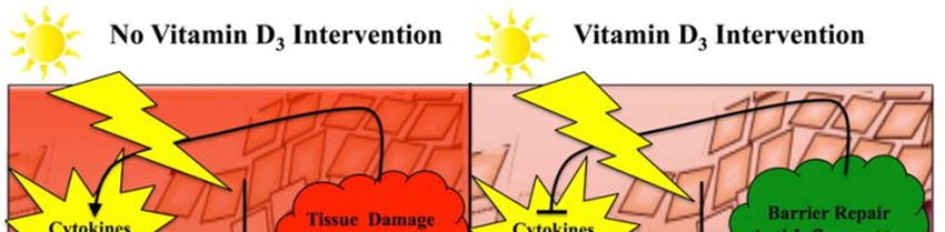
Preprints (www.preprints.org) | NOT PEER-REVIEWED | Posted: 30 April 2019 doi:10.20944/preprints201904.0327.v1
Peer-reviewed version available at ARC Journal of Pharmaceutical Sciences 2019, 5, 2; doi:10.20431/2455-1538.0502002
Exposure to UVR is not always considered bad. In fact, UVR has been found to be particularly
helpful in treating vitamin D deficiency, seasonal affective disorders, psoriasis, sarcoidosis,
mycosis fungoides, and numerous other cutaneous conditions. Within the epidermis, 7-
dehydrocholesterol is converted to vitamin D (cholecalciferol) by UVB light. The elderly and
young children are the ones who are particularly susceptible to vitamin D deficiency. Vitamin D
deficiency can lead to rickets in children, osteomalacia in adults, osteopenia/osteoporosis, and
factures in the elderly. The Institute of Medicine recommends the following vitamin D allowances:
400 IU for 0 to 12 months, 600 IU for 1 to 70 years, and 800 IU for greater than 70 years. Light
therapy is an inexpensive treatment and can be beneficial in treating certain diseases. Although it
has been known for the last 100 years that UV light (particularly UVC with a wavelength range of
240–280 nm) is highly germicidal, its use to treat wound or other localized infections remains at
an early stage of development. Candida auris is a globally emerging yeast, causing severe
infections in patients with underlying diseases. This yeast is responsible for several outbreaks
within healthcare facilities, where it can be found on hospital surfaces and patient care devices.
Spread from these fomites may be prevented by improving the decontamination of hospital
surfaces. UV-C decontamination may constitute an effective adjunct to routine room cleaning.
Additionally, difficult-to-treat psoriasis patients sometimes find relief with UVR. It is thought that
UVR has both antiproliferative and anti-inflammatory effects through downregulation of T-cell
response to antigens. Studies have also shown improvement of the cutaneous effects of sarcoidosis
with UVA-1 light and topical psoralen plus UVA (PUVA) therapy. PUVA and narrowband UVB
has been shown to induce and maintain remissions of mycosis fungoides. Phototherapy is the use
of light for reducing the concentration of bilirubin in the body of infants. However, the exposure
to UV in childhood has been established as an important contributing factor for melanoma risk in
adults and considering the high susceptibility to UV-induced skin damage of the newborn, related
to his pigmentary traits, the UV exposure of the infant during phototherapy should be "as low as
reasonably achievable," considering that it is unnecessary to the therapy. Protective eyewear can
be necessary during newborn assistance activities carried out in proximity of some sources. Most
people judge sun exposure in non-erythemic doses as pleasant. Exposure to sunlight has been
linked to improved energy and elevated mood. Seasonal affective disorder (SAD) is a seasonal
pattern of recurrent major depressive episodes that most commonly occurs during autumn or winter
and remits in spring. The prevalence of SAD ranges from 1.5% to 9%, depending on latitude.
Evidence on light therapy as preventive treatment for patients with a history of SAD is limited,
preventive treatment of SAD and the treatment selected should be strongly based on patient
preferencesLight therapy, an effective treatment for seasonal affective disorder (SAD), may also
be appropriate for MDD which is the second-ranked cause of disability worldwide. Bright light
treatment, both as monotherapy and in combination with fluoxetine, was efficacious and well
tolerated in the treatment of adults with nonseasonal MDD. The combination treatment had the
most consistent effects [77-83].
5. Sunburn
5.1. Sunburn and sunscreen facts
Sunburn and sun poisoning are forms of skin damage due to UV ray exposure. The symptoms
can range from mild to severe and may require treatment.
Sunburn might be sun poisoning if there is blisters, hives or rash, fever and chills, nausea,
dehydration, headache, pain and tingling, vision problems [1].
Preprints (www.preprints.org) | NOT PEER-REVIEWED | Posted: 30 April 2019 doi:10.20944/preprints201904.0327.v1
Peer-reviewed version available at ARC Journal of Pharmaceutical Sciences 2019, 5, 2; doi:10.20431/2455-1538.0502002
UV rays are most intense at noon and the hours immediately before and after (between 10AM
and 4PM) [2], [10].
Immediate symptoms of sunburn are hot, red, tender skin; pain when the skin is touched or
rubbed; and dehydration; several days after exposure the skin may swell, blister, and peel [3].
Most sunburns are mild and can be treated with home remedies such as applying damp cloths
or compresses to reduce the pain, soaking in a tepid bath (with no soap), gently patting the skin
dry, applying soothing creams or lotions, OTC pain relievers such as Tylenol or others, and
moisturizing the skin [4].
Sunburn may cause permanent skin damage and skin cancer (malignant melanoma, basal cell
carcinoma, squamous cell carcinoma) [5].
Persons with certain pigment disorders and individuals with fair skin are at most risk of
sunburn [6].
Certain diseases and conditions pose a higher risk of sunburn (for example, albinism, lupus,
porphyria, vitiligo, and xeroderma pigmentosum) [7].
Some medications may increase sensitivity to sunburn (photosensitivity) [8], [10].
Sun poisoning is caused by severe sunburn; its symptoms include fever, nausea, chills,
dizziness, rapid pulse, rapid breathing, dehydration, and shock [9].
UVA and UVB are mainly responsible for skin pathologies such as sunburns, cutaneous
degeneration, photosensitivity, phototoxicity, photo-aging, immunosuppression and skin
cancer [27].
Snow reflects up to 80% of the sun’s rays, sand reflects 15%, and grass, soil, and water reflect
10%. Due to these factors, an individual sitting under a solitary standard beach umbrella can
be exposed to up to 84% of the total UV radiation despite feeling adequately covered. For these
reasons, it is best to seek deep shade [28].
Less than 50% of the SPF number claimed on the label is spread on the consumer's skin,
meaning that a sunscreen with an SPF 30 will give the real protection of an SPF of 15.
Therefore, SPF 60 should be recommended if real protection of 30 is desired. Significant
injury, DNA damage, mutations, and carcinogenesis can and do occur also with cumulative
sub-erythemal UV exposure [29].
The SPF was created in 1956 by Schulze and it reflects the ratio between the lower amount of
UV energy required to produce a minimal erythema on sunscreen protected skin and the
amount of energy required to produce the same erythema on unprotected skin [31].
Since many of UV filters were shown to cross the blood-brain barrier (BBB), the risk for
neurotoxicity also occurs [37].
Sunscreen compounds might block vitamin D synthesis or act as endocrine disruptor and lead
to developmental toxicity. The effects of sunscreen on cutaneous synthesis of vitamin D
induced by sunlight have been a subject of debate for recent years, however the newest analysis
suggests, that normal usage of sunscreen by adults do not decrease cutaneous synthesis of
vitamin D [38].
The FDA is notoriously slow in approving sunscreen compounds, requiring exhaustive
evidence of their safety. Indeed, FDA has approved only 16 sunscreen ingredients (14 organic
filters and two nonorganic filters, including zinc oxide and titanium dioxide), while other areas
of the world, like the European Union and Australia, have approved nearly twice as many [39].
FDA approved sunscreen ingredients are Aminobenzoic acid, Avobenzone, Cinoxate,
Dioxybenzone, Homosalate, Meradimate, Octocrylene, Octinoxate, Octisalate, Oxybenzone,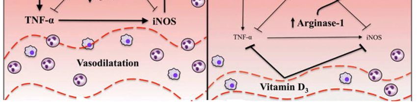
Preprints (www.preprints.org) | NOT PEER-REVIEWED | Posted: 30 April 2019 doi:10.20944/preprints201904.0327.v1
Peer-reviewed version available at ARC Journal of Pharmaceutical Sciences 2019, 5, 2; doi:10.20431/2455-1538.0502002
Padimate O, Ensulizole, Sulisobenzone, Titanium dioxide, Trolamine salicylate, Zinc oxide
[44].
Two of the 16 main ingredients used in OTC sunblock products are safe, the FDA said.
Moreover, the FDA is requesting more information on 12 ingredients among the 16 [45].
The FDA has changed its guidelines to address broad-spectrum sunscreen use, which involves
UVA and UVB coverage; water resistance, to indicate the time duration the sunscreen is
effective; and sun protection factor (SPF). SPF-15 or higher is recommended and can be
labeled as reducing the risk of skin cancer and early skin aging [40].
PABA and trolamine salicylate — are not GRASE for use in sunscreens, are no longer
permitted for use in OTC sunscreen products. No sunscreens sold in the United States contain
PABA or trolamine salicylate [41,42].
For a decade, Environmental Working Group (EWG) has worked to raise concerns about
sunscreens with oxybenzone, which is found in nearly all Americans, detected in breast milk
and potentially causing endocrine disruption [43].
Hawaii recently enacted legislation that will ban the use of two major ingredients -oxybenzone
and octinoxate-that have also been implicated in coral toxicity and will be banned. This creates
a healthcare dilemma: Will the protection of coral reefs result in an increase in human skin
cancers? [51].
5.2.Sunburn: Pathophysiology
Sunburn is a radiation burn to the skin caused by too much exposure to the sun’s UV rays or
artificial sources such as tanning beds. Chronic sun exposure creates premature cutaneous aging,
decreases immune response to environmental pathogens, and increases the risk for developing
premalignant and malignant neoplasms [77]. The biggest risk factors for sunburn is the amount of
time the skin is exposed to UV rays, plus the intensity. Many factors such as time of day,
medications, ozone depletion, high altitude, clear skies, and skin phototypes influence sunburns
[11]. Exposure to solar radiation has the beneficial effects of stimulating the cutaneous synthesis
of vitamin D and providing radiant warmth. Unfortunately, when the skin is subjected to excessive
radiation in the ultraviolet range, deleterious effects may occur. The most conspicuous is acute
sunburn or solar erythema. Initially, UVR causes vasodilation of cutaneous blood vessels, resulting
in the characteristic erythema. Within 1 hour of UVR exposure, mast cells release preformed
mediators including histamine, serotonin, and tumor necrosis factor, leading to prostaglandin and
leukotriene synthesis. Within 2 hours after UV exposure, damage to epidermal skin cells is seen.
Erythema usually occurs 3-4 hours after exposure, with peak levels at 24 hours [12]. Sunburns are
graded as pink, red, and blistering. In contrast, thermal burns are graded by degree (first, second,
and third), but this classification should not be applied to sunburns because thermal burns have
quite different sequelae, such as scarring and death, which are extremely rare consequences of
sunburn. Keratoconjunctivitis or ocular sunburn can also be caused by UV radiation, and it follows
a similar time course [13]. Studies have shown that UVA impairs the antigen presenting cell (APC)
activity of the epidermal cells and thereby causes immune suppression, thus contributing to the
growth of skin cancer [30]. In summary, UVA radiation can cause nuclear and mitochondrial DNA
damage, gene mutations and skin cancer, dysregulation of enzymatic chain reactions, immune
suppression, lipid peroxidation (membrane damage), and photoallergic and phototoxic effects.
On the molecular level, exposure to UV radiation can result in a covalent joining of pyrimidine
(usually thymine) dimers. DNA repair mechanisms, such as nucleotide excision repair, base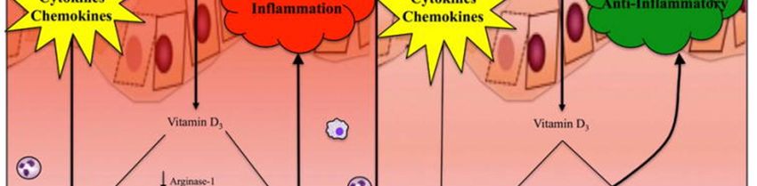
Preprints (www.preprints.org) | NOT PEER-REVIEWED | Posted: 30 April 2019 doi:10.20944/preprints201904.0327.v1
Peer-reviewed version available at ARC Journal of Pharmaceutical Sciences 2019, 5, 2; doi:10.20431/2455-1538.0502002
excision repair, or mismatch repair genes, do not recognize dimers, the mutations go uncorrected
to the cell cycle. When mutated genes reach the cell cycle, if not repaired by the induction of the
p53 pathway, a series of changes can result in malignant transformation and immunosupression.
UV-induced immunosuppression contributes to skin cancer due to damage to DNA and inhibition
of protective mechanism within the skin (Figure 2). A common type of sun-related skin damage
is actinic keratosis (AK). Age, Fitzpatrick skin type 1 or 2 (See exhibit 1), and UV light are the
major risk factors for developing AK. Most AKs do not progress into invasive squamous cell
carcinoma (SCC), but the risk is still present. The risk of malignant transformation of an AK to
SCC within one year is approximately 1 in 1,000. However, approximately 60% of invasive SCCs
of the skin probably arise from AKs. If not treated or protected against additional sun damage,
AKs may eventually progress to invasive SCC. Avoiding sun exposure and daily application of
sunscreen statistically decreases the number of AKs [77].
Exhibit 1. Fitzpatrick's Skin Phototypes [14]
Type I: Pale white skin, burns easily, does not tan
Type II: White skin, burns easily, tans with difficulty
Type III: White skin, may burn but tans easily
Type IV: Light brown/olive skin, hardly burns, tans easily
Type V: Brown skin, usually does not burn, tans easily
Type VI: Black skin, very unlikely to burn, becomes darker with UV radiation exposure
* Individuals with Type I to III Fitzpatrick skin phototypes were at increased risk for sunburn.
Preprints (www.preprints.org) | NOT PEER-REVIEWED | Posted: 30 April 2019                         doi:10.20944/preprints201904.0327.v1

         Peer-reviewed version available at ARC Journal of Pharmaceutical Sciences 2019, 5, 2; doi:10.20431/2455-1538.0502002

             Figure 2. Mechanism of UV induced immunosuppression [130], [139-141]. UVR, in particular the UVB range,
             suppresses the immune system in several ways. UVB inhibits antigen presentation, induces the release of
             immunosuppressive cytokines and causes apoptosis of leukocytes. UVB, however, does not cause general
             immunosuppression but rather inhibits immune reactions in an antigen-specific fashion. Application of contact
             allergens onto UV-exposed skin does not cause sensitization but induces antigen-specific tolerance since such an
             individual cannot be sensitized against the very same allergen later, although sensitization against other allergens is
             not impaired. This specific immunosuppression is mediated by antigen-specific suppressor/ regulatory T cells. UVB-
             induced DNA damage is a major molecular trigger of UV-mediated immunosuppression. Reduction of DNA damage
             mitigates UV-induced immunosuppression. Likewise, interleukin-12 which exhibits the capacity to reduce DNA
             damage can prevent UV-induced immunosuppression and even break tolerance. Presentation of the antigen by UV-
             damaged Langerhans cells in the lymph nodes appears to be an essential requirement for the development of regulatory
             T cells. Studies addressing the molecular mechanisms underlying UV-induced immunosuppression will contribute to
             a better understanding how UV acts as a pathogen but on the other hand can be also used as a therapeutic tool.

             The diagnosis of SCC has increased over the past 30 years. The most important risk factor for SCC
             is cumulative sun damage and age, the risk was greatest in those with more than 30,000 hours of
             cumulative lifetime sun exposure. UVA, UVB, PUVA, and tanning beds have been shown to
             increase the incidence of cutaneous SCC. Prevention of SCC includes protection from the sun,
             including the use of protective clothing and application of sunscreen. Basal cell carcinoma (BCC)
             is the most common skin cancer and occurs most frequently on the face and head. In Caucasians,
             the incidence of BCC has steadily increased and the lifetime risk of developing BCC is 30% [86].
Preprints (www.preprints.org) | NOT PEER-REVIEWED | Posted: 30 April 2019                      doi:10.20944/preprints201904.0327.v1

         Peer-reviewed version available at ARC Journal of Pharmaceutical Sciences 2019, 5, 2; doi:10.20431/2455-1538.0502002

             BCC arises from the basal layer of epidermis and its appendages. The most important risk factor
             is chronic UVR. Other known risk factors include fair skin, light eyes, red hair, chronic arsenic
             exposure, therapeutic radiation, immunosuppression, basal cell nevus syndrome, and various other
             genetic pre-dispositions. Primary prevention is protection from sun exposure beginning at an early
             age [77]. Other than UVR, exposure to arsenic, radiation, chronic inflammatory conditions in skin,
             and burns, scars, infections complications or even tattoos are also the contributing factors for BCC
             [96].

             Figure 3. UVB response in melanoma [85]. UVB exposure triggers macrophage and neutrophil infiltration into the
             skin. Upregulation of CCR2 and ATF2 in melanocytes promotes recruitment of macrophages into the skin, which in
             turn stimulates production of CCL2, MMP-9, and IFN-γ in macrophages. IFN-γ signaling from macrophages promotes
             a positive feedback loop between melanocytes and macrophages, in which melanocytes upregulate CCL8, a CCR2
             ligand, and promote further recruitment of macrophages. The inflammatory response created by macrophage and
             neutrophil recruitment promotes angiogenesis, as well as melanoma cell invasion, survival, and metastasis. UVB also
             independently regulates melanin production and MC1R signaling. Induction of pigmentation genes and subsequent
             increase in melanin production following UVB increases cell survival and NER, but decreases proliferation and
             ultimately, melanomagenesis. Signaling through MC1R is induced by UVB and activates DNA damage response.
             Signaling through αMSH and MC1R promotes phosphorylation of ATM and ATR, upregulates XPC, and promotes
             XPA recruitment to stimulate NER. αMSH also activates oxidative stress response genes to reduce oxidative stress in
             melanocytes/melanoma. UVB-induced expression of IL-23 also activates XPC and XPA to induce NER. IL-23
             signaling, melanin production, and MC1R signaling can all inhibit melanomagenesis induced by UVB. Abbreviation:
             Activating Transcription Factor 2 (ATF2); Chemokine (C-C motif) Receptor 2 (CCR2); Chemokine (C-C motif)
             Ligand 2 (CCL2); Matrix Metalloproteinase-9 (MMP-9); type II Interferon (IFN-γ); Interleukin-23 (IL-23);
             Nucleotide Excision Repair (NER); Melanocortin 1 Receptor (MC1R); Ataxia-Telangiectasia Mutated gene (ATM);
             α-Melanocyte-Stimulating Hormone (αMSH); Ataxia Telangiectasia and Rad3-related (ATR); Xeroderma
             Pigmentosum Group C (XPC); Xeroderma Pigmentosum group A (XPA).
Preprints (www.preprints.org) | NOT PEER-REVIEWED | Posted: 30 April 2019 doi:10.20944/preprints201904.0327.v1
Peer-reviewed version available at ARC Journal of Pharmaceutical Sciences 2019, 5, 2; doi:10.20431/2455-1538.0502002
Epidemiological analysis reveals that annually in the United States alone, ~5 million skin cancer
patients are treated, and that between 40 and 50% of Americans are susceptible to develop skin
cancer at least once by the age of 65 [96]. More than 90% of melanoma cases in the United States
are attributed to skin cell damage from UV radiation exposure. Skin cancer is the most common
form of cancer, representing 40–50% of all cancers diagnosed in the US. Melanoma is the most
serious form of skin cancer. Melanoma is responsible for the most skin cancer deaths, with about
9,000 persons dying from it each year [84]. Despite early screening and detection programs, the
overall mortality rate from melanoma has remained stable or continues to rise. The incidence of
melanoma has more than tripled in the Caucasian population in the United States over the past 30
years. An estimated 60–70% of cutaneous malignant melanomas are thought to be caused by UV
radiation exposure. Two types of UV radiation are primarily responsible for causing carcinogenic
skin damage: UVA (315 nm-400 nm) and UVB (280 nm-315 nm) [85]. Intense and intermittent
sun exposure at a young age increases a patient's risk of melanoma. Individuals with five or more
severe sunburns in childhood or adolescence have an estimated twofold greater risk of developing
melanoma. Melanoma is commonly found on areas sporadically exposed to UVR, such as the back
of the legs in women and the backs of men. UVB, UVA, and PUVA therapy all have been proven
to increase the risk of melanoma. Appropriate UVR protection decreases the risk of developing a
melanoma or having a secondary melanoma. Studies indicate that decreasing recreational sun
exposure following the diagnosis of primary melanoma can significantly decrease the chance of
developing a second melanoma [77].
Exhibit 2. Environmental Factors That Influence Ultraviolet Radiation [32-36]
Ozone layer: The main absorber of UVR, is produced for the most part in the terrestrial
stratosphere of the equatorial region of the planet. Ozone layer is the name given to the
region with high concentration of this gas in the Earth's atmosphere, located at a height
between 15 and 30 km. This layer contains between 80 and 90% of the total ozone in the
terrestrial atmosphere and is responsible for the intensive absorption of UVB radiation and
part of the extinction of UVC radiation.
Altitude: The higher the altitude of a location, the thinner is the atmosphere above it and,
consequently, the larger the quantity of UVR reaching the surface. In situations of clear and
cloudless skies, the UVR flux may increase between 5 and 10% for each 1000 m altitude.
Nevertheless, this altitude-related increase in URV may vary to values close to 20% per
kilometer, as it depends on a series of other factors, such as the quantity of ozone in the
lower layers of the atmosphere, the type of surface that reflects UV radiation, the
particulates present in the atmosphere and even the position of the sun.
The time of day: In a clear sky situation, the "higher" the Sun is in the sky, the higher the
levels of UV radiation are. This means that the farther the Sun is from the horizon, the
shorter the optical pathway the radiation has to cross in the atmosphere.
The season of the year: In the summer, the Sun reaches higher positions in relation to the
horizon than in the winter and, consequently, the UVR flux is more intense. The differences
between the seasons of the year become more relevant as the latitude becomes higher.
Clouds: Clouds can both attenuate and enhance UVB radiation, although attenuation is
generally the case. Completely overcast clouds always attenuate UVB rays, even up to 99%
of UVB radiation in extreme cases. Up to 50% enhancement of UVB radiation can occur
from broken clouds [39] or at elevated sites above clouds.Preprints (www.preprints.org) | NOT PEER-REVIEWED | Posted: 30 April 2019 doi:10.20944/preprints201904.0327.v1
Peer-reviewed version available at ARC Journal of Pharmaceutical Sciences 2019, 5, 2; doi:10.20431/2455-1538.0502002
Suspended Particles: Polluted environments or those with suspended dust may show UVR
attenuation in relation to clear sky situations. Some studies demonstrate that polluted
locations, such as São Paulo or Mexico City, may present situations with around 20% of
the incident UVR.
Surface reflection (albedo): The term 'albedo' is used to express the relationship between
the radiation reflected by a surface and the radiation such surface receives from the Sun.
Very light colored surfaces, such as freshly fallen snow, may reflect up to 90% of the
incident radiation; therefore, wearing adequate protection for the eyes and skin is required
in environments like mountains and ski tracks.
Outdoor Vs Indoor Working: Outdoor workers can get three to nine times as much solar
UVR exposure as indoor workers. Paradoxically, outdoor workers have a lower incidence
of cutaneous malignant melanoma compared to indoor workers. It is supposed that indoor
solar UVA exposure, which causes mutations, depletes vitamin D3 in the skin. Behavior
includes the time spent outdoors and the use of photoprotective agents. In adult life, a
British indoor worker in the UK might typically receive 30% of his or her annual UV
exposure from sun-seeking holidays, 40% from summer weekends, 20% from casual
weekday exposure between April and September and just 10% from sun exposure during
the 6 months period October to March.
5.3.Photoaging
Every organ experience aging in various ways. Skin aging can be categorized into three groups,
intrinsic, photo, and hormonal aging. The consequences of photo-aging are usually characterized
by morphological changes including wrinkle formation or by histological changes in connective
tissues. In order to generate more wrinkles, vasculature structures are needed to develop.
Numerous reports suggested that angiogenesis plays a significant role in inducing wrinkle
formation in photo-damaged skin [142]. Among harmful environmental factors that contribute to
extrinsic aging, long-term effects of repeated exposure to ultraviolet light are the most significant
and are referred to as photoaging. Premature skin aging and development of malignant cutaneous
tumors, melanoma and non-melanoma, are interrelated issues that are increasingly important
problems in the field of dermatology. Skin aging is important aesthetically, whereas skin cancer is
a direct threat to the health of the patient [143]. Photoaging is a multisystem degenerative process
that involves the skin and skin support system. It is a cumulative process and depends primarily
on the degree of sun exposure and skin pigment. The epidermis and dermis are both affected by
UVB, but the dermis is also affected to a significant extent by UVA. It has long been thought that
the majority of human photo-lesions due to UVB rays, now it is believed that UVA play a
substantial role in photoaging. Photoaging affects the sun-exposed areas and is characterized
clinically by fine and coarse wrinkling, roughness, dryness, laxity, teleangiectasia, loss of tensile
strength and pigmentary changes [144]. These changes are more severe in individuals with fair
skin and are further influenced by individual ethnicity and genetics. Photoaging may be prevented
and treated with a variety of modalities, including topical retinoids, cosmeceuticals, chemical
peels, injectable neuromodulators, soft tissue fillers, and light sources [145]. There is also an
increase in development of benign and malignant neoplasms on photoaged skin. During the years
the progress has been made in understanding the photoaging in human skin. UV irradiation invokes
a complex sequence of specific molecular responses that damage skin connective tissue [146]. In
severely damaged skin, there is loss of epidermal polarity (orderly maturation) and individual
keratinocytes may show atypia, especially the lower epidermal layers. More profound changesPreprints (www.preprints.org) | NOT PEER-REVIEWED | Posted: 30 April 2019 doi:10.20944/preprints201904.0327.v1
Peer-reviewed version available at ARC Journal of Pharmaceutical Sciences 2019, 5, 2; doi:10.20431/2455-1538.0502002
occur in the dermis, where photodamage is characterized by degeneration of collagen and
deposition of abnormal elastotic material, reflected by wrinkles, furrows, and yellow discoloration
of the skin. The greater the photodamage, the more the accumulation of thickened, tangled and
degraded elastic fibers [153]. The application of topical growth factors after microneedling can be
useful to reduce visual signs of facial photoaging by improving skin texture and minimizing the
appearance of fine lines and wrinkles [151]. Amongst retinoids, tretinoin is the most potent and
best-studied retinoid. However, its irritation potential has prompted dermatologists to switch over
to less irritating but comparably effective retinoids like adapalene and to some extent retinol and
retinaldehyde [154]. Bakuchiol (functional analogue of topical retinoids, as both compounds have
been shown to induce similar gene expression in the skin) is a phytochemical that has demonstrated
cutaneous antiageing effects when applied topically. Bakuchiol is promising as a more tolerable
alternative to retinol [152].
Figure 4. Mechanisms of Photoaging and Chronological Skin Aging [143], [147-150]. Photodamaged skin displays
variable epidermal thickness, dermal elastosis, decreased/fragmented collagen, increased matrix-degrading
metalloproteinases, inflammatory infiltrates and vessel ectasia. The main effects of acute and chronic exposure to UV
radiation are DNA damage, inflammation and immunosuppression. These effects are direct as well as indirect due to
ROS production. The ROS are particularly harmful in that they destabilize other molecules and promote chain
reactions that damage biomolecules rapidly, such as telomere shortening and deterioration, mitochondrial damage,
membrane degradation and oxidation of structural and enzymatic proteins. Collagen and elastin are the structural
proteins of the ECM. The deterioration/remodeling of the collagen and elastin fibers facilitates angiogenesis and
metastasis, and the damaged collagen and elastin proteins serve as additional sensitizers of photooxidative stress.
Accumulation of collagen fragments, which occurs with chronic UV exposure and the passage of time, impairs the
mechanical and functional properties of the dermal extracellular matrix. Elastic fibres, such as oxytalan fibres in
papillary dermis, are associated with not only skin resilience, but also skin surface texture, and elastic fibre formation
by fibroblasts is facilitated by increased expression of fibulin-5. Thus, induction of fibulin-5 expression is a damage-
repair mechanism, and fibulin-5 is an early marker of photoaged skin.Preprints (www.preprints.org) | NOT PEER-REVIEWED | Posted: 30 April 2019 doi:10.20944/preprints201904.0327.v1
Peer-reviewed version available at ARC Journal of Pharmaceutical Sciences 2019, 5, 2; doi:10.20431/2455-1538.0502002
Exhibit 3. Comparison between photoaging and intrinsic aging [130]
Particular Photoaging Intrinsic aging
Epidermal Thickness increases, acanthropic in early Thin epidermis
changes phase and atrophy in later stages
Proliferative rate is higher than normal Proliferative rate is lower than normal
Non-uniform and random distribution of Uniform and defined distribution of keratinocytes,
keratinocytes, polarity of cells is lost, polarity is maintained, usually atrophied
frequent enlargement
Diversifi ed melanosomes Uniformly distributed melanosomes
Increased number of stratum corneum Normal cell layer
(SC) cell layer
Vitamin A content is destroyed by sun Plasma content of retinol increases
exposure
Dermal Marked elastogenesis followed by massive Elastogenesis followed by elastolysis
changes degeneration
Massive increase in elastic fibers Gradual decline in production of dermal matrix
Increased lysozyme deposition on elastic Modest lysozyme deposition on elastic fibers
fibers
Decrease in amounts of mature collagen Mature collagen more stable in degradation
Increased mast cells Decreased mast cells
Vessels become dilated Micro vessels decrease
Pronounced inflammation No inflammatory response
Marked increase in glycosaminoglycans Slight decrease in glycosaminoglycans
Common Mild (age 28–35 years): Few wrinkles, no Fine wrinkles, thin and transparent skin
signs and keratoses Loss of underlying fat leading to hollowed cheeks and
symptoms Moderate (age 35–50 years): Early wrinkling, eye sockets with noticeable loss of firmness on the hands
sallow complexion with early actinic keratoses and neck
Advanced (age 50–60 years): Persistent Bones shrink away from the skin as a result of bone
wrinkling, discoloration of the skin with loss, which causes sagging of skin, dry skin with
telangiectases and actinic keratoses pruritus
Severe (age 65–70 years): Severe Inability to sweat sufficiently to cool the skin,
wrinkling, photo aging, gravitational and faster graying of hair
dynamic forces affecting the skin, actinic
keratoses with or without skin cancer
Exhibit 4. Exogenous antioxidants with photoprotective or damage protective effects [154]
Antioxidants Outcome of the study
Topical vitamin C 5% cream applied for six months led to clinical improvement in the
appearance of photoaged skin
Topical vitamin C stimulates the collagen-producing activity of the dermis
Ascorbic acid
Magnesium ascorbyl phosphate administration immediately after exposure in hairless
mice significantly delayed skin tumor formation and hyperplasia induced by chronic
exposure to UV radiationPreprints (www.preprints.org) | NOT PEER-REVIEWED | Posted: 30 April 2019 doi:10.20944/preprints201904.0327.v1
Peer-reviewed version available at ARC Journal of Pharmaceutical Sciences 2019, 5, 2; doi:10.20431/2455-1538.0502002
Exhibit 4. Exogenous antioxidants with photoprotective or damage protective effects [154]
Antioxidants Outcome of the study
Ascorbic acid was a photoprotectant when applied to mice and pig skin before
exposure to ultraviolet (UV) radiation
UV-induced vitamin E depletion
The interaction of vitamin E with the eicosanoid system may result in an anti-
Vitamin E inflammatory effect and thereby complement the photoprotective effects of other
antioxidants in the skin
Vitamin E has skin barrier-stabilizing properties
UV light decreased skin lycopene concentrations more so than skin β-carotene
Lycopene concentrations
Lycopene protects against UV-induced erythema in humans
Carotenoids are efficient in photoprotection, scavenging singlet oxygen, and peroxyl
radicals. Supplements or a carotenoid-rich diet decreased sensitivity against UV-
induced erythema
Supplementation with carotenoids contributes to basal protection of the skin but is not
sufficient to obtain complete protection against severe UV irradiation
Carotenoids (carotene, β-
carotene, and carotenoid Dietary beta-carotene has effect on wrinkles and elasticity, procollagen gene
mix) expression, and ultraviolet (UV)-induced DNA damage in human skin
Erythema-protective effect of a carotenoid mix inhibited serum lipid peroxidation
Presupplementation with β-carotene before and during sunlight exposure provides
protection against sunburn
Inhibition of UV-induced epidermal damage and tumor formation in mouse models
Topical tretinoin ameliorates the clinical signs of photoaging
The treatment of photodamaged skin with tretinoin increased collagen I formation.
Topical tretinoin is safe and effective in the treatment of photodamage
Tretinoin Improvement in photodamaged skin
Topical tretinoin reduced the effects of photoaging
Topical tretinoin in combination with sun protection as a useful approach to the
treatment of sun-damaged skin
Topical application of CoQ10 has the beneficial effect of preventing photoaging
Coenzyme Q10 protects against oxidative stress-induced cell death and enhances the
Coenzyme Q10 (CoQ10) synthesis of basement membrane components in dermal and epidermal cells
CoQ10 was shown to reduce UVA-induced MMPs in cultured human dermal
fibroblasts
Glutathione Glutathione is a photoprotective agent in skin cells
Zn-treated fibroblasts were more resistant to UVR than cells grown in normal medium
Zinc Zn can positively influence the effects of oxidative stress on cultured human retinal
pigment epithelial (RPE) cells
Application of resveratrol to the skin of hairless mice effectively prevented the UVB-
Resveratrol
induced increase in skin thickness and the development of the skin edema
Green tea polyphenols were shown to reduce UV light-induced oxidative stress and
Green tea
immunosuppressionPreprints (www.preprints.org) | NOT PEER-REVIEWED | Posted: 30 April 2019 doi:10.20944/preprints201904.0327.v1
Peer-reviewed version available at ARC Journal of Pharmaceutical Sciences 2019, 5, 2; doi:10.20431/2455-1538.0502002
Exhibit 4. Exogenous antioxidants with photoprotective or damage protective effects [154]
Antioxidants Outcome of the study
Topical treatment or oral consumption of green tea polyphenols (GTP) inhibits
chemical carcinogen- or UV radiation-induced skin carcinogenesis in different
laboratory animal models
Oral administration of green tea or caffeine in amounts equivalent to three or five cups
Green tea or caffeine of coffee per day to UVB-exposed mice increased levels of p53, slowed cell cycling,
and increased apoptotic sun burn cells in the epidermis
Silymarin strongly prevents both photocarcinogenesis and skin tumor promotion in
Sylimarin mice
Skin cancer chemopreventive effects
Genistein Antioxidant and anticarcinogenic effects on skin
Dietary flavanols from cocoa contribute to endogenous photoprotection, improve
dermal blood circulation, and affect cosmetically relevant skin surface and hydration
Cocoa variables
Photoprotection against UV-induced erythema
5.4.Sunburn Home Remedies and Treatment
Certain medical treatments have been tried and studied to treat sunburn. However, in general, most
remedies have not shown any clinically proven benefit as far as speeding the recovery or reversing
the damage. Therefore, most of the treatments available are only used to treat symptoms.
Nonsteroidal anti-inflammatory drugs (NSAIDs): Systemic and topical nonsteroidal anti-
inflammatory drugs, when used at dosages to achieve optimal serum levels for anti-
inflammatory effect, only result in an early and mild reduction of ultraviolet B-induced
erythema [15]. In oral (ibuprofen, Motrin, Naprosyn, Advil etc.) or topical diclofenac 0.1% gel
(Solaraze) forms have shown to reduce redness if applied before or immediately after UVB
exposure. This benefit may be diminished after 24 hours. These medications may also help
relieve the symptoms of sunburn such as pain and discomfort [4].
Topical steroid creams have not shown any significant improvement in sunburn symptoms.
Oral steroids such as prednisone have not proven to be beneficial and have been associated
with some significant side effects. Treatment with topical moderate-potency or high-potency
corticosteroids does not provide a clinically useful decrease in the acute sunburn reaction when
applied 6 or 23 hours after UV exposure [16].
Applying Aloe Vera gel: Topical application of Aloe Vera is not an effective prevention for
radiation-induced injuries and has no sunburn or suntan protection [17]. The Aloe Vera cream
was continuing applied at the test sites twice daily for the next three weeks. The results showed
no sunburn or suntan protection and no efficacy in sunburn treatment when compared to
placebo. The Aloe Vera cream has no bleaching effect too. However, this may be beneficial in
treating the symptoms [18].
Topical anesthetics: Advertised remedies such as topical anesthetics (benzocaine) may help
with symptoms of sunburn, however, very little clinical data is available to substantiate their
effectiveness. LMX is a topical liposomal formulation containing 4% or 5% lidocaine. LMX
4% is available OTC and is FDA-approved for temporary relief of pain and itching associated
with minor cuts, minor burns, sunburn, and insect bites [19].
Vitamin D Preparations: Vitamin D enables anti-inflammation to promote tissue repair in
response to injury. Mechanistically, vitamin D signaling activated M2-autophagy regulatorsPreprints (www.preprints.org) | NOT PEER-REVIEWED | Posted: 30 April 2019 doi:10.20944/preprints201904.0327.v1
Peer-reviewed version available at ARC Journal of Pharmaceutical Sciences 2019, 5, 2; doi:10.20431/2455-1538.0502002
Kruppel like factor 4 (KLF4), peroxisome proliferator activated receptor gamma (PPARG),
and arginase 1. Analysis of UV-exposed human skin biopsies detected a similar increase in
macrophage autophagy following vitamin D intervention, identifying an essential role for
autophagy in vitamin D-mediated protection of skin from UV damage [25].
Sunscreens: Sunscreens represent a practical approach to photoprotection for skin. The
importance of beginning sun protection at a young age cannot be overstated. In humans, the
regular use of sunscreens has been shown to reduce AKs, solar elastosis, UV-induced
immunosupression, and photosensitivities. Sunscreens also prevent the formation of SCCs in
animals. A thorough understanding of the mechanism of action of sunscreens, different
sunscreen vehicle choices, and adverse effects can help educate patients on their choice of
sunscreens [77].
Figure 5. The Effect of Oral Vitamin D3 Intervention on Skin Inflammation [26]. As depicted in panel A, levels
of vitamin D3 are at baseline levels in the absence of high dose oral vitamin D3 intervention. In this context, exposure
to erythemogenic doses of UVR results in sunburn and the release of pro-inflammatory cytokines and chemokines in
the skin, including TNF-α** and iNOS*, which further propagate tissue inflammation. Increased skin redness and
thickness are mediated by vasodilation, an influx of inflammatory cells, and vascular congestion within the skin. The
gene expression profile of skin at this time is characterized by increased expression of various pro-inflammatory genes.
As depicted in panel B, levels of vitamin D3 rapidly rise within the serum after high dose oral vitamin D3 intervention.
Arginase-1 is up regulated within the skin, and production of the pro-inflammatory mediator’s TNF-α and iNOS are
attenuated after sunburn. Reduced skin erythema and thickness are observed clinically. The gene expression profile
of skin at this time is characterized by increased expression of skin barrier genes, which help to repair the epidermal
barrier and attenuate the inflammatory insult. *iNOS is the synthase isoform most commonly associated withPreprints (www.preprints.org) | NOT PEER-REVIEWED | Posted: 30 April 2019 doi:10.20944/preprints201904.0327.v1
Peer-reviewed version available at ARC Journal of Pharmaceutical Sciences 2019, 5, 2; doi:10.20431/2455-1538.0502002
malignant disease.** TNF-α is a primary cytokine that can be induced in keratinocytes and in dermal fibroblasts by
UVB, synthesized in adipose tissue by adipocytes and other cells in the tissue matrix. TNF-α is mainly secreted by
activated macrophages immediately postburn. The host immune response is activated by TNF-α, as is the subsequent
release of cytokines following trauma and infection.
5.5.Medication responsible for increase skin sensitivity to sunlight
A large number of medications are known to increase skin sensitivity to sunlight and are called
photosensitive drugs or medications. Photo-sensitizing chemicals usually have a low molecular
weight (200 to 500 Daltons) and are planar, tricyclic, or polycyclic configurations, often with
heteroatoms in their structures enabling resonance stabilization. All absorb UV and/or visible
radiation, a characteristic that is essential for the chemical to be regarded as a photosensitizer.
Some of the common ones include:
NSAIDs (nonsteroidal anti-inflammatory drugs)
Antibiotics: Tetracyclines (tetracycline, doxycycline [Vibramycin]), Quinolone
(ciprofloxacin [Cipro], levofloxacin [Levaquin]), Sulfonamides (sulfamethoxazole and
trimethoprim; cotrimoxazole [Bactrim, Septra], sulfamethoxazole [Gantanol]).
Diuretics (water pills): thiazides (hydrochlorothiazide [Hydrodiuril], furosemide [Lasix])
Cardiac medications: amiodarone (Cordarone), quinidine
Diabetes drugs: sulfonylureas such as chlorpropamide (Diabinese), glyburide (Micronase,
DiaBeta, Glynase)
Psychiatric drugs: chlorpromazine (Thorazine), tricyclic antidepressants such as desipramine
(Norpramin) and imipramine (Tofranil)
Acne medications: isotretinoin (Accutane) [20-24], [34].
Exhibit 5. Cosmetic ingredients and cancer risk [34]
Cosmetic substance Risk
DEA (diethanolamine)
Can result in formation of carcinogenic nitrosamines
TEA (triethanolamine)
Bronopol (2-bromo-2- May break down into formaldehyde and also cause the
nitropropane-1,3-diol) formation of nitrosamines
1,2-Dioxane in
Contaminated with carcinogenic 1,4-dioxane
surfactants/detergents
Artificial colors (as Blue 1 and
Carcinogenic
Green 3)
Hair dyes Dark colors ingredients are carcinogenic
Can be contaminated with carcinogenic pesticides such as
Cosmetic lanolin DDT, dieldrin, and lindane, in addition to other neurotoxic
pesticides
Talc Carcinogenic
Silica May be contaminated with carcinogenic crystalline quartzPreprints (www.preprints.org) | NOT PEER-REVIEWED | Posted: 30 April 2019 doi:10.20944/preprints201904.0327.v1
Peer-reviewed version available at ARC Journal of Pharmaceutical Sciences 2019, 5, 2; doi:10.20431/2455-1538.0502002
Figure 6. Use of Sun Protective [178]. Public guidelines for sun protection have been updated as stakeholders warn
that Australians are using sunscreen as a “suit of armour”. Heather Walker, chair of Cancer Council Australia’s
National Skin Cancer Committee, said that recent research shows 85% of people do not apply sunscreen correctly.
The Australasian College of Dermatologists and Cancer Council updated the guidelines following concerns that many
Australians only use sunscreen to protect their skin. By not using clothing, a broad-brimmed hat, shade, sunglasses
and sunscreen correctly when the ultraviolet index reaches three or above, they are putting themselves at risk, the
organizations say.
Exhibit 6. Endocrine disrupting effects of the commonly used UV filters [74]
UV Filters Endocrine Disrupting Effects
Activation of ERα, ERβ; Inhibition of the activity of 17β-Estradiol;
Estrogenic disrupting
Induction of proliferation of MCF-7 cell; Induction of VTG in fathead
effects
minnows; Reduce of the uterine weight in immature Long-Evans rats
Antagonists of human AR transactivation; Repression of 4,5-
Androgenic disrupting
Benzophenones dihydrotestosterone-induced transactivational activity; Inhibition of
effects
testosterone formation in mice and rats
Disrupting effects
Inhibition of human recombinant TPO; Interference with THR;
toward other nuclear
Inhibition of TPO activity in rats; Antagonists of PR
receptors
Activation of ERα, ERβ; Inhibition of the activity of 17β-Estradiol;
Disrupting effects
Camphor Induction of proliferation of MCF-7 cell; Induction of pS2 protein in
toward estrogen
derivatives MCF-7 cells; Reduce of the uterine weight in rats; Induction of VTG
receptor
in fishPreprints (www.preprints.org) | NOT PEER-REVIEWED | Posted: 30 April 2019 doi:10.20944/preprints201904.0327.v1
Peer-reviewed version available at ARC Journal of Pharmaceutical Sciences 2019, 5, 2; doi:10.20431/2455-1538.0502002
Exhibit 6. Endocrine disrupting effects of the commonly used UV filters [74]
UV Filters Endocrine Disrupting Effects
Disrupting effects Repression of 4,5-dihydrotestosterone-induced transactivational
toward androgen activity; Inhibition of testosterone formation in HEK-293 cells;
receptor Antagonists of Human AR
Disrupting effects Antagonists of PR; Increase of PR mRNA levels in rats; Inhibition of
toward progesterone the expression of PR protein in rats; Disturbance of the expression of
receptor membrane-associate PR in insects
Disrupting effects Activation of ERα; Inhibition of the activity of 17β-Estradiol;
toward estrogen Induction of proliferation of MCF-7 cell; Reduce of the uterine
receptor weight in rats; Induction of VTG in fish
Disrupting effects
Cinnamate Decrease of T4 level; Inhibition of the conversion of T4 to
toward thyroid
derivatives triiodothyronine in rats
hormone receptor
Disrupting effects
Antagonists of PR and AR; Inhibition of 4,5-dihydrotestosterone
toward other nuclear
activity; Reduce of the prostate and testicular weight in rats
receptors
*AR: androgen receptor; ER: estrogen receptor alpha; PR: progesterone receptor; T4: thyroxine; THR: thyroid
hormone receptor; TPO: thyroid peroxidase; VTG: vitellogenin
5.6. Tanning/Pigmentation with or without Sun Exposure
Some Africans and Asians avoid sun and use bleaching products to lighten skin, while many
Caucasians seek the sun for tanning to achieve a bronze skin to “look good.” UV radiation from
the sun or from artificial sources increases skin pigmentation. Sunlight and indoor UV induced
tanning is a common behavior, especially among adolescents, young adults, and individuals with
lighter skin. Several health benefit claims such as improved appearance, enhanced mood, and
increased vitamin D levels have been attributed to tanning. The Indoor Tanning Association claims
that a base tan can act as “the body’s natural protection against sunburn. Sunless tanning products
may serve as a sensible, safer alternative for those who desire tanned skin [155]. There are three
phases of tanning: immediate pigment darkening (IPD), persistent pigment darkening (PPD) and
delayed tanning (DT). IPD occurs during the first minutes of exposure to UVA, and then fades
within few hours. PPD appears within hours of higher doses of UVA exposure and persist up to
several days or weeks. DT develops over 3–7 days after UVB exposure, and then remains for
weeks. The mechanisms of UVA- and UVB-induced pigmentation are different. UVA induces IPD
and PPD through oxidation of pre-existing melanin or melanogenic precursors. IPD is oxygen
dependent, and reactive oxygen radicals are considered to be responsible for this process. PPD is
also due to the upward movement of melanosomes toward the surface of the skin. Persons with
lightest skin (skin type I) do almost not tan, while IPD and PPD are strongest in moderately and
darkly pigmented skin. DT results from synthesis of melanin in the melanocytes, followed by
melanin distribution to neighboring keratinocytes [156]. UVA (320–400 nm) causes immediate
pigment darkening (IPD) as well as persistent pigment darkening (PPD) of skin within hours via
photooxidation and/or polymerization of existing melanin or melanogenic precursors due to the
generation of reactive oxygen species In contrast, UVB (280–320 nm) induces a slower but more
stable type of pigmentation termed delayed tanning (DT) which requires the increased synthesis
of melanin following the stimulation of tyrosinase activity and the entire melanogenic cascade
[157]. UVB-induced DT is photoprotective (it is estimated to have a SPF of 3)- while DT induced
by UVA is not considered to be photoprotective. DT is maximal from 10 days to 3-4 weeks,You can also read