Super-resolution Light Sheet Expansion Microscopy
←
→
Page content transcription
If your browser does not render page correctly, please read the page content below
CUSTOMER REFERENCE                                                                                 Kinetix Scientific CMOS Camera

Super-resolution Light Sheet Expansion
Microscopy
Prof. Ulrich Kubitscheck, Dr. Martin Schwarz, Mr Juan Eduardo Rodriguez-Gatica
Institute of Physical and Theoretical Chemistry, University of Bonn, Germany

                         The lab of Prof. Ulrich Kubitscheck works to analyse living as well as cleared biological cell systems,
                         developing novel quantitative light microscopy techniques. A combination of tissue clearing with light sheet
                         fluorescence microscopy (LSFM) is especially well suited to the fast analysis of complex arrangements of
                         large cleared cell clusters and thus allows fast light microscopic access into the complex 3D architecture
                         of neuronal tissue.

                         The limitations of LSFM and potential solutions to these were outlined by Prof. Kubitscheck: “LSFM cannot
                         reveal the very fine details of neuronal networks since these structures are well below the optical diffraction
                         limit and are of extreme complexity. A solution to this issue is realized by the combination of light sheet
BACKGROUND               fluorescence and expansion microscopy (LSFEM) allowing for the analysis of extended neural circuits in
                         super resolution… LSFEM is compatible with multicolour fluorescence imaging, thus enabling molecular
                         contrast for diverse neuronal populations and nanoscale resolution within a single, large tissue preparation.
                         LSFEM is optimally suited for the analysis of connectivity in large neuronal tissue regions.”

                         Together with Dr. Martin Scwarz from the University of Bonn Medical School, we developed a novel tissue
                         expansion pipeline and used LSFM and LSFEM to image very extended regions of mouse brain tissue in 3D.
                         We can zoom in and out from meso- to nanoscale resolution, which means to effective super resolution,
                         depicting the finest details of neuronal network parameters within larger context.

                         In this experiment, projections from the horizontal diagonal band of Broca (HDB) to the olfactory bulb (OB)
                         in the mouse brain are visualised using LSFEM and a novel clearing protocol.

Rev A2-06092021   ©2021 Teledyne Photometrics. All rights reserved.
CUSTOMER REFERENCE

“ The large sensor of the Kinetix greatly reduces the number of required
                         “
  image tiles to cover the field of view... the signal to noise ratio is
   excellent.

                          LSFEM involves huge specimen regions and consequently a large number of image tiles acquisitions needed
                          to cover the complete field. These images often need to be overlapping image fields to allow stitching,
                          which adds to both the time taken for an image to be produced, and the computational effort to perform
                          stitching of large number of image tiles. By using a camera with a very large field of view, a smaller number
                          of acquisitions would be needed, reducing both the time for acquisition and processing.
 CHALLENGE
                          The current imaging experiments can often have an exceedingly long duration, during which time the
                          sample is exposed to light and risks photobleaching. A highly sensitive camera with a low noise level is
                          needed in order to reduce the exposure required to image the sample, and avoid photodamage.

                          Finally, a small pixel size is also required in order to achieve diffraction limited resolution, this technique
                          combines the needs of light sheet and super-resolution imaging, meaning the camera also needs to operate
                          at a high resolution at certain magnifications.

                          The ground breaking Kinetix CMOS features a large field of view, a 10 megapixel sensor with small 6.5 µm
                          pixel size, and a high sensitivity thanks to a combination of back-illumination and low-noise imaging modes.
                          This makes the Kinetix an ideal solution for this demanding imaging application.

                          Prof. Kubitscheck told us about his experience with the Kinetix CMOS, “As demonstrated, the large sensor of
                          the Kinetix greatly reduces the number of required image tiles to cover the interesting field of view ranging
                          from the HDB to the olfactory bulb area of a 1.5x expanded and cleared mouse brain section. The signal to
 SOLUTION
                          noise ratio is excellent.”

                          “Using the new Kinetix camera in combination with novel tissue clearing protocols and LSFM we can show
                          in great detail and over extended regions that HDB neurons innervate different cell layers of the OB. Most
                          notably, we can, due to the large field of view, the exquisite resolution and the possibility to image ~2mm
                          thick sections, follow projections from individual HDB neurons up to their target region within the OB.”

                          “The new setup comprising the Kinetix camera will significantly improve our imaging capabilities and will
                          likely lead to novel insight into the HDB to OB synaptome.”

 Rev A2-06092021   ©2021 Teledyne Photometrics. All rights reserved.
CUSTOMER REFERENCE
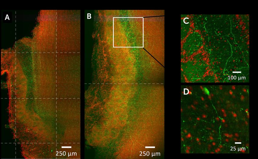
Figure 1: Optical section of a the olfactory bulb from a cleared sagittal slice of a ChAT Cre-mouse, expressing
endogenous EGFP (green) and nuclear staining with Hoechst (red). Acquired with a custom-built LSFM, Nikon
10x/0.3 N.A W objective. A) Image acquired with a typical CMOS, 12 separate acquisition tiles needed to construct.
B) Image acquired using Teledyne Photometrics Kinetix CMOS (on a similar sample), only 2 tiles were needed to
cover the same region as A. C) Single plane of the area marked in B at 208 μm depth. D) Single plane of a separate
area at 200 μm depth and higher magnification.
Images courtesy of Juan Eduardo Rodriguez-Gatica.
www.photometrics.com
info@photometrics.com / tel: +1 520.889.9933
Rev A2-06092021 ©2021 Teledyne Photometrics. All rights reserved.You can also read