SYMPTOMS AND FUNGI ASSOCIATED - WITH DECLINING MATURE GRAPEVINE PLANTS IN NORTHEAST SPAIN
←
→
Page content transcription
If your browser does not render page correctly, please read the page content below
013_JPP404Luque_381_COLORE        25-06-2009       11:55   Pagina 381

         Journal of Plant Pathology (2009), 91 (2), 381-390             Edizioni ETS Pisa, 2009                                         381

                                 SYMPTOMS AND FUNGI ASSOCIATED
                    WITH DECLINING MATURE GRAPEVINE PLANTS IN NORTHEAST SPAIN

                                          J. Luque1, S. Martos1, A. Aroca2, R. Raposo2 and F. Garcia-Figueres3
                 1 Institut
                          de Recerca i Tecnologia Agroalimentàries (IRTA). Carretera de Cabrils km 2, 08348 Cabrils, Barcelona, Spain
                                  2 CIFOR, Instituto Nacional Investigación y Tecnología Agraria y Alimentaria (INIA),

                                                   Carretera de La Coruña km 7.5, 28040 Madrid, Spain
                  3 Laboratori de Sanitat Vegetal (DAR), Via Circulació Nord, Tram VI, Carrer 3, Zona Franca, 08040 Barcelona, Spain

         SUMMARY                                                                sector, grapevine declines and their associated pathogens
                                                                                are poorly known in this country. In the last decade, stud-
            A field survey was carried out in Catalonia (northeast              ies on diseases and pathogenic mycoflora associated with
         Spain) to characterize the decline of mature grapevines.               rootstocks (Aroca et al., 2006), young vines (Armengol et
         The relationships of both external and internal symp-                  al., 2002; Giménez-Jaime et al., 2006), and mature vines
         toms of diseased plants and their associated mycoflora                 (Armengol et al., 2001a, 2001b; Úrbez-Torres et al.,
         were studied. Co-occurrence of different internal dis-                 2006a) have been carried out, but further studies are
         ease symptoms was frequently observed, since 44% of                    needed for a better insight in these complex diseases and
         the sampled plants had wood lesions commonly associ-                   an adequate estimation of their economic impact. Main
         ated with at least two of the following decline diseases:              decline diseases of mature grapevine observed in Spain
         eutypiose, black dead arm or esca. The results obtained                include esca, eutypiose, and black dead arm (BDA), as
         suggest that apoplexy might not be associated only with                reported by Armengol et al. (2001a, 2001b) and Úrbez-
         esca-affected plants, since 60% of surveyed vines show-                Torres et al. (2006a).
         ing apoplexy showed also V-shaped necroses, which are                      Esca is a complex disease where symptoms and their
         commonly associated with eutypiose and black dead                      expression over time are highly variable (Mugnai et al.,
         arm, and 20% were exclusively affected by V-shaped                     1999; Surico et al., 2006). Two main types of esca
         necroses. An experiment was conducted to establish the                 episodes can be defined: the chronic esca and the acute
         pathogenicity of the most representative fungi isolated                syndrome, the latter also known as apoplexy (Mugnai et
         from diseased tissues of declining plants, by artificially             al., 1999). Briefly, foliar symptoms of chronic esca are
         inoculating 1-year-old vines of cvs Macabeo and Tem-                   characterized by interveinal chlorosis or discolorations
         pranillo. As indicated by the extension of the vascular                (yellowish in white cultivars and reddish in red cultivars)
         lesions, pathogenicity was confirmed for most of the                   that later coalesce in large necrotic areas during summer.
         species tested, namely Botryosphaeria dothidea, Diplodia               Vine apoplexy usually occurs in mid summer, when leaves
         seriata, Eutypa lata, Neofusicoccum luteum, N. parvum                  of affected plants wither rapidly in a few days (Mugnai et
         and Phaeomoniella chlamydospora.                                       al., 1999). Despite of the external foliar symptoms of both
                                                                                esca types, several types of wood degradation have been
           Key words: black dead arm, esca, eutypiose, grapevine                described for esca, mainly including: (i) longitudinal
         decline, phytopathogenic fungi, Vitis vinifera.                        brown streakings that appear as necrotic black spots in
                                                                                cross sections, (ii) pink-brown or dark red-brown necrot-
                                                                                ic areas, and (iii) wood decay. Many fungi have been re-
         INTRODUCTION                                                           ported to be involved in the esca syndrome; several Basid-
                                                                                iomycetes species are responsible for the wood decay,
             The area given over to grapevine in Spain is about 1.2             with species in the genera Fomitiporia, Fomitiporella, and
         M ha, which makes this country the leading one in the                  Inocutis (Fischer, 2006), while vascular necroses are
         world for grape-growing, the third for wine production,                caused mainly by Phaeomoniella chlamydospora and sev-
         and the second for raisin production (Anonymous, 2006;                 eral Phaeoacremonium species (Surico et al., 2006).
         OIV data 2005, retrieved from http://www.oiv.org). Al-                     Eutypiose, also known as Eutypa dieback, is caused
         though ca. 97% of the total Spanish grapevine area is                  by the fungus Eutypa lata (Carter, 1988). The most rec-
         destined for wine production (Anonymous, 2006), thus                   ognized symptom of this disease is the stunted appear-
         viticulture is an essential component of the agricultural              ance of shoots at the early growth season, with small,
                                                                                cupped, and chlorotic leaves, and short internodes.
                                                                                Wood internal symptoms include characteristic V-
         Corresponding author: J. Luque
         Fax: +34. 937533954                                                    shaped necroses when affected arms and trunks are
         E-mail: jordi.luque@irta.es                                            cross-sectioned. Additionally, external cankers develop-
013_JPP404Luque_381_COLORE 25-06-2009 11:55 Pagina 382
382 Grapevine decline and associated pathogenic fungi Journal of Plant Pathology (2009), 91 (2), 381-390
ing from old pruning wounds can be observed. of the external symptoms was annotated and attributed
BDA was first described by Lehoczky (1974), who to known diseases: eutypiose, BDA or esca. Vines which
associated this disease with Botryosphaeria stevensii. showed stunted shoot growth in late spring, and V-
Several other species of Botryosphaeriaceae were later shaped wood necroses were classified as affected by eu-
found in BDA-affected vines, the most frequent being typiose/BDA during the field survey, since both diseases
B. dothidea, Diplodia seriata and Lasiodiplodia theobro- show similar symptoms (Castillo-Pando et al., 2001;
mae (Larignon et al., 2001; van Niekerk et al., 2006). Taylor et al., 2005). BDA foliar symptoms occurring in
Wood symptoms of BDA include V-shaped necroses, summer, as described by Larignon and Dubos (2001),
similar to those caused by E. lata, and longitudinal were not considered as these symptoms could be con-
brown streakings along the affected tissues. Occasional fused with those of esca (Lecomte et al., 2005; Surico et
stunted growth in early season was also reported for dis- al., 2006). Vines with characteristic interveinal chloroses
eases caused by Botryosphaeriaceae species (Castillo- and necroses, wood decay and vascular necroses differ-
Pando et al., 2001; Taylor et al., 2005), thus resembling ent from V-shaped ones were classified as esca-affected
the symptoms caused by E. lata. plants. Vines with sectorial necrosis and either one of
BDA foliar symptoms are controversial. While the esca-associated wood necroses were classified as af-
Lehoczky (1974) reported a slight diffuse chlorosis fol- fected by eutypiose/BDA and esca. Plants affected by
lowed by leaf wilting, Larignon and Dubos (2001) de- apoplexy whether partial (1 to several arms) or total
scribed an early red or yellow-orange patchy discol- (whole plant) were considered as a separate class from
oration of the leaves (in red- and white-berried grape the above diseases. Sections of trunks and arms, as well
varieties, respectively) that later developed into large as the whole plant when appropriate, were taken to the
marginal and interveinal necroses. However, Lecomte et laboratory for further examination and to conduct fun-
al. (2005) and Surico et al. (2006) have shown and dis- gal isolations.
cussed the similarity between these late BDA-associated
foliar symptoms and those typical of esca. Fungal isolation and identification. Cross and longi-
Additional more comprehensive information on the tudinal sections of arms and trunks of diseased vines
above diseases can be found in Carter (1988, 1991), Lar- were carefully examined and the type of wood necrosis
ignon et al. (2001), Lecomte et al. (2005), Mugnai et al. was recorded. Four types of wood alteration were con-
(1999), Surico et al. (2006) and van Niekerk et al. (2006). sidered: V-shaped necroses, irregular central necroses,
The present study aims at characterizing the decline black spots shown in cross sections, and wood decay
of mature grapevines in Catalonia, northeastern Spain, (Fig. 1, c to h). Wood pieces with each type of necrosis
by determining the relationship of both external and in- (approximately 10 cm in length) were excised every 20
ternal symptoms of diseased plants with the existing cm of affected arms and trunks, and processed separate-
mycoflora, and establishing the pathogenicity of fungi ly to isolate the fungi. Wood chips (about 5x5x5 mm;
isolated from diseased tissues of declining plants. minimum 15 pieces per sample and type of necrosis)
were surface-sterilized (3-4 min in 70% ethanol), blot-
ted on sterile filter paper, plated onto potato dextrose
MATERIALS AND METHODS agar (PDA, Difco, USA) amended with sulphate strep-
tomycin (Sigma-Aldrich, USA) at 100 units/ml (John-
Field survey. Seventy-nine vineyards known to be af- ston and Booth, 1983) and incubated at 25ºC in the
fected by decline diseases from previous field surveys dark. When necessary, sporulation was induced by incu-
were visited between 2003 and 2005 in Catalonia. Field bating fungal colonies in water agar with sterilized
data and plant samples were collected each year from grapevine wood chips at 25ºC and under near-UV
May to August. Eighteen grapevine varieties and three light/darkness for 12/12 h. Representative fungal iso-
rootstocks were surveyed, white-berried cultivars in- lates were maintained at 4ºC in sterile distilled water
cluded Chardonnay, Chenin Blanc, Garnatxa Peluda, tubes with mycelial plugs.
Macabeo, Parellada, Sauvignon Blanc, White Grenache, Isolated fungi were identified on the basis of morpho-
and Xarel·lo, whereas red-berried cultivars were Caber- logical characters of the colonies and reproductive struc-
net Sauvignon, Carignane, Merlot, Pinot Noir, Red tures. Identification was confirmed by analysing the
Grenache, Syrah, and Tempranillo, plus three unknown DNA sequences from selected regions: the internal tran-
varieties and three rootstocks (110R, 140Ru and SO4). scribed spacers ITS1 and ITS2 flanking the 5.8S rRNA
A total of 192 vines showing decline symptoms were gene (ITS), and parts of the translation elongation factor
surveyed from over 1500 inspected plants (about 20 de- 1-alfa (EF1-α) and the β-tubulin genes, when applicable.
cline-affected plants being examined per vineyard). Two Procedures of DNA extraction were as described by
to four affected plants were chosen from each vineyard Alves et al. (2004), and PCR amplifications were done ac-
for a careful symptom examination and wood sampling. cording to Alves et al. (2004) (ITS, for all fungi); Phillips
Declining vines were examined visually and the nature et al. (2005) (EF1-α, for Botryosphaeriaceae species), and013_JPP404Luque_381_COLORE 25-06-2009 11:55 Pagina 383
Journal of Plant Pathology (2009), 91 (2), 381-390 Luque et al. 383
Mostert et al. (2006) (β-tubulin, for Phaeoacremonium wards and downwards the inoculation site. Surface steril-
spp. and Botryosphaeriaceae species). DNA was se- ized wood pieces taken from necrotic tissues were plated
quenced as described by Alves et al. (2004). All regions on PDA to reisolate inoculated fungi so as to fulfill
were sequenced in both strands to clarify any nucleotide Koch’s postulates. The length of necroses was used as an
ambiguous position. BLAST searches in GenBank show- indicator of pathogenicity and was analyzed using ANO-
ing high identity levels with reference sequences (>97%) VA with the aid of the SPSS v.10 statistical package
were used to confirm the identifications. (SPSS Inc., USA), with ‘grapevine variety’ and ‘isolate’ as
independent factors. After ANOVA, mean values of each
Pathogenicity tests. Twenty-eight isolates represent- treatment (isolate) were compared against their respec-
ing 11 fungal taxa were chosen for pathogenicity trials, tive controls with the Dunnett two-tailed test. Additional
for which several isolates (2 to 5) were selected for each ANOVA followed by Tukey’s test were used to detect dif-
of the species that were more frequently encountered in ferences among isolates within a given species.
the field survey (Table 4). Artificial inoculations were
conducted in May 2004 on 1-year-old grapevine plants
of cvs Macabeo (white) and Tempranillo (red) cultivars RESULTS
grafted onto Richter 110 rootstocks. Plants were main-
tained in 3 liter pots filled with a 6:1 sand:peat mixture Field survey. A total of 192 diseased plants belonging
(Floratorf, Floragard, Germany) and watered regularly to 18 different grapevine varieties and three different
in a greenhouse. Plants were fertilised every two weeks rootstocks were visually analysed and sampled for labora-
with 10 ml of double-strength Hoagland-Arnon’s solu- tory analyses. The most surveyed white-berried varieties
tion (Hoagland and Arnon, 1950). Pathogenicity tests were the local cvs Macabeo (56 vines), Xarel·lo (24) and
were performed in a completely randomized experi- Parellada (11), whereas the red-berried varieties included
mental design, with 18 inoculated plants per cultivar Tempranillo (30), Red Grenache (17), Cabernet Sauvi-
and isolate. A superficial wound (15 x 5 mm, reaching gnon (14), and Carignane (11). As to the remaining vari-
into the xylem) was made on the stem of each plant eties and rootstocks, no more of 5 vines per source were
with a sterilized scalpel, 10 cm above the graft union. A checked. According to the external symptoms observed
mycelial plug (5 mm diameter) obtained from the mar- in the field, 58% of the surveyed vines were diagnosed as
gin of a fungal colony was placed in the wound with the affected by eutypiose/BDA, 19% by esca, and 14% by
mycelium facing the stem, and the wound was wrapped apoplexy. The remaining cases included dead plants
with Parafilm® (Pechiney Plastic Packaging, USA). (5%), uncertain diagnosis (1%), and vines showing both
Control plants were inoculated with sterile PDA plugs eutypiose/BDA and esca symptoms (3%).
instead of the fungal inoculum. Internal symptoms appeared to be the result of mul-
Nine months after inoculation, the length of the inter- tiple diseases that frequently coexisted in the same vine
nal vascular lesions was recorded, by removing the bark as shown in Table 1 and Fig. 1g, h. Of all sampled
from the stem and measuring the necrotic lesions up- plants, 44% (84 vines) showed internal symptoms typi-
Table 1. Percentage of declining grapevine plants showing different internal symptoms for a given exter-
nal symptomatology.
External symptoms Number Internal symptoms
of vines
Eutypiose/BDA 111 40% V-shaped necrosis
22% Black spots, central necroses, wood decay
38% V-shaped necrosis, black spots, central necroses, wood decay
Esca 37 7% V-shaped necrosis
37% Black spots, central necroses, wood decay
56% V-shaped necrosis, black spots, central necroses, wood decay
Eutypiose/BDA + Esca 5 100% V-shaped necrosis, black spots, central necroses, wood decay
Apoplexy 26 20% V-shaped necrosis
20% Black spots, central necroses, wood decay
60% V-shaped necrosis, black spots, wood decay013_JPP404Luque_381_COLORE 25-06-2009 11:55 Pagina 384
384 Grapevine decline and associated pathogenic fungi Journal of Plant Pathology (2009), 91 (2), 381-390
cal of each of the three main grapevine diseases. More- ber of isolates of E. lata and Botryosphaeriaceae species
over, 38% of vines with typical external symptoms of originated from other lesion types. Phaeomoniella
eutypiose/BDA (42 vines) showed also internal symp- chlamydospora was mostly isolated from the black spots,
toms of esca, 56% of vines with external symptoms of and was identified in 73.1% of all isolations (Table 3).
esca (21 vines) showed the typical V-shaped necrosis of Several fungal species were associated with central
eutypiose/BDA as well as internal esca symptoms, and necroses, with no single species clearly predominant.
60% of the apoplectic vines (16 vines) showed internal The fungi most frequently associated with these lesions
symptoms of both eutypiose/BDA and esca (Table 1). were Pa. chlamydospora (24.7%), D. seriata (20.6%),
In 19 vines (10% of the sampled plants), the internal Pm. aleophilum (12.4%), and E. lata (10.3%). Addition-
symptoms did not match the outward aspect of the dis- ally, about 10 more taxa were isolated from central
ease. Two of these vines showing external esca symp- necroses, but with a low frequency (Table 3). Fomitiporia
toms were free from internal wood lesions, whereas the mediterranea was predominant in decayed wood and was
remaining 17 plants showed stunted growth but only es- identified in 53.8% of the cases (Table 3). Other fungi
ca symptoms internally. (e.g. D. seriata, E. lata and Pa. chlamydospora) were iso-
Forty-five percent of the plants showing external lated from decayed wood with a low frequency.
symptoms of eutypiose/BDA exhibited at least two differ-
ent kinds of internal lesions (Table 2). The incidence of Pathogenicity tests. Control plants of both grapevine
two or more concomitant internal symptoms of esca, eu- cultivars grew normally during the experimental period.
typiose/BDA+esca, and apoplexy was even higher, name- Wounds of control plants healed successfully although
ly 80%, 100% and 81%, respectively (Table 2). On the some vascular discolourations were noticed (Table 4).
whole, an average of 63% of surveyed vines (121 vines) Isolations from control plants were negative. Only N.
showed at least two different types of internal lesions. luteum and N. parvum caused wilting in a variable num-
ber of inoculated vines (Table 4). While N. luteum
Fungal isolation and identification. Isolations were caused wilting of ten ‘Tempranillo’ and five ‘Macabeo’
attempted from 657 samples with V-shaped necroses, plants, N. parvum caused a higher proportion of wilted
314 with black spots, 297 with other necroses and 187 ‘Macabeo’ than ‘Tempranillo’ vines. No additional foliar
with wood decay, yielding 502 fungal isolates, i.e. 236 symptoms were observed among the remaining inocu-
from V-shaped necroses, 104 from black spots, 97 from lated plants that could be related to a potential patho-
other necroses and 65 from wood decay (Table 3). Fun- genic effect of the tested isolates.
gi were more frequently isolated from arms than trunks, ANOVA showed the significance of the factors ‘iso-
as shown by the number of taxa isolated from each late’ and ‘grapevine variety’ and their interaction (all
plant part and the number of isolates per taxon. P013_JPP404Luque_381_COLORE 25-06-2009 11:55 Pagina 385
Journal of Plant Pathology (2009), 91 (2), 381-390 Luque et al. 385
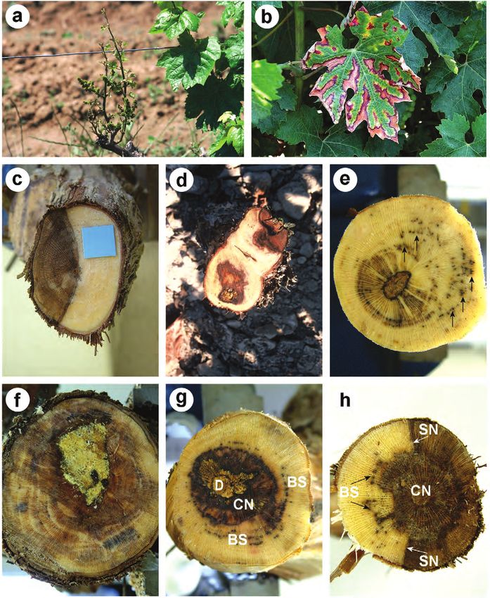
Fig. 1. Most common symptoms associated with grapevine declines: a. Reduced growth, and small, chlorotic leaves; b. Foliar
symptoms of esca in a red grape cultivar, characterised by the interveinal necroses and chloroses; c. V-shaped necrosis. d. Irregu-
larly-shaped necrosis around the pith (central necrosis); e. Black spots (shown by arrows), corresponding to vascular necroses; f.
Wood decay, characterised by a yellowish soft tissue; g and h. Co-occurrence of internal symptoms; BS, black spots, CN, central
necrosis, D, wood decay, SN, V-shaped necrosis. Symptoms “a” and “c” are usually associated with eutypiose and black dead arm,
whereas symptoms “b” and “d” to “f” are associated with esca.013_JPP404Luque_381_COLORE 25-06-2009 11:55 Pagina 386
386 Grapevine decline and associated pathogenic fungi Journal of Plant Pathology (2009), 91 (2), 381-390
Table 3. Number of fungi isolated from wood lesions of declining grapevines.
Lesion typea
Plant organ Fungal species V-shaped Black Central Wood Total
necroses spots necroses decay
Arms Acremonium sp. 0 0 1 1 2
Botryosphaeria dothidea 0 0 1 0 1
Cryptovalsa ampelina 0 0 1 0 1
Cylindrocarpon liriodendri 0 0 1 0 1
Diplodia seriata 91 8 17 5 121
Dothiorella viticola 1 0 0 0 1
Eutypa lata 47 1 8 3 59
Eutypa leptoplaca 1 0 0 0 1
Eutypella vitis 1 0 0 0 1
Fomitiporia mediterranea 3 1 2 19 25
Fusarium spp. 3 0 3 1 7
Neofusicoccum parvum 11 1 3 0 15
Phaeoacremonium aleophilum 2 4 11 1 18
Phaeoacremonium viticola 0 0 1 0 1
Phaeoacremonium sp. 1 0 0 0 1
Phaeomoniella chlamydospora 11 61 19 4 95
Phoma-like sp. 1 2 0 0 3
Phomopsis spp. 7 0 0 0 7
Stereum hirsutum 0 1 0 0 1
Unidentified Botryosphaeriaceae 6 0 1 0 7
Unidentified Diatrypaceae 1 0 1 1 3
Unidentified species 8 6 6 2 22
Trunk Acremonium sp. 0 1 0 0 1
Botryosphaeria dothidea 0 1 0 0 1
Cryptovalsa ampelina 3 0 0 1 4
Cylindrocarpon liriodendri 0 0 1 0 1
Diplodia seriata 15 0 3 3 21
Eutypa lata 8 1 2 2 13
Fomitiporia mediterranea 0 0 3 16 19
Fusarium spp. 1 0 3 0 4
Neofusicoccum luteum 1 0 0 0 1
Neofusicoccum parvum 5 1 0 0 6
Neofusicoccum vitifusiforme 1 0 0 0 1
Phaeoacremonium aleophilum 1 0 1 0 2
Phaeoacremonium sp. 1 0 0 0 1
Phaeomoniella chlamydospora 4 15 5 4 28
Phomopsis spp. 0 0 1 0 1
Unidentified Botryosphaeriaceae 1 0 1 0 2
Unidentified species 0 0 1 2 3
Totals 236 104 97 65 502
a
Number of lesions examined for isolations: V-shaped necroses, 657; black spots, 314; central necroses, 297; wood decay, 187.
probably due to the longer necroses (P013_JPP404Luque_381_COLORE 25-06-2009 11:55 Pagina 387
Journal of Plant Pathology (2009), 91 (2), 381-390 Luque et al. 387
seriata 421, F. mediterranea 356 and Phomopsis sp. 459 among isolates of D. seriata and N. parvum (Table 4).
did not cause any significant lesion on any cultivar. Reisolations from inoculated vines were successful
Fungal species with multiple isolates tested for patho- for all of the inoculated fungi, although percentages of
genicity showed some variability in the length of the le- positive reisolations were variable among the fungal
sions they caused. Thus lesions with a wide range of size species (Table 4). In general, reisolations from
were induced by N. parvum (maximum mean values ‘Macabeo’ plants were higher than those from ‘Tem-
about 2-3 fold greater than the minimum values), D. seri- pranillo’. Additionally, reisolation percentages were gen-
ata (2-3 fold), and E. lata (1.5-2 fold), whereas less varia- erally higher for the most virulent fungi (those causing
tion was observed for C. ampelina, F. mediterranea, Pa. longer necrosis, e.g. Botryosphaeriaceae species, and Pa.
chlamydospora and Pm. aleophilum (Table 4). Significant chlamydospora) than those from weak pathogens (e.g. C.
differences in the length of necroses were only observed ampelina and F. mediterranea).
Table 4. Number of wilted plants, length of vascular necroses and percentage of fungal recovery from grapevine
plants (n=18) of cvs Macabeo and Tempranillo inoculated with selected fungi isolated from declining grapevines.
‘Macabeo’ ‘Tempranillo’
Fungal species Wilted Necrosis Fungal Wilted Necrosis Fungal
plants (cm)a recovery plants (cm)a recovery
Isolate (%) (%)
Botryosphaeria dothidea 353 0 3.2 100 0 3.2 100
b
Diplodia seriata 398 0 2.4 a 100 0 3.5 a 67
D. seriata 421 0 0.8 c 83 0 1.9 b 28
D. seriata I-29 0 1.6 b 94 0 3.0 ab 78
D. seriata I-50 0 1.5 b 100 0 3.6 a 83
Dothiorella viticola 412 0 1.7 61 0 2.1 39
Cryptovalsa ampelina 413 0 1.5 50 0 2.5 39
C. ampelina 476 0 1.7 28 0 2.0 17
Eutypa lata 401 0 1.7 100 0 2.9 67
E. lata 411 0 1.9 94 0 4.0 50
E. lata 427 0 1.3 83 0 3.6 61
E. lata 438 0 1.4 100 0 2.3 50
E. lata 481 0 2.0 88 0 2.2 39
Fomitiporia mediterranea 356 0 0.7 33 0 1.5 11
F. mediterranea 452 0 1.4 33 0 2.5 22
F. mediterranea I-62 0 1.1 18 0 1.4 17
Neofusicoccum luteum 519 5 8.6 100 10 8.2 100
Neofusicoccum parvumb 387 0 4.0 c 94 0 5.6 b 100
N. parvum 396 5 10.8 a 100 1 13.8 a 78
N. parvum 434 5 12.7 a 100 0 11.6 a 100
N. parvum 444 1 6.7 b 100 0 6.9 b 94
Phaeoacremonium aleophilum 449 0 1.1 67 0 2.4 83
Pm. aleophilum 477 0 1.2 83 0 2.2 100
Pm. aleophilum I-10 0 1.4 89 0 1.9 94
Phaeomoniella chlamydospora 454 0 2.5 89 0 4.5 67
Pa. chlamydospora I-8 0 2.5 78 0 5.3 89
Pa. chlamydospora I-64 0 2.5 83 0 5.5 89
Phomopsis taxon 1 459 0 1.0 89 0 1.8 89
Control 0 0.7 0 0 1.4 0
a
Values in bold are significantly different from the corresponding mean value of controls according to the two-tailed
Dunnett’s test.
b
Mean values of necrosis lengths for D. seriata and N. parvum followed by different letters are significantly different
according to the Tukey’s test (P013_JPP404Luque_381_COLORE 25-06-2009 11:55 Pagina 388
388 Grapevine decline and associated pathogenic fungi Journal of Plant Pathology (2009), 91 (2), 381-390
DISCUSSION black spots, central necroses or wood decay, which are
usually associated with esca. Mugnai et al. (1999) re-
Results of field surveys confirmed the occurrence of ported that both D. seriata and E. lata, which are often
the three main decline diseases of adult grapevines in isolated from V-shaped necroses, can also be isolated
Catalonia: eutypiose, BDA and esca. External and inter- from esca-affected plants, which supports our field ob-
nal symptoms of both eutypiose and BDA, as recorded in servations. In this study, cross sections of arms and
May and June, looked very similar and thus were not reli- trunks of apoplectic vines showed a great percentage of
able for distinguishing the two diseases, as it has been re- dead, non-functional tissues. No quantitative data were
ported previously (Castillo-Pando et al., 2001; Taylor et recorded on the type and extension of these internal le-
al., 2005; Úrbez-Torres et al., 2006b). Eutypiose and sions. Further investigations are therefore needed to es-
BDA could only be differentiated after isolation of the re- tablish whether wood deterioration is related with
spective pathogens, E. lata and Botryosphaeriaceae spp. apoplexy. Furthermore, water relationships of apoplec-
Moreover, in our study, late BDA symptoms, as described tic plants should be studied to establish whether water
by Larignon et al. (2001a) (including leaf chlorosis and stress could be related to apoplexy.
necrosis, leaf shedding, wilting of cluster, and brown Fungal isolations of diseased wood showing a partic-
streaking of the wood), were occasionally seen in plants ular symptom indicated a general relationship between
affected by esca. Thus, external symptom expression of lesion type and isolated fungi. Thus, D. seriata and E. la-
BDA-affected plants would need further investigation for ta were mainly isolated from V-shaped necroses, Pa.
a clear-cut identification of this disease. Since several chlamydospora from black spots, and F. mediterranea
species of Botryosphaeriaceae are associated with BDA from decayed wood. Fungi isolated from central
(Larignon et al., 2001; Surico et al., 2006; van Niekerk et necroses included D. seriata, Pa. chlamydospora and Pm.
al., 2006), it would be also interesting to investigate the aleophilum, which is in accordance with previous re-
pathogenic role of each of these fungi and the particular ports (Mugnai et al., 1996; Larignon and Dubos, 1997;
symptoms they cause in adult vines. Mugnai et al., 1999; Serra, 1999). However, some re-
The present survey has also shown the frequent co- gional differences are observed in the distribution of
occurrence of internal symptoms associated with eutyp- some of these pathogens when our data are compared
iose, BDA and esca in the same plant, a condition al- with those from neighbouring regions. In France, Lar-
ready reported for esca and eutypiose (Mugnai et al., ignon and Dubos (1997) isolated E. lata more frequent-
1999), although we were unable to find any quantitative ly than any botryosphaeriaceous fungus from V-shaped
example in the extant literature. The occurrence of mul- necrosis, whereas our study and a previous one (Armen-
tiple lesion types in the same plant in northeast Spain, gol et al., 2001a) showed a greater incidence of D. seri-
which are especially frequent in the vine arms, may re- ata than E. lata in Spanish vineyards. Úrbez-Torres et al.
flect multiple infection events through pruning wounds. (2006b) reported the same from California. It is suggest-
In fact, it is widely accepted that most fungal pathogens ed that E. lata is probably less abundant in dryer
associated with grapevine decline are airborne Mediterranean climate countries as compared with oth-
pathogens that penetrate the plant through wounds er cooler and rainy regions, since E. lata dispersion is
caused by the annual pruning (Carter, 1988; Mugnai et enhanced when mean annual rainfall exceeds 350 mm
al., 1999; Surico et al., 2006; van Niekerk et al., 2006). (Carter, 1991; Mugnai et al., 1999).
Apoplexy is characterized by the sudden wilting and Most of the species tested for pathogenicity showed
death in midsummer of vines or their organs, including significant longer necrotic lesions than those in
clusters. Apparently healthy leaves may rapidly wilt ‘Macabeo’ and ‘Tempranillo’ controls. Only N. luteum
basipetally and dry in a few days (Mugnai et al., 1999). and N. parvum caused wilting of inoculated plants but no
Weather conditions are thought to influence this phe- external disease symptoms were recorded for any other
nomenon, since the apoplectic events often occur in hot fungus-plant combination during the experimental peri-
summers, when rainfall is followed by dry, hot weather od. Some influencing factors have been suggested to ex-
(Mugnai et al., 1999). Apoplexy has been frequently de- plain this phenomenon, which include the short experi-
scribed as a severe form of esca or specifically as an mental period, and other unsuitable experimental condi-
“acute esca syndrome” (Larignon and Dubos, 1997; tions such as the use of young, potted plants, and the in-
Mugnai et al., 1999; Graniti et al., 2000; Surico, 2001; oculation of fungi into green, non-lignified plant tissues.
Surico et al., 2006). However, the results of our study Moreover, our pathogenicity tests were done using
suggest that apoplectic events might not be restricted mycelium instead of spores as inoculum sources, which
only to esca-affected plants, since a significant percent- does not correspond to natural conditions for fungal in-
age of the surveyed vines showing apoplexy (60%; 15 fection. Additionally, it has been reported that some fun-
vines) were also affected by V-shaped necroses, which gi (e.g. F. mediterranea) are only able to colonize
are commonly associated with eutypiose and BDA grapevine tissues previously damaged by other fungi
(Carter, 1988; van Niekerk et al., 2006). Moreover, 20% (Larignon and Dubos, 1997). A combination of the
of apoplectic plants (5) had V-shaped necroses but no above factors could lead to unsuccesful fungal coloniza-013_JPP404Luque_381_COLORE 25-06-2009 11:55 Pagina 389
Journal of Plant Pathology (2009), 91 (2), 381-390 Luque et al. 389
tion of inoculated plant tissues, as shown by the low re- pathogen (Sparapano et al., 2001). Foliar symptoms of
covery of some fungi (e.g. C. ampelina, F. mediterranea) esca were reproduced on ‘Thompson seedless’ vines 6
and the short, non-significant necroses recorded occa- months after inoculation with Pm. aleophilum and Pa.
sionally in pathogenicity tests. chlamydospora (W.D. Gubler, personal communication).
Lack of foliar symptom in plants artificially inoculat- The remaining fungal species tested in our study were
ed with known grapevine pathogens has occasionally considered as non-pathogenic (Phomopsis sp. taxon 1)
been reported (Larignon and Dubos, 1997; Mugnai et or weakly pathogenic (C. ampelina, F. mediterranea and
al., 1999). Since foliar symptom expression often fails to Pm. aleophilum), based on the size of the necrotic le-
occur in artificial inoculations, the pathogenicity and sions caused. However, a pathogenic behaviour was re-
virulence of fungi have often been evaluated analysing ported for the latter two species (Adalat et al., 2000; Es-
the necrotic lesions caused by fungi in the plant vascular kalen et al., 2001; Sparapano et al., 2001).
tissues, as reported in previous works (Mugnai et al., Several fungal pathogens occur at the same time in
1999; Van Niekerk et al., 2004; Surico et al., 2006). the same grapevine, each one causing a particular wood
Pathogenicity has been reported previously for several lesion. This may lead to complex relationships among
Botryosphaeriaceae species (van Niekerk et al., 2004; Tay- these pathogens and the host plant. This study has
lor et al., 2005; van Niekerk et al., 2006), E. lata (Carter et shown that co-occurrence of internal disease symptoms
al., 1985; Carter, 1991; Péros et al., 1999; Sosnowski et al., and their associated fungi is frequent in northeast Spain,
2007), F. mediterranea (Sparapano et al., 2001), and Pm. and that the relationships between visual external symp-
aleophilum and Pa. chlamydospora (Adalat et al., 2000; Es- toms and inferred internal lesions are often misleading.
kalen et al., 2001; Sparapano et al., 2001; Halleen et al., This makes field diagnosis of these wood diseases diffi-
2007). Neofusicoccum luteum and N. parvum were the cult when only the external symptoms are considered.
most virulent pathogens tested in our study. While N.
parvum was proven to be a virulent pathogen by van
Niekerk et al. (2004), pathogenicity of N. luteum seems ACKNOWLEDGEMENTS
controversial. Van Niekerk et al. (2004) considered this
species as a low virulent pathogen since it caused no sig- Research financed by the “Instituto Nacional de In-
nificant necroses on inoculated mature canes of cvs vestigación y Tecnología Agraria y Alimentaria” (INIA)
Chardonnay and Cabernet sauvignon in South Africa. under project RTA03-058-C2-1. Soledad Martos was
However, in our study N. luteum was clearly pathogenic. supported by the “Departament d’Educació i Universi-
Pathogenicity of D. seriata has also been debated, as tats de la Generalitat de Catalunya” (Regional Govern-
summarized by Úrbez-Torres et al. (2006b). This fungus ment of Catalonia, Spain) and the European Social
was considered as a weak pathogen in Portugal Fund. The authors would like to thank V. Barnés and O.
(Phillips, 2002), but virulent in Chile (Auger et al., Jurado for their help in laboratory work, and Dr. W.D.
2004), South Africa (van Niekerk et al., 2004) and Aus- Gubler (University of California, Davis, USA) for his
tralia (Castillo-Pando et al., 2001), although Taylor et al. helpful comments on an early version of the manuscript.
(2005), in Australia, reported no significant vascular le-
sions caused by D. seriata to inoculated grapevine cut-
tings. In accordance with previous reports (Larignon et REFERENCES
al., 2001; van Niekerk et al., 2004), our field observa-
tions and the results of pathogenicity tests indicate that Adalat K., Whiting C., Rooney S., Gubler W.D., 2000. Patho-
D. seriata is pathogenic, though to a variable extent. genicity of three species of Phaeoacremonium spp. on
Eutypa lata is a widely-known pathogen of grapevine grapevine in California. Phytopathologia Mediterranea 39:
(Carter, 1988, 1991; Dubos, 1996). In our study, all five 92-99.
isolates tested for pathogenicity caused significant Alves A., Correia A., Luque J., Phillips A., 2004.
Botryosphaeria corticola, sp. nov. on Quercus species, with
necrotic lesions on ‘Macabeo’ vines while those in ‘Tem-
notes and description of Botryosphaeria stevensii and its
pranillo’ were significant only for three fungal isolates anamorph, Diplodia mutila. Mycologia 96: 598-613.
(401, 411 and 427). Although these findings may be in-
Anonymous, 2006. Anuario de Estadística Agroalimentaria
dicative of variability in pathogen virulence, more iso- 2004. Ministerio de Agricultura, Pesca y Alimentación,
lates should be tested to confirm this hypothesis. Vari- Madrid, Spain (ISBN 978-84-491-0712-2).
ability in virulence of E. lata has been shown previously Armengol J., Vicent A., García-Jiménez J., 2002. El de-
(Péros et al., 1999; Sosnowski et al., 2007). caimiento y muerte de vides jóvenes (enfermedad de Petri)
Our study confirmed the pathogenicity of Pa. en España. Phytoma España 138: 91-93.
chlamydospora although no foliar symptoms were ob- Armengol J., Vicent A., Torné L., García-Figueres F., García-
served during the experimental period. Foliar symptom Jiménez J., 2001a. Fungi associated with esca and
expression due to infection by Pa. chlamydospora only grapevine declines in Spain: A three-year survey. Phy-
were observed after long inoculation periods (2-3 years) topathologia Mediterranea 40: S325-S329.
in mature plants artificially inoculated with this Armengol J., Vicent A., Torné L., García-Figueres F., García-013_JPP404Luque_381_COLORE 25-06-2009 11:55 Pagina 390
390 Grapevine decline and associated pathogenic fungi Journal of Plant Pathology (2009), 91 (2), 381-390
Jiménez J., 2001b. Hongos asociados a decaimientos y Lehoczky J., 1974. Black dead-arm disease of grapevine caused
afecciones de madera en vid en diversas zonas españolas. by Botryosphaeria stevensii infection. Acta Phytopathologica
Boletin de Sanidad Vegetal, Plagas 27: 137-153. Academiae Scientarum Hungaricae 9: 319-327.
Aroca A., García-Figueres F., Bracamonte L., Luque J., Ra- Mostert L., Groenewald J.Z., Summerbell R.C., Gams W.,
poso R., 2006. A survey of trunk disease pathogens within Crous P.W., 2006. Taxonomy and pathology of Togninia
rootstocks of grapevines in Spain. European Journal of (Diaporthales) and its Phaeoacremonium anamorphs. Stud-
Plant Pathology 115: 195-202. ies in Mycology 54: 1-115.
Auger J., Esterio M., Ricke G., Pérez I., 2004. Black dead arm Mugnai L., Graniti A., Surico G., 1999. Esca (black measles)
and basal canker of Vitis vinifera cv. Red globe caused by and brown wood-streaking: two old and elusive diseases of
Botryosphaeria obtusa in Chile. Plant Disease 88: 1286. grapevines. Plant Disease 83: 404-418.
Carter M.V., 1988. Eutypa dieback. In: R. C. Pearson, A. C. Mugnai L., Surico G., Esposito A., 1996. Micoflora associata
Goheen (eds.). Compendium of Grape Diseases, pp. 32- al mal dell’esca della vite in Toscana. Informatore Fitopato-
33. APS Press, St. Paul, MN, USA. logico 46(11): 49-55.
Carter M.V., 1991. The status of Eutypa lata as a pathogen. Péros J.-P., Jamaux-Despréaux I., Berger G., Gerba D., 1999.
Phytopathological Papers No. 32. CAB International, Oxon, The potential importance of diversity in Eutypa lata and
UK. co-colonising fungi in explaining variation in development
Carter M.V., Bolay A., English H., Rumbos I.C., 1985. Varia- of grapevine dieback. Mycological Research 103: 1385-
tion in the pathogenicity of Eutypa lata (= E. armeniacae). 1390.
Australian Journal of Botany 33: 361-366. Phillips A.J.L., 2002. Botryosphaeria species associated with dis-
Castillo-Pando M., Somers A., Green C.D., Priest M., eases of grapevines in Portugal. Phytopathologia Mediter-
Sriskanthades M., 2001. Fungi associated with dieback of ranea 41: 3-18.
Semillon grapevines in the Hunter Valley of New South Phillips A., Alves A., Correia A., Luque J., 2005. Two new
Wales. Australasian Plant Pathology 30: 59-63. species of Botryosphaeria with brown, 1-septate ascospores
Dubos B., 1996. L’eutypiose de la vigne, Eutypa lata and Dothiorella anamorphs. Mycologia 97: 513-529.
(Pers.:Fr.) Tul. Comptes Rendus de l’Academie d’Agricul-
Serra S., 1999. Relazione tra sintomatologia fogliare, alter-
ture de France 82: 21-30.
azioni e micoflora del legno in viti affette da mal dell’esca
Eskalen A., Gubler W.D., Khan A., 2001. Rootstock susceptibil- ed eutipiosi. Informatore Fitopatologico 49(6): 30-34.
ity to Phaeomoniella chlamydospora and Phaeoacremonium
Sosnowski M.R., Lardner R., Wicks T.J., Scott E.S., 2007. The
spp. Phytopathologia Mediterranea 40: S433-S438.
influence of grapevine cultivar and isolate of Eutypa lata
Fischer M., 2006. Biodiversity and geographic distribution of on wood and foliar symptoms. Plant Disease 91: 924-931.
Basidiomycetes causing esca-associated white rot in
grapevine: a worldwide perspective. Phytopathologia Medi- Sparapano L., Bruno G., Graniti A., 2001. Three-year observa-
terranea 45: S30-S42. tion of grapevines cross-inoculated with esca-associated fun-
gi. Phytopathologia Mediterranea 40: S376-S386.
Giménez-Jaime A., Aroca A., Raposo R., García-Jiménez J.,
Armengol J., 2006. Occurrence of fungal pathogens associ- Surico G., 2001. Towards commonly agreed answers to some
ated with grapevine nurseries and the decline of young basic questions on esca. Phytopathologia Mediterranea 40:
vines in Spain. Journal of Phytopathology 154: 598-602. S487-S490.
Graniti A., Surico G., Mugnai L., 2000. Esca of grapevine: a Surico G., Mugnai L., Marchi G., 2006. Older and more re-
disease complex or a complex of diseases?. Phytopatholo- cent observations on esca: a critical overview. Phy-
gia Mediterranea 39: 16-20. topathologia Mediterranea 45: S68-S86.
Halleen F., Mostert L., Crous P.W., 2007. Pathogenicity test- Taylor A., Hardy G.E.St.J., Wood P., Burgess T., 2005. Identi-
ing of lesser known vascular fungi of grapevines. Aus- fication and pathogenicity of Botryosphaeria species associ-
tralasian Plant Pathology 36: 277-285. ated with grapevine decline in Western Australia. Aus-
Hoagland D.R., Arnon D.I., 1950. The water culture method tralasian Plant Pathology 34: 187-195.
for growing plants without soil. California Agricultural Ex- Úrbez-Torres J.R., Gubler W.D., Peláez H., Santiago Y., Martín
periment Station, Circular No. 347. University of Califor- C., Moreno C., 2006a. Occurrence of Botryosphaeria obtusa,
nia, Berkeley, CA, USA. B. dothidea, and B. parva associated with grapevine trunk
Johnston A., Booth C. (eds.), 1983. Plant Pathologist’s Pock- diseases in Castilla y Leon region, Spain. Plant Disease 90:
etbook. 2nd Ed. CAB, Slough, UK. 835.
Larignon P., Dubos B., 1997. Fungi associated with esca dis- Úrbez-Torres J.R., Leavitt G.M., Voegel T.M., Gubler W.D.,
ease in grapevine. European Journal of Plant Pathology 103: 2006b. Identification and distribution of Botryosphaeria
147-157. spp. associated with grapevine cankers in California. Plant
Larignon P., Dubos B., 2001. Le black dead arm. Maladie Disease 90: 1490-1503.
nouvelle à ne pas confondre avec l’esca. Phytoma - La Van Niekerk J.M., Crous P.W., Groenewald J.Z., Fourie P.H.,
Défense des Végetaux 538: 26-29. Halleen F., 2004. DNA phylogeny, morphology and patho-
Larignon P., Fulchic R., Cere L., Dubos B., 2001. Observation genicity of Botryosphaeria species on grapevines. Mycologia
on black dead arm in French vineyards. Phytopathologia 96: 781-798.
Mediterranea 40: S336-S342. Van Niekerk J.M., Fourie P.H., Halleen F., Crous P.W., 2006.
Lecomte P., Leyo M., Louvet G., Corio-Costet M.F., Gaudil- Botryosphaeria spp. as grapevine trunk disease pathogens.
lère J.P., Blancard D., 2005. Le Black dead arm, genèse des Phytopathologia Mediterranea 45: S43-S54.
symptômes. Phytoma-La Défense des Végetaux 587: 29-37.
Received October 15, 2008
Accepted January 22, 2009You can also read