The effect of time since stroke, gender, age, and lesion size on thalamus volume in chronic stroke: a pilot study - Nature
←
→
Page content transcription
If your browser does not render page correctly, please read the page content below
www.nature.com/scientificreports
OPEN The effect of time since stroke,
gender, age, and lesion size
on thalamus volume in chronic
stroke: a pilot study
Lisa C. Krishnamurthy1,2,3*, Gabriell N. Champion1,4, Keith M. McGregor1,5,
Venkatagiri Krishnamurthy1,3,5, Aaminah Turabi1, Simone R. Roberts1,4,5, Joe R. Nocera1,5,6,
Michael R. Borich3,6, Amy D. Rodriguez1,5, Samir R. Belagaje5,6, Rachael M. Harrington1,3,4,
Michelle L. Harris‑Love7, Stacy M. Harnish8, Jonathan H. Drucker1,5, Michelle Benjamin9,
M. Lawson Meadows1, Lauren Seeds9, Zvinka Z. Zlatar10, Atchar Sudhyadhom11,
Andrew J. Butler12, Amanda Garcia13, Carolynn Patten14, Jonathan Trinastic15,
Steven A. Kautz16,17, Chris Gregory17 & Bruce A. Crosson1,3,4,5
Recent stroke studies have shown that the ipsi-lesional thalamus longitudinally and significantly
decreases after stroke in the acute and subacute stages. However, additional considerations in the
chronic stages of stroke require exploration including time since stroke, gender, intracortical volume,
aging, and lesion volume to better characterize thalamic differences after cortical infarct. This cross-
sectional retrospective study quantified the ipsilesional and contralesional thalamus volume from
69 chronic stroke subjects’ anatomical MRI data (age 35–92) and related the thalamus volume to
time since stroke, gender, intracortical volume, age, and lesion volume. The ipsi-lesional thalamus
volume was significantly smaller than the contra-lesional thalamus volume (t(68) = 13.89, p < 0.0001).
In the ipsilesional thalamus, significant effect for intracortical volume (t(68) = 2.76, p = 0.008), age
(t(68) = 2.47, p = 0.02), lesion volume (t(68) = − 3.54, p = 0.0008), and age*time since stroke (t(68) = 2.46,
p = 0.02) were identified. In the contralesional thalamus, significant effect for intracortical volume
(t(68) = 3.2, p = 0.002) and age (t = − 3.17, p = 0.002) were identified. Clinical factors age and intracortical
volume influence both ipsi- and contralesional thalamus volume and lesion volume influences the
ipsilesional thalamus. Due to the cross-sectional nature of this study, additional research is warranted
to understand differences in the neural circuitry and subsequent influence on volumetrics after stroke.
The thalamus acts not only as a relay from the periphery to the cortex, but also as a higher order relay from one
cortical area to a nother1. In the context of these higher order relays, the role of the thalamus has been impli-
cated as a platform to stabilize signals in various cortical processing streams2,3 and is particularly vulnerable to
diaschisis after stroke, the effects of which remain poorly understood. Recent stroke studies have shown that the
ipsi-lesional thalamus longitudinally and significantly decreases after stroke in the acute and subacute stages,
1
Center for Visual and Neurocognitive Rehabilitation, Atlanta VA Health Care System, 1670 Clairmont Rd,
Decatur, GA 30033, USA. 2Department of Physics and Astronomy, Georgia State University, Atlanta, GA,
USA. 3Center for Advanced Brain Imaging, Georgia State University and Georgia Institute of Technology, Atlanta,
GA, USA. 4Department of Psychology, Georgia State University, Atlanta, GA, USA. 5Department of Neurology,
Emory University, Atlanta, GA, USA. 6Department of Rehabilitation Medicine, Emory University, Atlanta, GA,
USA. 7Center for Brain Plasticity and Recovery, Georgetown University, Washington, DC, USA. 8Department of
Speech and Hearing Science, Ohio State University, Columbus, OH, USA. 9Department of Physical Therapy, Brooks
Rehabilitation Center, Jacksonville, FL, USA. 10Department of Psychiatry, University of California San Diego, La
Jolla, CA, USA. 11Brigham and Women’s Hospital, Dana Farber Cancer Institute, Harvard Medical School, Boston,
MA, USA. 12School of Health Professions, University of Alabama Birmingham, Birmingham, AL, USA. 13Clinical
and Health Psychology, University of Florida, Gainesville, FL, USA. 14Department of Physical Medicine and
Rehabilitation, University of California Davis, Sacramento, CA, USA. 15Data Science, Duke Energy, Charlotte, NC,
USA. 16Ralph H. Johnson VA Medical Center, Charleston, SC, USA. 17Department of Health Sciences and Research,
Medical University of South Carolina, Charleston, SC, USA. *email: lkrishnamurthy@gsu.edu
Scientific Reports | (2020) 10:20488 | https://doi.org/10.1038/s41598-020-76382-x 1
Vol.:(0123456789)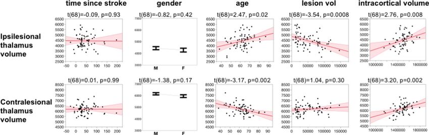
www.nature.com/scientificreports/
Figure 1. Representative freesurfer recon-all segmentation and Iglesias segmentation on original T1w contrast
and squared T1w contrast.
but the contra-lesional thalamus remains relatively unchanged4,5. However, additional considerations require
exploration including time since stroke in the chronic stages, gender, intracortical volume, aging, and lesion
volume to better characterize thalamic differences after cortical infarct.
In the context of additive factors that may influence thalamus volume, it is well accepted that gender and
intracortical volume influences the quantification of brain volumetrics6, including thalamus volume and lesion
volume. Further, neuroimaging studies of the human brain have shown that thalamic volume reduces with age
and is associated with aging-related cognitive and motor differences7,8. These studies found a direct relationship
between decrease in thalamic volume and decreased density of thalamo-cortical projections, providing evidence
that aging-related cortical atrophy could explain differences in thalamic volume. Thus, if gender and intracortical
volume differences and aging-related atrophy have effects on the thalamus volume in healthy cohorts, then it is
important to understand how stroke’s effect on thalamus volume relates to time since stroke, gender, intracortical
volume, age, and lesion size to accurately design rehabilitation treatments based on these clinical factors. In this
pilot study we investigate these effects on both ipsilesional and contralesional thalamus volume. The hypoth-
eses for this cross-sectional cohort are: (1) that we will identify a negative relationship between total thalamus
volume and time since stroke, (2) that gender effects on thalamus volume will be evident in both contra- and
ipsilesional thalamus, (3) that age has a negative relationship with thalamus volume, and (4) that lesion volume
will have a negative relationship with ipsi-lesional thalamus volume but no relationship with contra-lesional
thalamus volume.
Methods
Subjects. This retrospective cross-sectional study combined deidentified T1-weighted (T1w) anatomical
imaging data from three sites in accordance with relevant guidelines and regulations approved by the joint
Atlanta VA/Emory University institutional review board, University of Florida institutional review board, or
Georgetown University institutional review board. All participants provided informed consented to participate
in a magnetic resonance imaging (MRI) study that incorporated a T1w anatomical image. A total of 69 chronic
stroke participants (5–207 months post-stroke, 28 Female, 41 Male, age = 35–92, and lesion volume = 0.25–
172 cc) were identified to have usable T1w images, relevant demographic information, and intact thalami in
both hemispheres. Either the left or right hemisphere was lesioned in each participant, but not both.
MRI processing. T1w images were inspected for motion artifacts, d enoised9,10, bias field c orrected11, and
then squared to increase the signal contrast between grey and white matter, thereby improving segmentation
results of the subcortical regions. The squared T1w image was processed through Freesurfer’s cross-sectional
‘recon-all’ pipeline12 followed by the Iglesias thalamic nuclei segmentation13. In effect, the thalamus segmenta-
tion was optimized with a two-pronged approach: (1) by squaring the T1w signal to improve signal contrast and
(2) by applying the Iglesias segmentation algorithm which uses Bayesian inference initialized by the location of
the neighboring subcortical brain structures (Fig. 1).
Automatic stroke lesion segmentations were generated using LINDA14 on the non-squared T1w images,
followed by manual touch-up with itksnap15 to exclude healthy areas falsely identified as lesion and to include
damaged regions falsely identified as non-lesioned. For each individual subject, the thalamus volume and lesion
volume were extracted for further statistical tests. The thalamus is designated as either ipsi-lesional (same hemi-
sphere as the lesion) or contra-lesional (opposite side to the lesion) to allow combination between left and right
hemisphere stroke participants. A paired student’s t-test is used to determine if contra- and ipsilesional thalamus
volume are significantly different.
Scientific Reports | (2020) 10:20488 | https://doi.org/10.1038/s41598-020-76382-x 2
Vol:.(1234567890)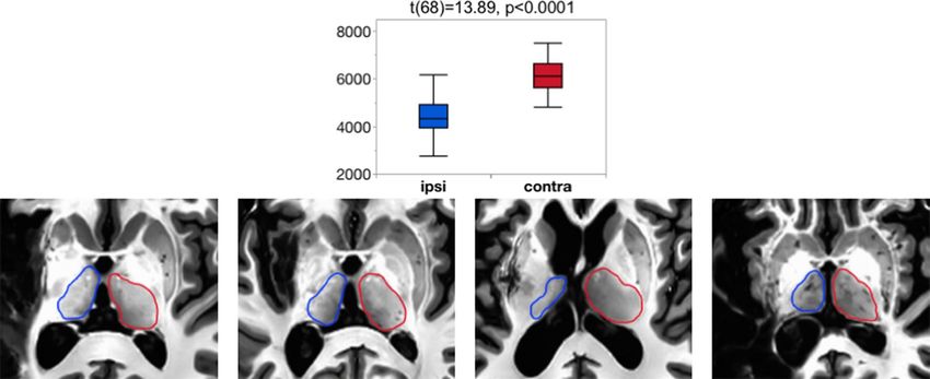
www.nature.com/scientificreports/

                                  Figure 2.  Ipsilesional thalamus volume is significantly smaller than contralesional thalamus volume. This effect
                                  can be identified visually from the T1w images. Note: blue = ipsilesional thalamus, red = contralesional thalamus.

                                  Figure 3.  Modeling results of ipsilesional and contralesional thalamus volume. T-statistic and probability are
                                  shown above each plot.

                                  Model fit of time since stroke, gender, intracortical volume, age, and lesion volume.                    We com-
                                  pleted an ANOVA in JMP Pro15 to test if (time since stroke), (gender), (intracortical volume), (age), (lesion vol-
                                  ume), and cross terms (time since stroke*age), (time since stroke*lesion volume), (intracortical volume*lesion
                                  volume), (gender*intracortical volume), and (age*lesion volume) explain ipsilesional and contralesional thala-
                                  mus volume. The results of the model are reported with F-statistic and subsequent t-tests are performed to
                                  determine which terms had significant effect.

                                  Results
                                  The ipsi-lesional thalamus volume was confirmed to be smaller than the contra-lesional thalamus volume
                                  (t(68) = 13.89, p < 0.0001) using a two-tailed paired Student’s t-test in JMP Pro15. These effects can be identified
                                  in the T1w images with visual inspection, especially in the participants with larger lesions (Fig. 2).

                                  Effect of time since stroke, gender, intracortical volume, age, and lesion volume. The whole
                                  model for ipsilesional thalamus volume was significant (F(10,58) = 3.05, p = 0.004). Subsequent t-tests revealed
                                  a significant effect for intracortical volume (t(68) = 2.76, p = 0.008), age (t(68) = 2.47, p = 0.02), lesion volume
                                  (t(68) = − 3.54, p = 0.0008), and cross-term age*time since stroke (t(68) = 2.46, p = 0.02). Time since stroke
                                  (t(68) = − 0.09, p = 0.93) nor gender (t(68) = − 0.82, p = 0.42) were significantly related to in ipsilesional thala-
                                  mus volume with this model. The whole model fit for contralesional thalamus volume was also significant
                                  (F(10,58) = 7.04, p < 0.0001). Subsequent t-tests revealed significant effect for intracortical volume (t(68) = 3.2,
                                  p = 0.002) and age (t = − 3.17, p = 0.002). Although adding both gender and intracranial volume may lead to
                                  multicollinearity in the model, the variance inflation factor for each variable was below 3 and therefore does not
                                  warrant corrective measures. A graphical summary of the effects can be seen in Fig. 3.

Scientific Reports |   (2020) 10:20488 |               https://doi.org/10.1038/s41598-020-76382-x                                                    3

                                                                                                                                                Vol.:(0123456789)
www.nature.com/scientificreports/
Discussion
In this cross-sectional pilot study of chronic stroke participants, we tested five demographic factors that may
have influence on ipsi-lesional and contra-lesional whole thalamus volume: (1) time since stroke, (2) gender, (3)
intracortical volume, (4) age, and (5) lesion volume. We showed that thalamus volume in the chronic stages of
stroke is related to intracortical volume, age, and lesion volume. These findings will require replication in the field.
We found that time since stroke does not describe thalamus volume in either ipsi- or contralesional hemi-
spheres, which was unexpected as previous longitudinal studies have identified a significant negative longitudinal
relationship in the acute and subacute stages4,5. Thus, hypothesis 1 was not confirmed. There could be two reasons
for this negative finding in our study: (1) structural thalamus changes due to loss of input/output may be limited
to the acute and subacute stages, or (2) the thalamus changes in the chronic stage related to time post onset are
too subtle to study using current MRI techniques or in a cross-sectional manner. Additional study is warranted
to validate these findings in a larger study cohort. Interestingly, this particular cohort showed that ipsilesional
thalamus volume had a significant relationship with the age*time since stroke interaction term. This may be
because older chronic stroke participants may have a longer time since stroke, while younger participants have
a shorter time since stroke, and may be a product of the cross-sectional nature of this dataset.
There was no significant effect of gender in thalamus volume. Thus, Hypothesis 2 was not confirmed. Instead,
the dominant and significant effect was with intracranial volume, which showed that a larger head size corre-
sponds to a larger thalamus volume, regardless of ipsi- or contralateral placement. It is well accepted that males,
on average, have larger head sizes than females, which is the case in our dataset as well. Although adding two
correlated variables into the model to describe thalamus volume could lead to multicollinearity, no correction
was warranted due to the small variance inflation factor of each variable. Thus, intracranial volume may be a
more important factor to consider in modeling volumetrics than gender and may have statistical advantages in
modeling and prediction due to its continuous nature.
As expected, the contra-lesional thalamus volume has a negative relationship with age. Such findings have
previously been identified in healthy aging cohorts in both hemispheres7,8. Interestingly, the ipsi-lesional thala-
mus volume has a significantly positive relationship with age. Thus, Hypothesis 3 was only partially confirmed.
It is unclear why the ipsi-lesional thalamus is larger in older chronic stroke participants compared to younger
stroke participants. It may be that the longitudinal trajectory in thalamus decline differs in young and old, and
that the cross-sectional nature of this dataset cannot adequately describe the effects of age-by-disease interaction
in this manner. Importantly, the finding that the contralesional has a negative relationship with age and that the
ipsilesional thalamus has a positive relationship with age requires replication and further scrutiny.
Finally, we showed that the ipsilesional thalamus volume has a significant negative relationship with lesion
volume. This is likely due to loss of input or output into the thalamus of the lesioned hemisphere but remains to
be tested as the quantification of structural or functional connections to the thalamus are beyond the scope of this
work. In line with previous work, we found that the contralesional thalamus volume does not have a relationship
with lesion volume. Thus, Hypothesis 4 was confirmed.
This pilot study had limitations and its findings should be replicated in a larger and preferably longitudinal
cohort. First, although previous evidence points to a similarity in trajectories quantified from longitudinal and
cross-sectional data in brain volume decline with healthy a ging16, such similarities may not hold for stroke related
changes. This may be the case in our lack of relationship between thalamus volume and time since stroke. Second,
the retrospective nature of this study precluded acquisition of consistent demographic/functional descriptors
such as NIH stroke scale and comorbidities across all participants and represents a major limitation. NIHSS
assessments describe the stroke severity and are an important descriptor of stroke recovery and outcome. Fur-
ther, Comorbidities such as diabetes, high blood pressure, history of smoking, heart disease, and other vascular
risk factors stemming from lifestyle factors could cause small-vessel d isease17,18 and reduce blood supply to the
thalamus, in turn influencing the thalamus volume. The complex interplay of different comorbidities on stroke
outcome and recovery is thought to influence the effect of neuroprotective drugs and their outcome in clini-
cal trials19. Future studies identifying volumetric brain difference should also take these factors into account,
including dietary and exercise habits, and may benefit from using large well-characterized datasets20,21. Finally,
this study falls short of relating the thalamus volume to functional outcomes. That is likely the next important
step to identify if the thalamus volume is relevant in predicting functional outcomes or rehabilitation potential
after stroke.
In conclusion, we identified important demographic factors that influence the ipsi-lesional and contra-lesional
thalamus volume, including intracortical volume, age, and lesion volume in a relatively small cross-sectional
cohort of chronic stroke participants. The ipsi- and contra-lesional thalamus have unique relationships with age
and requires further exploration.
Received: 3 May 2020; Accepted: 9 September 2020
References
1. Usrey, W. M. & Sherman, S. M. Corticofugal circuits: Communication lines from the cortex to the rest of the brain. J. Comp. Neurol.
527, 640–650. https://doi.org/10.1002/cne.24423 (2019).
2. Kawaguchi, Y. Pyramidal cell subtypes and their synaptic connections in layer 5 of rat frontal cortex. Cereb. Cortex 27, 5755–5771.
https://doi.org/10.1093/cercor/bhx252 (2017).
3. Crosson, B. The role of cortico-thalamo-cortical circuits in language: recurrent circuits revisited. Neuropsychol. Rev. https://doi.
org/10.1007/s11065-019-09421-8 (2019).
4. Liu, G. et al. Regional shape abnormalities in thalamus and verbal memory impairment after subcortical infarction. Neurorehabil.
Neural Repair. 33, 476–485. https://doi.org/10.1177/1545968319846121 (2019).
Scientific Reports | (2020) 10:20488 | https://doi.org/10.1038/s41598-020-76382-x 4
Vol:.(1234567890)www.nature.com/scientificreports/
5. Haque, M. E. et al. Ongoing secondary degeneration of the limbic system in patients with ischemic stroke: a longitudinal MRI
study. Front. Neurol. 10, 154. https://doi.org/10.3389/fneur.2019.00154 (2019).
6. Ritchie, S. J. et al. Sex differences in the adult human brain: evidence from 5216 UK biobank participants. Cereb. Cortex 28,
2959–2975. https://doi.org/10.1093/cercor/bhy109 (2018).
7. Hughes, E. J. et al. Regional changes in thalamic shape and volume with increasing age. Neuroimage 63, 1134–1142. https://doi.
org/10.1016/j.neuroimage.2012.07.043 (2012).
8. Serbruyns, L. et al. Subcortical volumetric changes across the adult lifespan: subregional thalamic atrophy accounts for age-related
sensorimotor performance declines. Cortex 65, 128–138. https://doi.org/10.1016/j.cortex.2015.01.003 (2015).
9. Coupe, P. et al. An optimized blockwise nonlocal means denoising filter for 3-D magnetic resonance images. IEEE Trans. Med.
Imaging 27, 425–441. https://doi.org/10.1109/TMI.2007.906087 (2008).
10. Coupe, P. et al. Robust Rician noise estimation for MR images. Med. Image. Anal. 14, 483–493. https://doi.org/10.1016/j.media
.2010.03.001 (2010).
11. Zhang, Y., Brady, M. & Smith, S. Segmentation of brain MR images through a hidden Markov random field model and the
expectation-maximization algorithm. IEEE Trans. Med. Imaging 20, 45–57. https://doi.org/10.1109/42.906424 (2001).
12. Fischl, B. et al. Whole brain segmentation: automated labeling of neuroanatomical structures in the human brain. Neuron 33,
341–355 (2002).
13. Iglesias, J. E. et al. A probabilistic atlas of the human thalamic nuclei combining ex vivo MRI and histology. Neuroimage 183,
314–326. https://doi.org/10.1016/j.neuroimage.2018.08.012 (2018).
14. Pustina, D. et al. Automated segmentation of chronic stroke lesions using LINDA: Lesion identification with neighborhood data
analysis. Hum. Brain Mapp. 37, 1405–1421. https://doi.org/10.1002/hbm.23110 (2016).
15. Yushkevich, P. A. et al. User-guided 3D active contour segmentation of anatomical structures: significantly improved efficiency
and reliability. Neuroimage 31, 1116–1128. https://doi.org/10.1016/j.neuroimage.2006.01.015 (2006).
16. Scahill, R. I. et al. A longitudinal study of brain volume changes in normal aging using serial registered magnetic resonance imag-
ing. Arch. Neurol. 60, 989–994. https://doi.org/10.1001/archneur.60.7.989 (2003).
17. Hermann, D. M., Doeppner, T. R. & Popa-Wagner, A. Opportunities and limitations of vascular risk factor models in studying
plasticity-promoting and restorative ischemic stroke therapies. Neural Plast. 2019, 9785476. https://doi.org/10.1155/2019/97854
76 (2019).
18. Popa-Wagner, A. et al. Dietary habits, lifestyle factors and neurodegenerative diseases. Neural Regen. Res. 15, 394–400. https://
doi.org/10.4103/1673-5374.266045 (2020).
19. Sandu, R. E., Buga, A. M., Uzoni, A., Petcu, E. B. & Popa-Wagner, A. Neuroinflammation and comorbidities are frequently ignored
factors in CNS pathology. Neural Regen. Res. 10, 1349–1355. https://doi.org/10.4103/1673-5374.165208 (2015).
20. Warach, S. J. et al. Acute stroke imaging research roadmap iii imaging selection and outcomes in acute stroke reperfusion clini-
cal trials: consensus recommendations and further research priorities. Stroke 47, 1389–1398. https://doi.org/10.1161/STROK
EAHA.115.012364 (2016).
21. Corbetta, M. et al. Common behavioral clusters and subcortical anatomy in stroke. Neuron 85, 927–941. https://doi.org/10.1016/j.
neuron.2015.02.027 (2015).
Acknowledgements
This work was supported by the Veterans Affairs Rehabilitation Research & Development Service (USA) Grant
IK1 RX002629.
Author contributions
Conception or design of the work—L.C.K., G.N.C., K.M.M., V.K., B.A.C. Acquisition of data—L.C.K., K.M.M.,
V.K., S.R.R., J.R.N., M.B., A.D.R., S.R.B., R.M.H., M.L.H., S.M.H., J.H.D., M.B., L.S., Z.Z.Z., A.S., A.J.B., A.G., C.P.,
J.T., S.K., C.G. Analysis of data—L.C.K., G.N.C., K.M.M., V.K., M.L.M. Interpretation of data—L.C.K., K.M.M.,
V.K., B.A.C. Manuscript writeup and editing—L.C.K., G.N.C., K.M.M., V.K., A.T., B.A.C.
Competing interests
The authors declare no competing interests.
Additional information
Correspondence and requests for materials should be addressed to L.C.K.
Reprints and permissions information is available at www.nature.com/reprints.
Publisher’s note Springer Nature remains neutral with regard to jurisdictional claims in published maps and
institutional affiliations.
Open Access This article is licensed under a Creative Commons Attribution 4.0 International
License, which permits use, sharing, adaptation, distribution and reproduction in any medium or
format, as long as you give appropriate credit to the original author(s) and the source, provide a link to the
Creative Commons licence, and indicate if changes were made. The images or other third party material in this
article are included in the article’s Creative Commons licence, unless indicated otherwise in a credit line to the
material. If material is not included in the article’s Creative Commons licence and your intended use is not
permitted by statutory regulation or exceeds the permitted use, you will need to obtain permission directly from
the copyright holder. To view a copy of this licence, visit http://creativecommons.org/licenses/by/4.0/.
© The Author(s) 2020
Scientific Reports | (2020) 10:20488 | https://doi.org/10.1038/s41598-020-76382-x 5
Vol.:(0123456789)You can also read