The multilevel human brain atlas in EBRAINS - Timo Dickscheid Forschungszentrum Jülich
←
→
Page content transcription
If your browser does not render page correctly, please read the page content below
The multilevel human brain
atlas in EBRAINS

Timo Dickscheid
Forschungszentrum Jülich
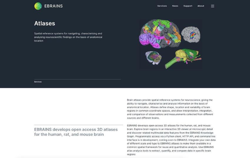
2
https://ebrains.eu/services/atlases
3
https://ebrains.eu/services/atlases
Basic principle of a brain atlas

Reference space   Map of regions                      Taxonomy
                  (defined in the coordinate space)   Names and relationships of regions
                                                                                           4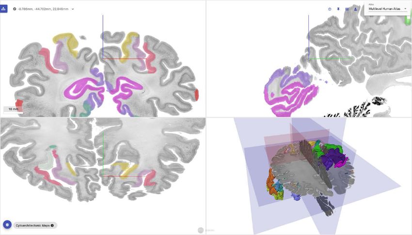
Concept of the EBRAINS
multilevel human brain atlas

                               6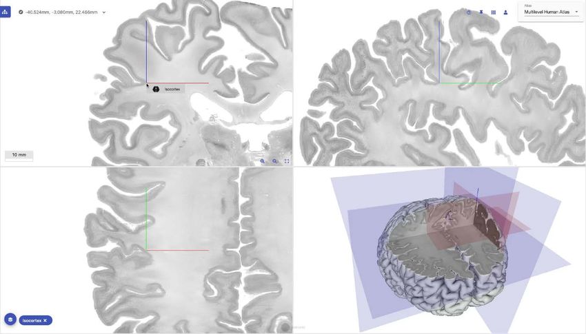
Aim: Capture the many facets of human
brain organization in a common framework
                • Multiple scales
                 Link the cellular scale to the
                 macroscopic scale

                • Multiple maps
                 Provide complementary
                 brain parcellations

                • Multimodal features
                 Provide a framework for linking
                 data features to brain regions
                                                   7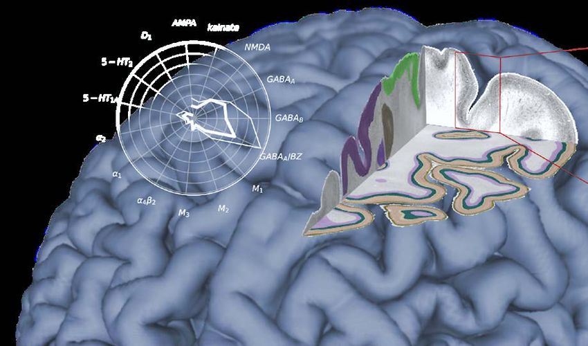
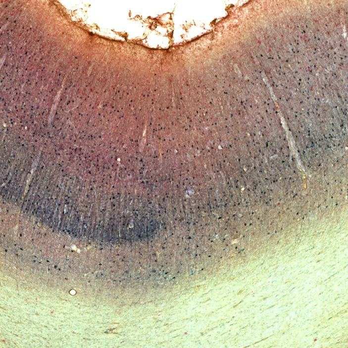
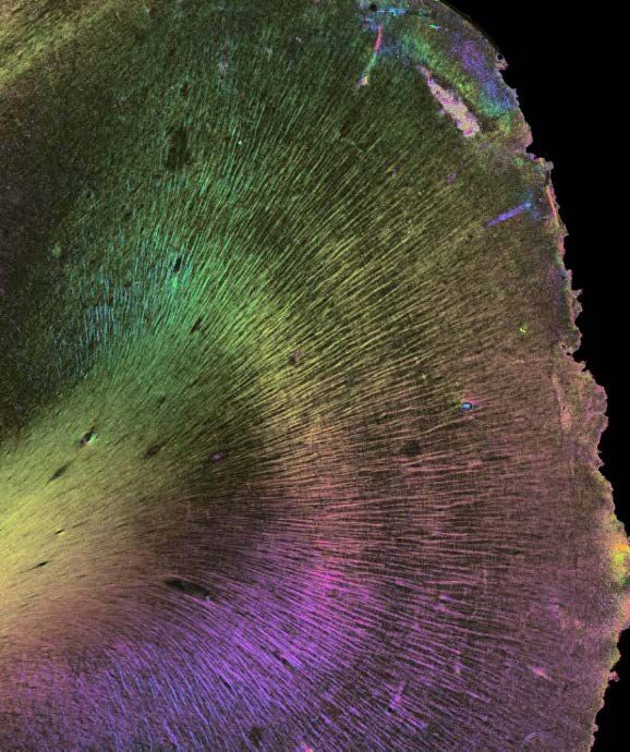
1 micrometer resolution
Cortical structure
1-20 micron resolution

 Amunts, K. and K. Zilles, Architectonic
 Mapping of the Human Brain beyond
   Brodmann. Neuron 2015. 88(6)
3D reconstructed from
7400 tissue sections

                        12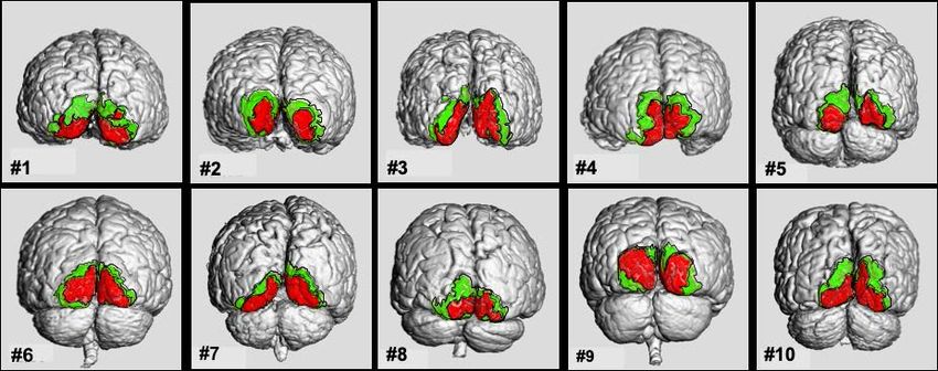
13
15
Not one brain resembles another
Amunts, Zilles et al.:
Brodmann's Areas 17 and 18
Brought into Stereotaxic Space
—Where and How Variable?
NeuroImage, Volume 11, Issue
1, 2000, Pages 66-84
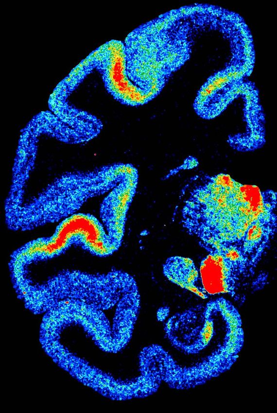
16Julich-brain probabilistic
cytoarchitectonic maps
Bludau et al. 2014
Individual Probabilistic map Maximum
delineations in ~10 (mm scale) probability map
brains, projected to
MNI reference space
Katrin Amunts, Hartmut Mohlberg, Sebastian Bludau, Karl Zilles:
Julich-Brain – a 3D probabilistic atlas of human brain’s cytoarchitecture.
Science (First Release), DOI: 10.1126/science.abb4588
17Linking the scales
Corres-
ponding MNI Colin27
regions
FS Surface
BigBrain
MNI ICBM 152
18Complementary maps of brain regions
Julich-Brain cytoarchitectonic maps
(Amunts et al.)
Maps of fibre bundles
(Mangin et al.)
Dictionaries of functional modes
(Thirion et al.)
Maps of BigBrain cortical layers
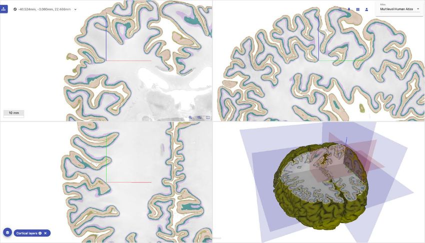
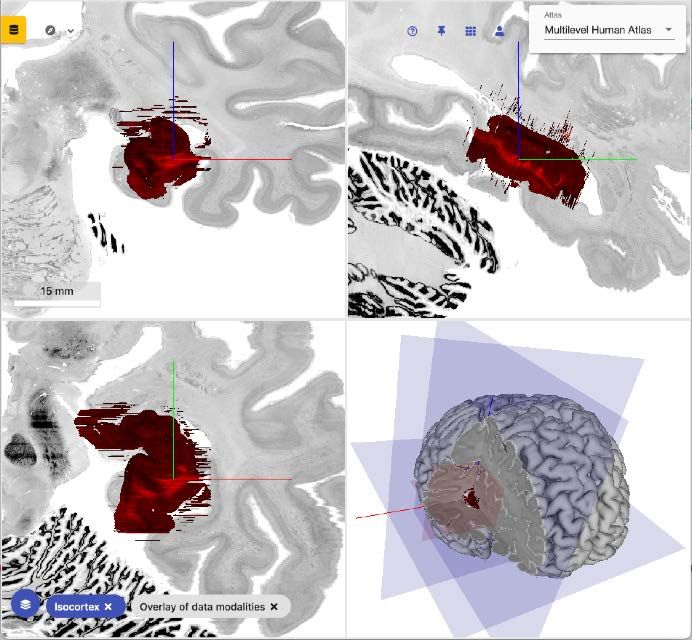
(Wagstyl et al.) 20EBRAINS Interactive Atlas Viewer:
Accessing regional features
22A „shopping cart“ for data downloads
23Infrastructure embedding
in EBRAINS
24ebrains.eu/service/share-data/
EBRAINS
curation services
search.kg.ebrains.eu fenix-ri.eu
EBRAINS Federated High
Knowledge Graph Performance Computing
EBRAINS
Atlas services
ebrains.eu/services/atlases
25EBRAINS
Atlas services
ebrains.eu/services/atlases
26Some use cases
27Some usecases
• Experimental neuroscience: Integrate data from
experiments into a common reference space
• Data analysis: Use atlases and data features to run
reproducible neuroscience experiments
• Brain simulation and biologically inspired AI:
Understanding the structure of biological networks
• Hospitals: Planning surgeries, comparing diseased to
healthy brains, anatomical location assignment
• Education: Studying brain anatomy
28Integrating data to a common
reference space Connectivity
• Many labs analyze
high-resolution VOIs,
but not the whole brain Cyto-
architecture
• BigBrain is a natural
reference space for
such data
• No standard workflows
to anchor partial Receptor
Function
volumes architecture
29A volume of interest
30Available in Matlab, interactive viewer plugin,
and Python:
• Matlab: Information page of the original
authors
• https://ebrains.eu/service/jugex
• Example Python notebook
31Describing the structure of biological
networks
Nerve fibers Distributions of cells Distributions of Cell types Cell morphologies
(3D PLI, M. Axer et al.) (K. Amunts et al) neurotransmitter receptors (R. Koijmans et al.) (H. Mansvelder et al.)
32
(N. Palomero-Gallagher et al.)What’s next?
33High-level roadmap
Today 2023 Beyond 2023
Easy open access to maps and data A community-driven
High coverage of data features
features from large and long-term reference framework at
from high-resolution data
projects single cell resolution
• ~250 cytoarchitectonic maps from • A unique multi-scale connectome • A software ecosystem for lively
~80.000 delineations in >20 brains linked with the atlas community contributions in terms
• Probabilistic maps of ~1000 fibre • Many ultra-high-resolution maps of data and software plugins
bundles extracted from X individual available for BigBrain • A Petabyte-scale data resource
subjects • Cell densities, axon densities, fibre connected to web frontends and
• Maps of functional modes extracted orientations from high-resolution HPC systems
from millions of fMRI scans from 27 data for most atlas regions
studies and a total size of 2.4TB • Whole-brain distributions of selected
• Multimodal data features linked to receptor transmitters
many brain regions • In-vivo receptor PET/fMRI dataHelmholtz International
BigBrain Analytics Learning
Laboratory (HIBALL)
Alan Evans (McGill) Human Brain Project
Paule-J Toussaint (McGill) Jan Bjaalie
Konrad Wagstyl (UCL) Trygve Leergard
Claude Lepage (McGill) Oliver Schmid
Blake Richards (MILA) Marc Morgan
… Viktor Jirsa
Big Data Analytics group Jean-Francois Mangin
Christian Schiffer Bertrand Thirion
Hannah Spitzer Rainer Goebel
Xiao Gui
Pavel Chervakov
Daviti Gogshelidze
Stefan Köhnen
Thank you
Vadim Marcenko
Lyuba Zehl
Sara Zafarnia
Anna Hilverling
Susanne Wenzel INM, Jülich
Katrin Amunts Heinrich Heine University
Markus Axer Jülich Supercomputing Düsseldorf
Sebastian Bludau Center Stefan Harmeling
Simon Eickhoff Thomas Lippert
Svenja Caspers Morris Riedel
Hartmut Mohlberg Jenia Jitsev
… Dirk Pleiter
35You can also read