Treatment Update for Intertrochanteric Fractures
←
→
Page content transcription
If your browser does not render page correctly, please read the page content below
UPDATE

Treatment Update for Intertrochanteric
Fractures
Sebastián Pereira, Gabriel Vindver, Fernando Bidolegui
Orthopedics and Traumatology Service, Hospital Sirio Libanés, ECICARO (Buenos Aires, Argentina)

Abstract
Intertrochanteric fractures represent 50% of all proximal femur fractures and their incidence is increasing due to the greater life
expectancy of the population. Reduction and fixation with a proximal femoral nail is the treatment of choice. However, the failure of
osteosynthesis generates an increase in morbidity and mortality, especially elderly patients. Numerous studies indicate that main
factors of failure are related to errors in fracture reduction and incorrect implant placement. These errors can occur at different
steps of the surgical technique: preoperative planning, patient positioning, visualization and reduction of the fracture; location of the
entry point and positioning of the cephalic element (screw or plate). Therefore, based on the existing literature and the experience
of more than 1000 intertrochanteric fractures treated with proximal femoral nails from April 2002 to May 2020, we set to describe
possible errors during the surgical technique and provide a systematic guide to avoid them. Conclusion: Despite implant design
improvements in recent years, the main factors that determine the final result of the fixation of intertrochanteric fractures are the
quality of reduction and the correct positioning of the implant. Awareness of the different errors that may occur at each step of the
surgical technique is essential to avoid them.
Key words: Intertrochanteric fracture; proximal femur nail; hip fracture.
Level of Evidence: V

Actualización del tratamiento de las fracturas intertrocantéricas

Resumen
Las fracturas intertrocantéricas representan el 50% de todas las fracturas del fémur proximal y su incidencia aumenta debido a la
mayor expectativa de vida de la población. La reducción y fijación con un clavo de fémur proximal es el tratamiento de elección.
Sin embargo, la falla de la osteosíntesis genera un aumento en la morbilidad y mortalidad, especialmente en el grupo de pacientes
más añosos. Numerosos estudios señalan que los principales factores predictivos de falla están relacionados con errores de re-
ducción de la fractura o con una incorrecta colocación del implante. Estos errores pueden ocurrir en distintas etapas de la técnica
quirúrgica, como la planificación preoperatoria, la ubicación del paciente, la visualización y la reducción de la fractura, la ubicación
del punto de ingreso y la colocación del clavo, y el posicionamiento del elemento (tornillo o lámina) cefálico. Por lo tanto, sobre
la base de la bibliografía disponible y las más de 1000 fracturas intertrocantéricas tratadas con clavos de fémur proximal desde
abril de 2002 hasta mayo de 2020, nos proponemos describir los posibles errores durante la técnica quirúrgica y ofrecer una guía
sistematizada para evitarlos. Conclusión: A pesar del gran avance y desarrollo de implantes en los últimos años, los principales
factores determinantes del resultado final de la fijación de las fracturas intertrocantéricas siguen siendo la calidad de la reducción
y el correcto posicionamiento del implante. Conocer los diferentes errores que se pueden producir durante cada uno de los pasos
de la técnica quirúrgica resulta indispensable para poder evitarlos.
Palabras clave: Fracturas intertrocantéricas; clavo de fémur proximal, fractura de cadera.
Nivel de Evidencia: V

 Received on August 23rd, 2020. Accepted after evaluation on: September 11th, 2020 • Sebastián Pereira, MD • sebopereira@hotmail.com               ID https://orcid.org/0000-0001-9475-3158
 How to cite this article: Pereira S, Vindver G, Bidolegui F. Treatment Update for Intertrochanteric Fractures. Rev Asoc Argent Ortop Traumatol 2021;86(2):253-262. https://doi.org/10.15417/
 issn.1852-7434.2021.86.2.1192

            This Journal is licensed under Attribution-NonCommercial-ShareAlike 4.0 International
            Creative Commons (CC-BY-NC-SA 4.0).
                                                                                                                Rev Asoc Argent Ortop Traumatol 2021; 86 (2): 253-262 • ISSN 1852-7434 (online)   253
S. Pereira et al.
Introduction
Intertrochanteric fractures represent 50% of proximal femur fractures and their incidence is increasing due to
the greater life expectancy of the population.1 Reduction and fixation with a proximal femoral nail is the treatment
of choice; however, the failure of osteosynthesis generates an increase in morbidity and mortality, especially in
elderly patients.2 Numerous studies indicate that the main predictive factors for failure are related to fracture re-
duction errors or incorrect implant placement.3 These errors can occur at different stages of the surgical technique,
such as preoperative planning, patient placement, fracture visualization and reduction, entry point location, nail
placement, and cephalic element positioning (screw or plate).
Therefore, based on the available literature and our personal experience, we propose to update the concepts of
the treatment of intertrochanteric fractures with intramedullary nails, as well as to describe possible errors during
the surgical technique and to offer a systematic guide to avoid them.4
1. Preoperative planning
The key aspects that should be anticipated during preoperative planning are: the cervico-diaphyseal angle of the
femur, the diameter of the femoral canal and the presence of any deformity that may prevent implant placement.
For this, it is essential to have an radiograph of both hips so as to evaluate the healthy contralateral proximal femur,
as well as a complete radiograph of the affected femur. Occasionally, a traction radiograph of the proximal femur
may facilitate the interpretation of the fracture pattern.5
In most cases, an implant with an angle of 130º will be adequate. Although in elderly patients, due to the loss of
cortical thickness, the femoral canal is usually larger than that of young patients; occasionally, it may be neces-
sary to ream the canal to insert the nail without subjecting the femur to forces that could generate an iatrogenic
fracture.
2. Positioning of the patient
Positioning the patient supine on a traction table is the most commonly used position. Although it requires
extra time and is not free of perineal complications, it offers the advantages of facilitating a complete visual-
ization of the proximal femur (especially in the profile) and the reduction and maintaining it throughout the
procedure. The key points are: the correct fixation of the foot to the boot stirrup that guarantees an effective
traction; careful padding of the perineal post to avoid injury to the pudendal nerve, vulva or scrotum6 and, lastly,
management of the contralateral lower limb. Regarding this last point, if the patient presents normal mobility of
the contralateral hip, we prefer to abduct the limb to facilitate the visualization of the proximal femur in profile
(Figure 1A). If the mobility of the contralateral hip is limited, we always adopt the “scissors” position, in both
cases, exerting traction on the limb to counteract the traction generated on the affected leg and thus avoid the
pelvic tilt (Figure 1B).
Another alternative is to position the patient supine on a common radiolucent table (Figure 1C). The main
advantage is to keep the limb free, allowing mobility in all planes during the procedure; however, it requires
manual traction by an extra assistant and makes viewing with the C-arm more difficult.
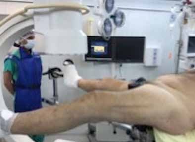
254 Rev Asoc Argent Ortop Traumatol 2021; 86 (2): 253-262 • ISSN 1852-7434 (online)Intertrochanteric fractures
A
C
Figure 1. A. Supine decubitus position on the
traction table with the contralateral limb abducted
and tractioned. B. Supine decubitus position on the
traction table with the contralateral limb in hip and knee
extension (“scissors”). C. Dorsal decubitus position on
a common table. An enhancement is placed under the
affected limb to facilitate lateral vision with the C-arm.
B
3. Visualization of the proximal femur
Correct visualization and optimal fracture reduction are closely related. The probability of poor reduction or
improper implant placement is high if visualization is not correct.7 The structures that must be clearly identified in
both the frontal and profile views are: the femoral head and joint space, the femoral neck, both trochanters, and the
proximal two-thirds of the femur. The true lateral view should consider the anteversion of the femoral neck. For
this, the image intensifier beam should be located between 0 ° and 20 ° with respect to the horizontal plane until
a straight line is drawn from the center of the head, parallel to the femoral neck and the femoral shaft (Figure 2).7
Once the desired images are achieved, it is useful to make some marks on the floor to indicate the place where the
C-arm should be repositioned after placing the fields (Figure 3).
Rev Asoc Argent Ortop Traumatol 2021; 86 (2): 253-252 • ISSN 1852-7434 (online) 255S. Pereira et al.
A B
Figure 2. A. Lateral image of the proximal femur (C-arm at 180º) B. Lateral image
contemplating the anteversion of the femoral neck (C-arm at 160º).
Figure 3. References on the floor to reposition the
C-arm.
256 Rev Asoc Argent Ortop Traumatol 2021; 86 (2): 253-262 • ISSN 1852-7434 (online)Intertrochanteric fractures
4. Fracture reduction
In recent years, implants have undergone design improvements, but none are a substitute for a proper reduction
technique. It is worth noting that, while many articles emphasize the importance of the quality of the reduction
as the main predictor of failure, few actually describe the reduction technique. In 2020, Yoon et al. reported that,
in more than 50% of the 322 intertrochanteric fractures analyzed, closed reduction was insufficient and required
percutaneous reduction efforts to achieve optimal reduction.8
a. Coronal plane reduction
While it is true that the internal rotation traction maneuver generally results in adequate reduction; on some
occasions, especially in unstable patterns, it may be insufficient. Ottolenghi et al. warned about some intertro-
chanteric fractures, which they classified as “extradigital”, because they maintained the insertion of the external
rotator muscles, which require external rotation of the diaphyseal fragment for their reduction.9 If, after traction
on the axis and the internal or external rotation maneuver, the desired cervico-diaphyseal angle is not achieved,
a slight abduction of the limb can help. It should be anticipated that the position of the abducted leg may make it
difficult to locate the nail entry point correctly. For this reason, in these situations it is advisable to temporarily
fixate the fracture with pins so that the leg can then be repositioned, avoiding loss of reduction.
In the anteroposterior projection, the continuity of the calcar should also be evaluated, since, on occasions,
despite having recovered the cervico-diaphyseal angle, a translation of the cortices may persist. In this case,
the use of a collinear clamp or a bone hook on the medial cortex of the neck or the distal fragment may reduce
displacement.
b. Sagittal plane reduction
In certain fracture patterns, the proximal fragment tends to flex and the shaft to fall. The first maneuver to
reduce this displacement in the sagittal plane is usually the placement of a support on the posterior aspect
of the thigh (fist of the hand, hammer, crutch, etc.). However, this is often insufficient. For these situations,
some percutaneous reduction maneuvers have been described. In 2011, Chun et al. described a technique that
consists of the percutaneous insertion of a 4.2 mm Steinmann pin in the anteromedial cortex of the neck and,
by the exertion of a posterior force, reducing the flexion of the proximal fragment (Figure 4).10 Yoon et al.
introduced, through a small anterolateral approach proximal to the lesser trochanter in the thigh, a long hemo-
static clamp and, by generating upward traction, the defect of flexion and rotation of the proximal fragment
was corrected.11
In 2007, Carr described a displacement pattern in comminuted intertrochanteric fractures, in which the di-
aphyseal fragment shortened and externally rotated, and the proximal fragment was displaced in varus and
impacted within the distal fragment, generating an overlap of the anterior cortices. He proposed, then, to insert
a Steinmann pin between the two fragments to lift the proximal fragment and disimpact it from the distal frag-
ment (Figure 5).12
5. Location of entry point
Defining an ideal entry point may be impossible, since it depends on the anatomical variations of the greater
trochanter and the variations in the angle of the different types of nails. In a cadaveric study using different
nail designs, Osrtum et al. defined the tip of the greater trochanter 2 to 3 mm medial to it as the “universal”
entry point in the coronal plane. They also mentioned that the lateral entry point should be avoided, because
it inevitably generates a varus displacement of the proximal fragment.13
The profile entry point has been less studied; however, an incorrect entry in this plane can cause an iatro-
genic fracture of the proximal femur; thus, an entry point 5 mm posterior to the tip of the trochanter is recom-
mended.
Rev Asoc Argent Ortop Traumatol 2021; 86 (2): 253-252 • ISSN 1852-7434 (online) 257S. Pereira et al.
A B
C D
Figure 4. A. Flexion displacement of the proximal fragment. B and C. Reduction of displacement
with scissors through a percutaneous approach. D. Fluoroscopic image of the reduction achieved in
the front.
Once the ideal entry point is defined, all the factors that may interfere with its correct positioning must be
recognized. In this sense, the situations that can cause a lateralization of the entry point are: the comminution
of the fracture that extends towards the tip of the trochanter, the incorrect placement of the patient on the trac-
tion table or surgery table, the incorrect preparation of the the fields that interfere with the approach or errors in
the approach. Another time when the entry point may be inadvertently lateralized is during reaming. Reaming
should not be started until close contact of the reamer with the bone to avoid eccentric reaming and thus cause
a lateral displacement of the entry canal.13
6. Nail placement
Once the entry canal has been reamed, we proceed to insert the nail. It is usually possible to insert it manually,
with gentle rotational movements. Only once the nail is aligned with the femoral canal can gentle hammer blows be
applied—if necessary—to achieve the desired position. If, despite this, its descent does not progress, it is advisable
to remove the nail and ream the canal to avoid an iatrogenic fracture of the femur.
After inserting the nail in the desired position—especially if the entry point was lateral to the tip of the greater
trochanter—a secondary varus displacement of the proximal fragment and lateralization of the diaphysis (“wedge
258 Rev Asoc Argent Ortop Traumatol 2021; 86 (2): 253-262 • ISSN 1852-7434 (online)Intertrochanteric fractures
A B
C D E
Figure 5. A. Lateral fluoroscopic image showing the posterior fall of the proximal fragment and the overlapping
of the cortices, which makes closed reduction impossible. B and C. Inserting a 3mm pin into the site and reducing
displacement. D. Transient stabilization with two anterior pins, ensuring that they do not interfere with the introduction
of the nail. E. Definitive fixation with the cephalic screw.
effect”) can be generated.14 It is important to detect this situation before placing the cervicocephalic guide pin. The
solution is usually to apply more traction to the axis or, in some circumstances, to apply a slight abduction on the
limb. If, despite this, the desired reduction is not achieved, the conflict is probably caused by a lateral entry point.
In that case, it is advisable to reposition the entry point. The first thing we do, after extracting the nail, is to check
that the soft tissues are not interfering with the access. If this happens, we extend the approach proximally to allow
an entry more aligned with the femoral canal. If, despite this, the comminution of the trochanter or the previously
generated canal causes the pin to move laterally, re-reaming the entrance canal exerting force medially to remove
the superolateral cortex of the head-neck fragment will grant more space to the proximal end of the nail, thus pre-
venting the nail from causing varus displacement.15
Although rare, a valgus displacement of the fracture may occur after inserting the nail, this is the “reverse wedge
effect”.16 This type of secondary displacement is described mainly in basicervical fractures. In this situation, it is
possible to insert a bone hook or collinear clamp and, in this way, generate lateral traction from the medial cortex
of the femoral neck.16
Rev Asoc Argent Ortop Traumatol 2021; 86 (2): 253-252 • ISSN 1852-7434 (online) 259S. Pereira et al.
7. Cephalic element positioning
In terms of the position of the screw or plate in the femoral head, there is considerable consensus that the ideal
position in the profile is the center.6 However, in the frontal image, like us, there are those who defend the central
location6 and those who defend the lower location.17 In practice, a screw in an inferior position with good clinical
results was often associated with a tip-apex index> 25 mm; therefore, it was necessary to describe a new predic-
tive failure index that favors this inferior screw / plate position. The “Cal TAD” index, described by Kuzyk et
al. in 2012, differs from the tip-apex index, described by Baumgaertner,6 only in the frontal image. It uses a line
parallel to the femoral neck and adjacent to the calcar, rather than running down the center of the neck.17
In any situation, the concept of the tip-apex index is to position the cephalic element in the area of the best bone
to guarantee the best possible fixation and reduce the chances of cut-out.
Once the desired position in the head is established, the insertion of the plate or screw may cause rotational
displacement of the fracture. This is more likely in basicervical or comminuted fractures. One way to avoid this
displacement is to temporarily fixate the reduction achieved initially with 3.2 mm pins outside the nail guide sys-
tem. However, if the fracture was not initially stabilized and rotation occurs, it is indicated to remove the screw
and reinsert it after stabilization, monitoring with the image intensifier. If a rotational defect is observed when the
desired position is reached with the tip of the screw, the screw should be removed as much as necessary to correct
the deformity observed in the image intensifier.
Another possibility is that, due to the traction necessary to achieve the reduction or due to the introduction of
the screw or plate, a distraction of the fracture site is generated. If this occurs, after inserting the plate or screw,
the traction on the limb should be released and, by means of the compression system, the reduction of the fracture
should be completed.
8. Short nail vs. long nail
Since the vast majority of intertrochanteric fractures occur in elderly patients with osteoporosis, it is reasonable
to think that a longer implant will protect the femur from a second fracture. In contrast, Curtis et al. reported a
higher incidence of fractures around the implant in the metaphyseal region than in the diaphyseal region.18 How-
ever, the results of a 2019 meta-analysis show that the risk of secondary fracture, osteosynthesis failure, nonunion
or infection was similar between long and short nails. Only a longer surgical time to place the long nails was
significant due to the need to ream and drill distally, freehand.19 Our practice of using short nails, as a routine,
is not only due to the shorter surgical time, but also to the fact that, if a distal fracture occurs, the exchange of a
short nail for a longer one would be simpler than the treatment of a supracondylar fracture with a distal femural
plate over the nail.
9. Plate vs. screws
Adequate fixation of the implant to the femoral head is a determining factor for the success of osteosynthesis.
Some authors argue that it is the rotational forces that cause a loss of screw fixation, the subsequent varus collapse
of the femoral head and finally the “cut-out”.20 In biomechanical studies, it has been shown that plates are more
resistant to rotational forces, due to their geometry and their radially compacting entry into the bone.21 Clinical
studies have supported the laboratory results. In a 2015 meta-analysis, Shuang Li et al. showed that the risk of
cut-out is significantly lower in the plate group than in the screw group.22 On the other hand, in 2019, Ibrahim et
al. did not find statistically significant differences in the failure rate between the plate group and the screw group.
However, they mention a difference in the failure pattern between both groups: axial migration and intra-articular
penetration were more frequent with the plate.23
10. Distal locking
Load transfer in the proximal femur after osteosynthesis with a proximal femoral nail will depend on the frac-
ture pattern and the quality of reduction. In unstable fractures, all loads will be transmitted to the distal femur by
the nail lock until union or rupture. Whereas, in stable fractures, if the reduction is adequate, the intimate corti-
cal contact will transmit the loads directly to the distal femur. For this reason, some authors have questioned the
usefulness of distal locking in stable intertrochanteric fractures.24 In 2019, Yan et al. conducted a meta-analysis
260 Rev Asoc Argent Ortop Traumatol 2021; 86 (2): 253-262 • ISSN 1852-7434 (online)Intertrochanteric fractures
on the need to perform the distal locking of the nail in stable fractures and found that the surgical time and the
exposure time to fluoroscopy were shorter, and that there was less bleeding and thigh pain in the postoperative
period in the group of nails without distal locking. At the same time, the functional results of both groups were
similar.At the same time, the functional results of both groups were similar.25 As the additional irradiation time
is minimal with the use of the guides, our routine practice is to perform the distal locking. With nails that offer
the possibility of two distal locks, we always choose the most proximal to reduce the stress concentration on the
nail tip.
Conclusions
Despite the great progress and development of implants in recent years, the main determinants of the final out-
come of the fixation of intertrochanteric fractures continue to be the quality of the reduction and the correct posi-
tioning of the implant. Awareness of the different errors that can be made during each of the steps of the surgical
technique is essential to avoid them.
––––––––––––––––––
Conflict of interests: The authors declare they do not have any conflict of interests.
G. Vindver ORCID ID: https://orcid.org/0000-0003-3858-6687
F. Bidolegui ORCID ID: https://orcid.org/0000-0002-0502-2300
References
1. Radaideh AM, Qudah HA. Functional and radiological results of proximal femoral nail antirotation (PFNA)
osteosynthesis in the treatment of unstable pertrochanteric fractures. J Clin Med 2018;7(4):78.
https://doi.org/10.3390/jcm7040078
2. Hardy DC, Descamps PY, Krallis P, Fabeck L, Smets P, Bertens CL, et al. Use of an intramedullary hip- screw
compared with a compression hip-screw with a plate for intertrochanteric femoral fractures: a prospective,
randomized study of one hundred patients. J Bone Joint Surg Am 1998;80(5):618-30.
https://doi.org/10.2106/00004623-199805000-00002
3. Baumgaertner MR, Curtin SL, Lindskog DM, Keggi JM. The value of the tip-apex distance in predicting failure
of fixation of peritrochanteric fractures of the hip. J Bone Joint Surg Am 1995;77(7):1058-64.
https://doi.org/10.2106/00004623-199507000-00012
4. Bidolegui F, Vindver G, Di Stefano C. Manejo de las fracturas inestables del fémur proximal con el clavo PFN de la
AO/ASIF Evaluación de una serie prospectiva de 100 casos. Rev Asoc Argent Ortop Traumatol 2008;73(1):55-62.
Disponible en: http://www.aaot.org.ar/revista/2008/n1_vol73/art09.pdf
5. Koval KJ, Oh CK, Egol KA. Does a traction-internal rotation radiograph help to better evaluate fractures of the
proximal femur? Bull NYU Hosp Jt Dis 2008;66(2):102-6. PMID: 18537778
6. Lyon T, Koval KJ, Kummer F, Zuckerman JD. Pudendal nerve palsy induced by fracture table. Orthop Rev
1993;22(5):521-5. PMID: 8316416
7. Rikli D, Goldhahn S, Blauth M, Mehta S, Cunningham M, Joeris A, PIP Study group. Optimizing intraoperative
imaging during proximal femoral fracture fixation – a performance improvement program for surgeons. Injury
2018;49(2):339-44. https://doi.org/10.1016/j.injury.2017.11.024
8. Yong-Cheol Yoon, Chang-Wug Oh, Jae-Ang Sim, Jong-Keon Oh. Intraoperative assessment of reduction quality
during nail fixation of intertrochanteric fractures. Injury 2020;51(2):400-6.
https://doi.org/10.1016/j.injury.2019.10.087
9. Ottolenghi CE, Japas LM. Lateral fractures of the femur neck: the extradigital type. Rev Chir Orthop Reparatrice
Appar Mot 1964;50:389-98. PMID: 14186381
Rev Asoc Argent Ortop Traumatol 2021; 86 (2): 253-252 • ISSN 1852-7434 (online) 261S. Pereira et al.
10. Young Soo Chun, Hyunsup Oh, Yoon Je Cho, Kee Hyung Rhyu. Technique and early results of percutaneous
reduction of sagittally unstable intertrochateric fractures. Clin Orthop Surg 2011;3(3):217-24. https://doi.
org/10.4055/cios.2011.3.3.217
11. Yong-Cheol Yoon, Ashutosh Jha, Chang-Wug Oh, Senthil Kumar Durai, Young-Woo Kim, Jong-Hoon Kim,
et al. The pointed clamp reduction technique for spiral subtrochanteric fractures: A technical note. Injury
2014;45(6):1000-5. https://doi.org/10.1016/j.injury.2014.01.007
12. Carr JB. The anterior and medial reduction of intertrochanteric fractures: A simple method to obtain a stable
reduction. J Orthop Trauma 2007;21:485-9. https://doi.org/10.1097/BOT.0b013e31804797cf
13. Ostrum RF, Marcantonio A, Marburger R. A critical analysis of the eccentric starting point for trochanteric
intramedullary femoral nailing. J Orthop Trauma 2005;19:681-6.
https://doi/org/10.1097/01.bot.0000184145.75201.1b
14. O’Malley MJ, Kang KK, Azer E, Siska PA, Farrell DJ, Tarkin IS. Wedge effect following intramedullary hip screw
fixation of intertrochanteric proximal femur fracture. Arch Orthop Trauma Surg 2015;135(10):1343-7.
https://doi.org/10.1007/s00402-015-2280-0
15. Hak DJ, Bilat C. Avoiding varus malreduction during cephalomedullary nailing of intertrochanteric hip fractures.
Arch Orthop Trauma Surg 2011;131:709-10. https://doi.org/10.1007/s00402-010-1182-4
16. Yu Zhang, Jun Hu, Xiang Li, Xiaodong Qin. Reverse wedge effect following intramedullary nailing of a
basicervical trochanteric fracture variant combined with a mechanically compromised greater trochanter. BMC
Musculoskelet Disord 2020;21(1):195. https://doi.org/10.1186/s12891-020-03212-6
17. Kuzyk PR, Zdero R, Shah S, Olsen M, Waddell JP, Schemitsch EH. Femoral head lag screw position for
cephalomedullary nails: a biomechanical analysis. J Orthop Trauma 2012;26:414-21.
https://doi.org/10.1097/BOT.0b013e318229acca
18. Curtis R, Goldhahn J, Schwyn R, Regazzoni P, Suhm N. Fixation principles in metaphyseal bone—a patent based
review. Osteoporos Int 2005;16(Suppl 2):S54-S64. https://doi.org/10.1007/ s00198-004-1763-6
19. Pernille Bovbjerg, Lonnie Froberg, Hagen Schmal. Short versus long intramedullary nails for treatment of
intertrochanteric femur fractures (AO 31‐A1 and AO 31‐A2): a systematic review. Eur J Orthop Surg Traumatol
2019;29(8):1823-31. https://doi.org/10.1007/s00590-019-02495-3
20. Nobuaki Chinzei, Takafumi Hiranaka, Takahiro Niikura, Mitsuo Tsuji, Ryosuke Kuroda, Minoru Doita, et al.
Comparison of the sliding and femoral head rotation among three different femoral head fixation devices for
trochanteric fractures. Clin Orthop Surg 2015;7(3):291-7. https://doi.org/10.4055/cios.2015.7.3.291
21. Sommers MB, Roth C, Hall H, Kam BCC, Ehmke LW, Krieg JC, et al. A laboratory model to evaluate cutout
resistance of implants for pertrochanteric fracture fixation. J Orthop Trauma 2004;18:361-8.
https.//doi.org/10.1097/00005131-200407000-00006
22. Shuang Li, Shi‐Min Chang, Wen‐Xin Niu, Hui Ma. Comparison of tip apex distance and cut‐out complications
between helical blades and lag screws in intertrochanteric fractures among the elderly: a meta‐analysis. J Orthop Sci
2015;20:1062-9. https://doi.org/10.1007/s00776-015-0770-0
23. Ibrahim I, Appleton PT, Wixted JJ, DeAngelis JP, Rodriguez EK. Implant cut-out following cephalomedullary
nailing of intertrochanteric femur fractures: Are helical blades to blame? Injury 2019;50(4):926-30.
https://doi.org/10.1016/j.injury.2019.02.015
24. Caiaffa V, Vicenti G, Mori C, Panella A, Conserva V, Conserva V, et al. Is distal locking with short intramedullary
nails necessary in stable pertrochanteric fractures? A prospective, multicentre, randomised study. Injury
2016;47(Supp4):S98-S106. https//doi.org/10.1016/j.injury.2016.07.038
25. Wen-Shan Yan, Wei-Li Cao, Ming Sun, Deng-Yue Ma, Peng Zhang. Distal locked or unlocked nailing for stable
intertrochanteric fractures? A meta-analysis. ANZ J Surg 2020;90(1-2):27-33. https://doi.org/10.1111/ans.15232
262 Rev Asoc Argent Ortop Traumatol 2021; 86 (2): 253-262 • ISSN 1852-7434 (online)You can also read