Maternal and fetal outcome in placenta accreta spectrum (PAS) associated with placenta previa: a retrospective analysis from a tertiary center
←
→
Page content transcription
If your browser does not render page correctly, please read the page content below
JOURNAL of MEDICINE and LIFE

JML | ORIGINAL ARTICLE

Maternal and fetal outcome in placenta accreta spectrum (PAS)
associated with placenta previa: a retrospective
analysis from a tertiary center
Valentin Nicolae Varlas 1, 2 *, Roxana Georgiana Bors 1, Simona Birsanu 1, Bogdan Maxim 1, Eliza Clotea 1, Maria Mihailov 1

Author Affiliations:                                                                                * Corresponding Author:
1. Department of Obstetrics and Gynaecology, Filantropia Clinical Hospital,                         Valentin Nicolae Varlas,
   Bucharest, Romania                                                                               Department of Obstetrics
2. Department of Obstetrics and Gynaecology,                                                        and Gynaecology, Carol Davila
   Carol Davila University of Medicine and Pharmacy, Bucharest, Romania                             University of Medicine and
                                                                                                    Pharmacy, 37 Dionisie Lupu St.,
                                                                                                    020021, Bucharest, Romania.
                                                                                                    E-mail: valentin.varlas@umfcd.ro

                                                                                                    DOI
ABSTRACT                                                                                            10.25122/jml-2021-0134
Accreta placenta spectrum is a complex obstetrical condition of abnormal placental
                                                                                                    Dates
invasion associated with severe maternal morbidity. This study aimed to analyze our                 Received: 11 April 2021
therapeutic management and counseling of the cases with placenta accreta spec-                      Accepted: 31 May 2021
trum (PAS) associated with placenta previa. We performed a retrospective study of
pregnant women with PAS associated with placenta previa at the Filantropia Clinical
Hospital between January 2017–April 2021. In these cases, the earlier diagnosis was
realized by an ultrasonographic scan and was confirmed by histopathological find-
ings after the surgical treatment. The conservative management was obtained in one
case at
JOURNAL of MEDICINE and LIFE

       A                                            B                                               C

      Figure 1. Placenta accreta spectrum (PAS), with all grades of abnormal placentation (A-accreta, B-increta and C-percreta).

  cornual resection of ectopic pregnancy is most frequently associated with PAS. Other factors that may be involved in the development
  of PAS are previous curettage, submucous fibroids, or uterine malformations [6].

  The most important risk factor for the development of a PAS is placenta previa after a prior cesarean delivery/section. The difficult
  diagnosis of this pathology explains the variation in placenta accreta prevalence between 1 in 300 and 1 in 2000 pregnancies [3]. In
  the case of women with a prior single cesarean delivery, the presence of placenta previa is associated with a 3% risk of PAS, while the
  absence of placenta previa is associated with a 0.03% risk of PAS. In a recent meta-analysis, Jauniaux et al. indicate a PAS prevalence
  between 0.01% and 1% [7]. This difference of risk is even more evident in women with a history of multiple cesarean sections. Women
  over 35 years or with a personal history of pelvic irradiation, manual removal of the placenta, endometritis, or infertility have a higher
  PAS risk compared to control groups [8].

  The increased rate of cesarean sections led to a higher prevalence of PAS. Progress was made both in the detection and treatment of
  these cases. Most patients with PAS can be detected prenatally, and appropriate management can be planned. The cesarean delivery
  should take place in a tertiary center with the participation of a multidisciplinary team that includes a neonatologist, obstetrician, urol-
  ogist and anesthesiologist [1].

  MATERIAL AND METHODS

  We performed a retrospective study of 12 pregnant women with PAS and placenta previa identified and treated at the Filantropia
  Clinical Hospital, Bucharest, from January 2017 to April 2021. The diagnosis was suspected after the ultrasonographic scan and was
  confirmed after surgery by the intraoperative findings and by the pathological report. In some cases where bladder invasion was sus-
  pected, magnetic resonance imaging (MRI) was used to assess the degree of bladder involvement. The ultrasound evaluation of the
  interface between the placenta and myometrium was done at a gestational age between 18 and 24 weeks. As a result, an early diagnosis
  of suspicion was made in 75% of cases (Figure 2).

  In 2 (16.6%) cases, the suspicion diagnosis was made in the third trimester when the patients presented to the hospital with symptoms
  such as bleeding, uterine contractions, or reduced fetal movement. In one (8.3%) case, the diagnosis of PAS was made during the cesare-
  an section for placenta previa. The patients with placenta previa associated with PAS were divided into two groups according to the PAS
  risk factors: group A (83.33%) had only one prior cesarean delivery, and group B (16.67%) had two cesarean deliveries. We compared
  the fetal and maternal outcomes after the surgical intervention.

  RESULTS

  Among the 12 patients, the mean age was 34±3.44 years. The analysis of the risk factors for placenta previa associated with PAS shows
  that the most important risk factor for PAS is the presence of a prior cesarean section (all cases). The risk for PAS is higher in the group
  of patients with multiple uterine surgical interventions (16.6% of cases) compared with those with only one prior cesarean section

                                                                 © 2021 JOURNAL of MEDICINE and LIFE. VOL: 14 ISSUE: 3 MAY-JUNE 2021
368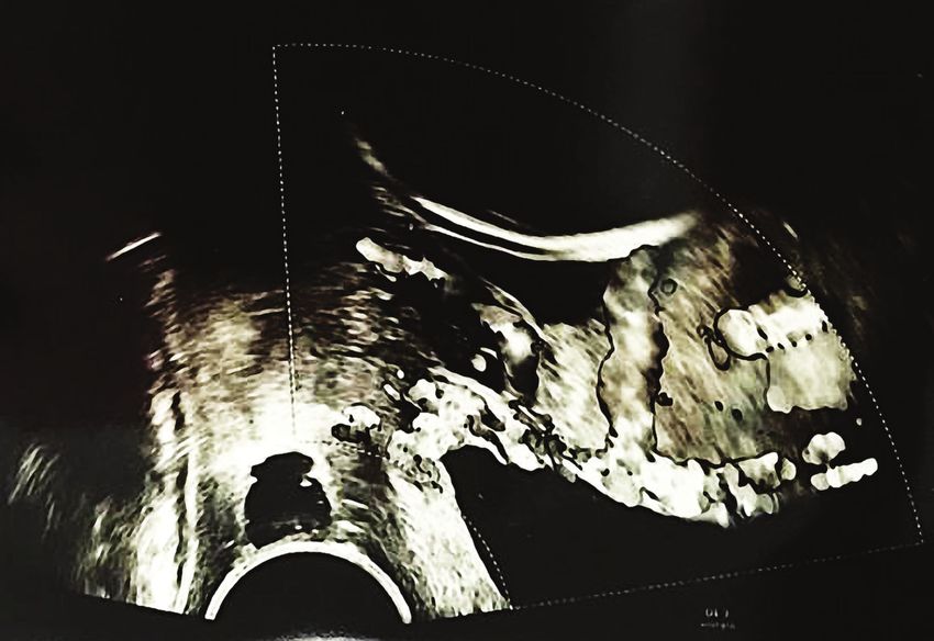
JOURNAL of MEDICINE and LIFE
A B
C
Figure 2. Ultrasound findings: (A) anterior placenta previa with abnormal lacunae, neovascularity, (B) bulge sign, no myometrium detect-
able, no clear zone, hypervascularity; (C) neovascularity with abnormal lacunae.
(83.3% of cases). The demographic and obstetric characteristics collected from the observation sheet of the patient were analyzed using
Excel 2017 for Windows and are presented below (Table 1).
Most women (91.6%) underwent planned cesarean delivery at the mean gestational age of 36.4±0.9 weeks. In only one case, the patient
underwent an emergency cesarean section for intense abdominal pain and severe bleeding. Fetal Apgar scores were not significantly
different in the group of patients who underwent planned cesarean section and did not depend on the risk factors for PAS (the mean
fetal Apgar score was 9). However, the patient with emergency cesarean section had a lower fetal Apgar score, namely 7. This fact shows
the importance of an early diagnosis and a close follow-up of the patients with this pathology. Fetal Apgar scores were not significantly
affected by the depth of placental invasion but were influenced by the gestational age at the moment of delivery.
In our study, the uterus was preserved in only one case (8.3%). In this case, the pregnancy was full term (37 weeks of gestation), fetal Ap-
gar score was 9 at 1 minute after birth and 10 at 5 minutes after birth. The patient had a history of one prior cesarean section and was
suspected of having placenta accreta in the current pregnancy. A fundal uterine incision was made, extended to the posterior uterine
wall of approximately 8–10 cm in length. No placental separation signs were observed after infant delivery. There was no active bleed-
ing, and the decision to leave the placenta in situ was made. Blood loss was estimated to be around 500 mL. The complete resorption
of the placenta occurred in the following months with no significant complications associated.
Conservative management by abandoning the placenta in situ was also tried in the case of a placenta accreta at a pregnancy of 37
weeks. However, an important postpartum hemorrhage 4 days after the cesarean section imposed an emergency hysterectomy with left
ovary removal.
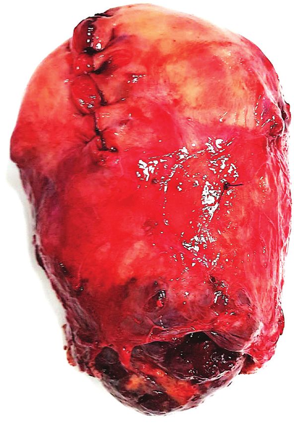
In the great majority of cases (83.4%), a cesarean hysterectomy with preservation of ovaries was performed (Figures 3, 4). The mean
maternal blood loss during surgical treatment was 2175±1440 ml.
© 2021 JOURNAL of MEDICINE and LIFE. VOL: 14 ISSUE: 3 MAY-JUNE 2021
369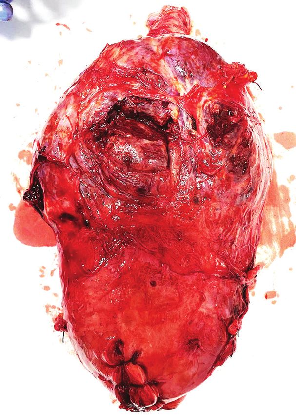
JOURNAL of MEDICINE and LIFE

      Table 1. Demographic and obstetric characteristics of the patients        In the emergency cesarean section case, the patient present-
      and neonates.                                                             ed at the hospital for intense abdominal pain, which did not
                                                                                subside under tocolysis and analgesia. The patient had a
                                         High-risk pregnant women with          history of prior cesarean section. The gestational age of the
      Demographic data
                                          placenta previa and PAS (n=12)        current pregnancy was 29 weeks and one day. The patient
                                                                                was taken up for emergency laparotomy, and a hemoperi-
      Maternal age, mean (SD) years                  34±3.4
                                                                                toneum of 1000 ml was found along with a uterine midline
      Area of residence                                                         and inferior defect, where the placenta was emerging. The
                                                                                fetus was extracted, weighing 1500 g and receiving an Ap-
      Urban (n)                                     9 (75%)                     gar score of 6 at 1 minute after birth and 7 at 5 minutes.
      Rural (n)                                      3 (25%)                    Bladder invasion was also documented. Suture of the uter-
                                                                                us was attempted but could not be performed due to the
      Parity                                                                    fragile uterine wall penetrated by the placenta. A total hys-
      Primiparous (n)                                   0                       terectomy was performed to stop the massive hemorrhage.
                                                                                When separation of the bladder from the uterine wall was
      Multiparous (n)                               12 (100%)                   attempted, bladder injury occurred and cystorrhaphy had
                                                                                to be carried out under difficult circumstances. Due to the
      Patients with previous cesarean section
                                                                                massive hemorrhage (5500 ml), the patient suffered a hem-
      1                                            10 (83.3%)                   orrhagic shock. She was transfused several times intraoper-
                                                                                atively and postoperatively. She was intensively monitored
      2                                             2 (16.7%)
                                                                                postoperatively and was transferred on the second postop-
      Gestational age                               35.8 ± 2.3                  erative day to a multidisciplinary hospital for hemodialysis
                                                                                and observation.
      Placenta previa topography

      Anterior                                      9 (75%)                     Blood loss differed significantly in the group with only one
                                                                                prior cesarean section (1800±1300 ml) compared with
      Posterior                                      3 (25%)                    the group with multiple cesarean sections (3400±100 ml).
      Blood loss, mean (SD)                                                     However, intraoperative blood loss was higher in the case
                                                                                of non-conservative management (2000±1400 ml) com-
      500–1000 ml                                    3 (25%)                    pared with conservative surgery (500 ml). Bladder injury
      1000–1500 ml                                  1 (8.3%)
                                                                                was reported in 3 out of 12 cases (25%), only in cases of
                                                                                non-conservative surgical treatment. Bladder invasion was
      1500–2000 ml                                   3 (25%)                    suspected preoperatively and documented intraoperatively
                                                                                in one case (8.3%).
      2000–2500 ml                                  1 (8.33%)

      >2500 ml                                      4 (33.3%)                   Short-term complications were reported more often in the
                                                                                group of patients with conservative management com-
      Neonatal outcomes
                                                                                pared to those who underwent hysterectomy and included
      Birth weight (grams), mean (SD)             2696.3±466.3                  delayed hemorrhage. No severe maternal outcome was re-
                                                                                ported after conservative management of placenta previa
      NICU admission, n (%)                         1 (8.33%)                   associated with PAS.
      NICU – neonatal intensive care unit.

                                                                                DISCUSSION

                                                                                 The highest incidence of PAS cases is in the lower uterine
  wall in the area of the post-cesarean scar, involving the cervix in cases of coexisting placenta previa.

  The increase in cesarean rates in most middle‐ and high‐income countries led to an increase in the prevalence of PAS. Following a single
  cesarean, the risk of placenta previa is 50% higher. Studies suggest that elective cesarean deliveries may be associated with a higher PAS
  risk compared to emergent cesarean deliveries [3, 9]. Existing data show that a prior myomectomy is associated with a very low risk of
  PAS. Possible factors that may induce a false conclusion include the surgical techniques used for cesarean delivery and myomectomy. For
  example, myomectomy with entry into the uterine cavity and a large myometrial scar may influence the risk of PAS [10].

  The surgical technique used for closing the uterus during cesarean delivery could play a role in the etiology of PAS. Studies suggest
  that a single‐layer uterine closure compared with a multiple-layer uterine closure, locked versus interrupted suturing or suture materials
  could influence the risk of PAS in future pregnancies [11]. However, single‐layer closure of the uterine incision is associated with a
  reduction in mean blood loss and duration of the operative procedure [12]. More studies are necessary to assess the impact of surgical
  techniques used during cesarean delivery on the risks of PAS [10].

  Ultrasound can assess the topography of the placental invasion, the degree of vascularization in the lower uterine segment and the
  depth of the area of abnormal adhesion, as well as the invasion of other structures determining factors of maternal morbidity [13].

                                                                 © 2021 JOURNAL of MEDICINE and LIFE. VOL: 14 ISSUE: 3 MAY-JUNE 2021
370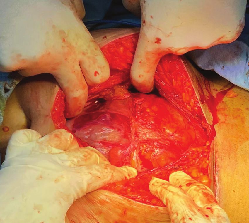
JOURNAL of MEDICINE and LIFE

 A                                                B                                               C

 D                                                E                                                F

Figure 3. Different uterus specimens after hysterectomy: (A) placenta accreta, (B) placenta percreta with bulge, (C) placental bulge, neo-
vascularity, invasive increta/percreta, (D) anterior placenta previa with placenta increta, (E) placental bulge under intact serosa, neovas-
cularity, (F) placenta increta, with neovascularity and no macroscopic invasion into uterine serosa.

In the second and third trimesters of pregnancy, the following ultrasound markers have been associated with PAS: placental lacunae
(large, irregular intraplacental sonolucent spaces in the center of a cotyledon) with high-velocity feeder vessels, disruption of the bladder
wall-uterine serosa interface (the “bladder line”), disruption of the normal hypoechoic area behind the placenta (known as the “clear
space”), abnormal vascularity (vessels that extend from the placenta through the myometrium into the bladder or the serosa), placental
bulge, and a focal exophytic mass (most often seen inside a filled urinary bladder) [14, 15]. The “clear space” can be obscured at an
advanced gestational age by pressure from the ultrasound probe and bladder filling or by posterior placental location. Color Doppler
is helpful in conjunction with the other ultrasound findings. Doppler markers of PAS include diffuse or focal placental lacunar flow,
vascular lakes with turbulent flow, and hypervascularity of the serosa-bladder interface [16].

The accuracy of first-trimester ultrasound evaluation for the diagnosis of PAS is less documented. PAS should be suspected if ultra-
sound examination during the first trimester reveals a gestational sac in the lower uterine segment in the proximity of a uterine scar or
a very thin (
JOURNAL of MEDICINE and LIFE
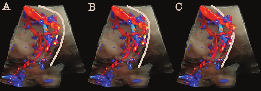
Figure 4. Placenta accreta spectrum (PAS), with all grades of abnormal placentation (A-accreta, B-increta and C-percreta).
with a full bladder (approximately 200–300 mL) [8]. The positive predictive value of ultrasonography for placenta accreta ranges from
65% to 93%. This is why it is recommended to wait for spontaneous placental separation after delivery of the infant during the surgical
intervention. The absence of placenta separation confirms placenta accreta clinically [4]. The use of Doppler ultrasound markers has
increased the sensitivity of ultrasound examination to around 90%. The prenatal detection rates depend on the ultrasound signs used,
operator's experience, scanning conditions, equipment used, and gestational age [7].
The solution of these cases must be done in a tertiary center with interventional experience, with an intensive care unit for premature
babies, and possibly with interventional radiology. In practice, we frequently encounter two scenarios; first, the case of a patient that is
hemodynamically stable without bleeding episodes, in which case a solution plan can be established in case of emergency and second,
the case of an unstable patient with different degrees of bleeding up to hemorrhagic shock, a situation in which the patient's manage-
ment plan will be individualized and made ad hoc depending on the location and degree of placental invasion (Figure 5).
Recommendations for treating placenta previa associated with PAS vary widely and are primarily based on case series and reports, per-
sonal experience, expert opinion, and clinical judgment. All women in our study received antenatal corticosteroids between 23 and 34
weeks of gestation. Delivery was planned at 37 weeks of gestation with the availability of blood products and a multidisciplinary team
was ensured [2]. However, hysterectomy of necessity following a planned cesarean section at 34–35 weeks with placental abandonment
in situ is the ACOG-recommended procedure for PAS [19].
Management of the placenta with abnormal adherence is mainly radical by performing a peripartum hysterectomy. In recent years,
taking into account the increasing incidence of this condition secondary to the increase in the number of cesarean sections, studies have
led to the identification of conservative techniques, such as intentional placental abruption, partial myometrial excision and the “Triple
P procedure”. “The triple P procedure” includes the following steps: preoperative placental topography to identify the upper edge of
the placenta; use of occlusal balloons in the iliac arteries to reduce blood flow to the placental bed and resection of the placenta with
the underlying adherent myometrium without separation, followed by suture of the myometrium [20].
The recommended management of placenta accreta is planned preterm cesarean hysterectomy with the placenta left in situ [21]. The
removal of the placenta is associated with significant hemorrhagic risk. Extraction of the newborn, unaccompanied by the placenta
separation, requires leaving the placenta in place, hysterorrhaphy, and performing a hysterectomy, with a low risk of bleeding. There
are situations during hysterectomies in which a deliberate cystotomy can be considered to separate the placental tissue from the bladder.
The RCOG and ACOG protocols recommend judicious transfusion monitoring of blood loss, correction of coagulation disorders, as
well as hydroelectrolytic disorders. Follow-up of the patient postoperatively in the intensive care unit continues the correction of vital
parameters.
However, there are many women who want to maintain fertility. In these cases, this approach may not be the first-line treatment. Thus,
conservative or expectant management can be considered. In the case of reduced areas of abnormal adhesion, the uteroplacental tissue
can be resected using the uterus-sparing surgical technique. Another procedure involves the use of the Bakri balloon [17]. In the case of
a large area of abnormal adhesion, the placenta is usually left in situ. Therefore, the therapeutic decision should be individualized [4].
The prognosis is better in patients who have placenta accreta without a placenta previa due to a higher risk of bleeding and hysterec-
tomy, respectively. An additional risk is presented by patients with the placenta percreta, which may be associated with an increased
incidence of urological lesions, and sometimes massive blood loss.
© 2021 JOURNAL of MEDICINE and LIFE. VOL: 14 ISSUE: 3 MAY-JUNE 2021
372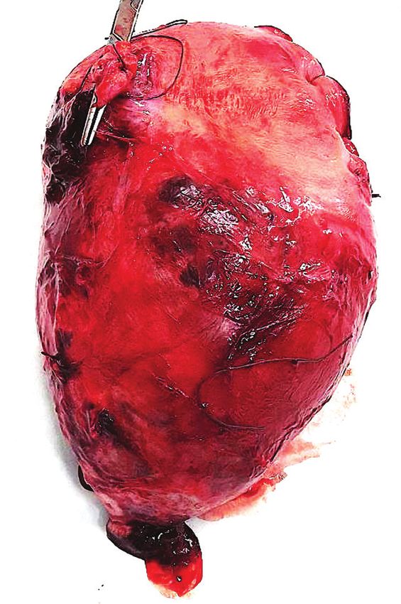
JOURNAL of MEDICINE and LIFE

                                                                PAS disorders

                                        Ultrasound, MRI identification of topography, depth of
                                                       invasion, and hypervascularity

              Prenatal diagnosis                                                                            Postnatal diagnosis

  Planned cesarean

                        Conservative –                           Vaginal delivery
                         reconstructive
                            tehniques

                                                           Leaving placenta in situ

                                                                  Spontaneously                 Curettage
                                                                      placental
                                                                      delivery

                                          Delayed
    Hysterectomy                       hysterectomy                      Uterus sparing tehniques

Figure 5. Flow diagram of PAS management with or without placenta previa.

In a multicentre retrospective case series on 452 patients on the management of invasive placenta, Palacios et al. showed that when using
the resection-reconstruction approach (one-step conservative surgery), the uterus can be preserved with minimal morbidity and blood
loss in almost 80% of cases [22].

Subsequent pregnancies after conservative technique (resection-reconstruction) for PAS have shown similar perinatal outcomes to ce-
sarean delivery, with no differences regarding the pregnancy course [23].

The main risk associated with PAS is severe obstetric hemorrhage, which causes secondary complications including coagulopathy, mul-
tisystem organ failure, and death. Surgical risks increase with the depth of placental invasion. Studies report the following incidence
of surgical complications in women with PAS – bladder injury: 5–40%, ureteral injury: 0–18%, bowel injury/obstruction: 2–4%,
venous thromboembolism: 4%, surgical site infection: 18–32%, maternal mortality: 1–7%, large‐volume blood transfusions: 5–40%
[24]. Many studies have evaluated the role of prophylactic placement of balloon occlusion catheters to reduce bleeding at the time of
cesarean hysterectomy for PAS. However, there is insufficient evidence to demonstrate the real safety and efficacy of these devices and
establish which groups of patients with PAS would have more benefit from their use. Other studies analyzed the advantage of ligating
the internal iliac arteries and concluded that the efficacy is similar to balloon occlusion devices [25, 26].

When placenta previa associated with PAS is suspected, surgeons should consider uterine incision at a site distant from the placenta and
should deliver the baby without disturbing the placenta in order to enable conservative management or elective hysterectomy with a

© 2021 JOURNAL of MEDICINE and LIFE. VOL: 14 ISSUE: 3 MAY-JUNE 2021
                                                                                                                                        373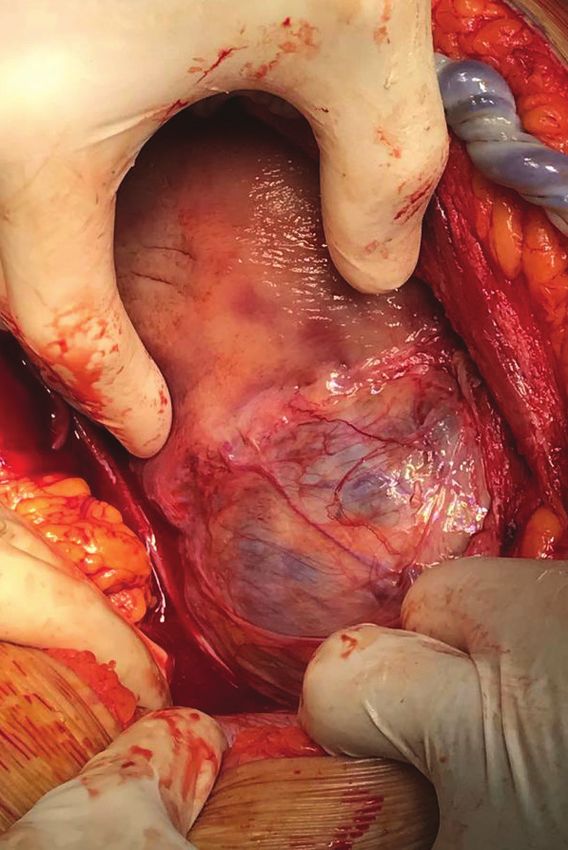
JOURNAL of MEDICINE and LIFE

  reduced blood loss [3, 4]. The abdominal incision must allow sufficient access to the uterus, and hysterotomy should be above the upper
  placental margin. Preoperative or intraoperative ultrasound examination enables a good visualization of the upper placental margin,
  and as a result, helps the surgeons to establish both the abdominal and uterine incision [8]. The choice of skin and uterine incisions
  required to avoid the placenta depends on the placenta's location. A Pfannenstiel skin incision allows good visualization of the lower
  uterine segment and is recommended if the upper margin of the anterior aspect of the placenta does not rise above the lower segment
  of the uterus. When the placenta extends above the lower uterine segment, a midline skin incision is needed to allow a high longitudinal
  uterine incision.

  Women who are candidates for conservative management should be warned of the risks of bleeding and infection. Studies failed to con-
  firm the benefit of methotrexate or arterial embolization to reduce these risks, and as a result, these treatments are not recommended by
  the current guidelines [3, 4]. A progressive decrease in blood circulation within the uterus and the placenta with necrosis of the villous
  tissue and a progressive detachment of the placenta from the uterus is expected [3].

  There is insufficient evidence to recommend magnetic resonance imaging and/or serial evaluation of serum beta-human chorionic
  gonadotropin (β‐hCG) to monitor cases that were managed conservatively. Patients should undergo weekly follow‐up visits during the
  first two months and then monthly visits until complete resorption of the placenta. The follow‐up should include a clinical examination
  (bleeding, temperature, pelvic pain), ultrasound (size of retained tissue), and laboratory tests (hemoglobin and leukocytes count, vaginal
  sample for bacteriological analysis). Postoperative antibiotic therapy (amoxicillin and clavulanic acid) should be administered to reduce
  the risk of infection. Subsequent pregnancies are at increased risk for recurrent PAS, uterine rupture, postpartum hemorrhage, and
  peripartum hysterectomy. The average risk of recurrence of PAS is approximately 22–29%, and the risk of early postpartum hemor-
  rhage is approximately 8.6–19%. Long‐term complications include intrauterine adhesions and secondary amenorrhea with secondary
  infertility [2].

  CONCLUSIONS

  Prenatal diagnosis and leaving the placenta in situ may be associated with reduced maternal morbidity. In this retrospective study, we
  identified a low successful uterine preservation rate, a low maternal complication rate, and a relatively good fetal outcome. After very
  careful prenatal counseling, we recommend that hysterectomy should be used as the treatment of choice for morbidly adherent placenta
  associated with placenta previa. Conservative management should be reserved for women with a strong fertility desire and women with
  an extensive disease that precludes primary hysterectomy due to surgical difficulty. Early identification of the risk factors and strategic
  management may improve maternal and fetal outcomes.

  ACKNOWLEDGMENTS
  Ethical approval
  The approval for this study was obtained from the Ethics Committee of the Filantropia Clinical Hospital (approval ID: 11040/13.10.2020).

  Consent to participate
  Written informed consent was obtained from the participants.

  Conflict of interest
  The authors declare that there is no conflict of interest.

  REFERENCES                                                    4. American College of Obstetricians and Gynecologists,
                                                                Society for Maternal-Fetal Medicine (2018) Obstetric Care
                                                                                                                               spectrum: a systematic review and meta-analysis. Am J Obstet
                                                                                                                               Gynecol 221:208–218.
                                                                Consensus No. 7: Placenta Accreta Spectrum. Obstet             https://doi.org/10.1016/j.ajog.2019.01.233
                                                                Gynecol 132:e259–e275.
                                                                https://doi.org/10.1097/AOG.0000000000002983                   8. Jauniaux E, Bhide A, Kennedy A, et al (2018) FIGO
  1. Eshkoli T, Weintraub AY, Sergienko R, Sheiner E (2013)                                                                    consensus guidelines on placenta accreta spectrum
  Placenta accreta: risk factors, perinatal outcomes, and       5. Jauniaux E, Grønbeck L, Bunce C, et al (2019)               disorders: Prenatal diagnosis and screening,. Int J Gynecol
  consequences for subsequent births. Am J Obstet Gynecol       Epidemiology of placenta previa accreta: a systematic review   Obstet 140:274–280. https://doi.org/10.1002/ijgo.12408
  208:219.e1–7. https://doi.org/10.1016/j.ajog.2012.12.037      and meta-analysis. BMJ Open 9:e031193.
                                                                https://doi.org/10.1136/bmjopen-2019-031193                    9. Previous Cesarean Delivery and Risks of Placenta Previa
  2. Jauniaux E, Ayres-de-Campos D, Langhoff-Roos J, et                                                                        and Placental Abruption. Obstet Anesth
  al (2019) FIGO classification for the clinical diagnosis of   6. Collins SL, Alemdar B, van Beekhuizen HJ, et al             Dig (2006) 26:138–139
  placenta accreta spectrum disorders. Int J Gynaecol Obstet    (2019) Evidence-based guidelines for the management of
  Off Organ Int Fed Gynaecol Obstet 146:20–24.                  abnormally invasive placenta: recommendations from the         10. Jauniaux E, Chantraine F, Silver RM, Langhoff-Roos
  https://doi.org/10.1002/ijgo.12761                            International Society for Abnormally Invasive Placenta. Am J   J (2018) FIGO consensus guidelines on placenta accreta
                                                                Obstet Gynecol 220:511–526.                                    spectrum disorders: Epidemiology,. Int J Gynecol Obstet
  3. Jauniaux E, Alfirevic Z, Bhide AG, et al (2019) Placenta   https://doi.org/10.1016/j.ajog.2019.02.054                     140:265–273. https://doi.org/10.1002/ijgo.12407
  Praevia and Placenta Accreta: Diagnosis and
  Management: Green-top Guideline No. 27a. BJOG Int J           7. Jauniaux E, Bunce C, Grønbeck L, Langhoff-Roos J (2019)
  Obstet Gynaecol 126:e1–e48.                                   Prevalence and main outcomes of placenta accreta
  https://doi.org/10.1111/1471-0528.15306

                                                                                 © 2021 JOURNAL of MEDICINE and LIFE. VOL: 14 ISSUE: 3 MAY-JUNE 2021
374
JOURNAL of MEDICINE and LIFE

11. Roberge S, Chaillet N, Boutin A, et al (2011)                 17. Shepherd AM, Mahdy H (2021) Placenta Accreta.               23. Palacios-Jaraquemada JM, Basanta N, Labrousse C,
Single- versus double-layer closure of the hysterotomy            In: StatPearls. StatPearls Publishing, Treasure Island (FL)     Martínez M (2021) Pregnancy outcome in women with
incision during cesarean delivery and risk of uterine rupture.                                                                    prior placenta accreta spectrum disorders treated with
Int J Gynecol Obstet 115:5–10.                                    18. Jauniaux E, Ayres-de-Campos D (2018) FIGO consensus         conservative-reconstructive surgery: analysis of 202 cases. J
https://doi.org/10.1016/j.ijgo.2011.04.013                        guidelines on placenta accreta spectrum disorders:              Matern-Fetal Neonatal Med Off J Eur Assoc Perinat Med Fed
                                                                  Introduction,. Int J Gynecol Obstet 140:261–264.                Asia Ocean Perinat Soc Int Soc Perinat Obstet 1–5.
12. Dodd JM, Anderson ER, Gates S, Grivell RM (2014)              https://doi.org/10.1002/ijgo.12406                              https://doi.org/10.1080/14767058.2021.1910671
Surgical techniques for uterine incision and uterine closure at
the time of caesarean section. Cochrane Database Syst Rev         19. Obstetric Care Consensus No. 7 Summary: Placenta            24. Allen L, Jauniaux E, Hobson S, et al (2018) FIGO
CD004732.                                                         Accreta Spectrum. Obstet Gynecol (2018) 132:1519–1521.          consensus guidelines on placenta accreta spectrum
https://doi.org/10.1002/14651858.CD004732.pub3                    https://doi.org/10.1097/AOG.0000000000002984                    disorders: Nonconservative surgical management,. Int J
                                                                                                                                  Gynecol Obstet 140:281–290.
13. Yu FNY, Leung KY (2021) Antenatal diagnosis of                20. Chandraharan E, Rao S, Belli A-M, Arulkumaran S
                                                                                                                                  https://doi.org/10.1002/ijgo.12409
placenta accreta spectrum (PAS) disorders. Best Pract Res         (2012) The Triple-P procedure as a conservative surgical
Clin Obstet Gynaecol 72:13–24.                                    alternative to peripartum hysterectomy for placenta percreta.   25. Dai YM, Wei J, Wang ZQ , et al (2020) [Intrauterine
https://doi.org/10.1016/j.bpobgyn.2020.06.010                     Int J Gynaecol Obstet Off Organ Int Fed Gynaecol Obstet         balloon tamponade combined with temporary abdominal
                                                                  117:191–194. https://doi.org/10.1016/j.ijgo.2011.12.005         aortic balloon occlusion in the management of women with
14. Jha P, Pōder L, Bourgioti C, et al (2020) Society of                                                                          placenta accreta spectrum:a randomized controlled trial].
Abdominal Radiology (SAR) and European Society of                 21. Society of Gynecologic Oncology, American College
                                                                                                                                  Zhonghua Fu Chan Ke Za Zhi 55:450–456.
Urogenital Radiology (ESUR) joint consensus statement for         of Obstetricians and Gynecologists and the Society for
                                                                                                                                  https://doi.org/10.3760/cma.j.cn112141-20200225-00135
MR imaging of placenta accreta spectrum disorders. Eur            Maternal–Fetal Medicine, Cahill AG, et al (2018) Placenta
Radiol 30:2604–2615.                                              Accreta Spectrum. Am J Obstet Gynecol 219:B2–B16.               26. Shamshirsaz AA, Fox KA, Erfani H, et al (2018)
https://doi.org/10.1007/s00330-019-06617-7                        https://doi.org/10.1016/j.ajog.2018.09.042                      Outcomes of Planned Compared With Urgent Deliveries
                                                                                                                                  Using a Multidisciplinary Team Approach for Morbidly
15. Jha P, Rabban J, Chen L-M, et al (2019) Placenta accreta      22. Palacios Jaraquemada JM, Pesaresi M, Nassif JC,
                                                                                                                                  Adherent Placenta. Obstet Gynecol 131:234–241.
spectrum: value of placental bulge as a sign of myometrial        Hermosid S (2004) Anterior placenta percreta: surgical
                                                                                                                                  https://doi.org/10.1097/AOG.0000000000002442
invasion on MR imaging. Abdom Radiol N Y 44:2572–2581.            approach, hemostasis and uterine repair. Acta Obstet
https://doi.org/10.1007/s00261-019-02008-0                        Gynecol Scand 83:738–744.
                                                                  https://doi.org/10.1111/j.0001-6349.2004.00517.x
16. Philips J, Gurganus M, DeShields S, et al (2019)
Prevalence of Sonographic Markers of Placenta Accreta
Spectrum in Low-Risk Pregnancies. Am J Perinatol
36:733–780. https://doi.org/10.1055/s-0038-1676488

© 2021 JOURNAL of MEDICINE and LIFE. VOL: 14 ISSUE: 3 MAY-JUNE 2021
                                                                                                                                                                                              375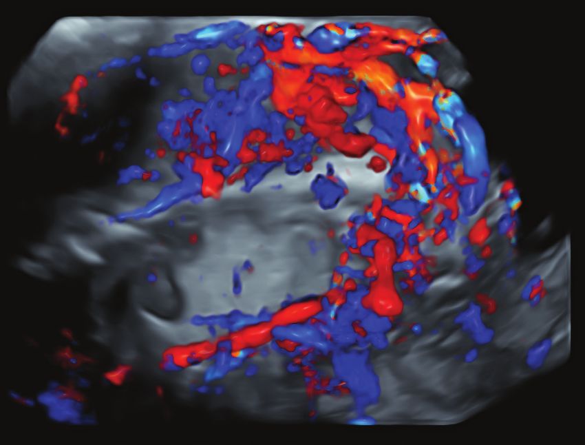
You can also read