Visualization of intestinal infections with astro- and sapovirus in mink (Neovison vison) kits by in situ hybridization
←
→
Page content transcription
If your browser does not render page correctly, please read the page content below
FEMS Microbes, 2, 2021, xtab005

                                                                                         doi: 10.1093/femsmc/xtab005
                                                                                         Advance Access Publication Date: 16 April 2021
                                                                                         Research Article

R E S E A R C H A R T I C L E – Microbes & Disease

                                                                                                                                                       Downloaded from https://academic.oup.com/femsmicrobes/article/doi/10.1093/femsmc/xtab005/6228835 by guest on 09 October 2021
Visualization of intestinal infections with astro- and
sapovirus in mink (Neovison vison) kits by in situ
hybridization
Julie Melsted Birch1,2, *,† , Mikael Leijon3,‡ , Søren Saxmose Nielsen4 ,
Tina Struve2 and Henrik Elvang Jensen1
1
 Section for Pathobiological Sciences, Department of Veterinary and Animal Sciences, University of
Copenhagen, 1870 Frederiksberg C, Denmark, 2 Kopenhagen Fur, 2600 Glostrup, Denmark, 3 Department of
Microbiology, National Veterinary Institute, 751 89 Uppsala, Sweden and 4 Section for Animal Welfare and
Disease Control, Department of Veterinary and Animal Sciences, University of Copenhagen, 1870
Frederiksberg C, Denmark
∗
  Corresponding author: Section for Pathobiological Sciences, Department of Veterinary and Animal Sciences, University of Copenhagen, Ridebanevej 3,
1870 Frederiksberg C, Denmark. Tel: +45-35-33-47-28; E-mail: jubi@sund.ku.dk
Editor: Kimberly Kline
†
  Julie Melsted Birch, http://orcid.org/0000-0002-3579-327X
‡
  Mikael Leijon, http://orcid.org/0000-0001-8701-9887

ABSTRACT
Clarification of the infection microbiology remains a challenge in the pre-weaning diarrhea (PWD) syndrome in farmed
mink (Neovison vison). Duodenal, jejunal and colon sections from 36 mink kits with PWD were systematically examined by
chromogen in situ hybridization targeting two incriminated viruses: mink astrovirus and mink sapovirus. Using the
RNAscope   R
              2.5 HD Duplex Assay, astrovirus and sapovirus were visualized and simultaneously demonstrated in the gut
tissue. Both viruses infect enterocytes in the small intestine with a specific localization pattern; astrovirus affects the two
apical thirds of the villi, whereas sapovirus generally affects the basal parts of the villi. Furthermore, we demonstrated that
astrovirus in mink does not target the goblet cells. This is the first time astro- and calicivirus have been visualized in mink
kit gut tissue, and these findings might be important in clarification of the impact of these viruses in the PWD syndrome.

Keywords: mink; sticky kits; diarrhea; intestine; virus; in situ hybridization

INTRODUCTION                                                                  ‘wet kits’ among mink farmers. The syndrome is multifacto-
                                                                              rial and several risk factors concerning parity profile, manage-
The aetiology of pre-weaning diarrhea (PWD) in mink kits has
                                                                              ment and feeding of the dams have been found (Chriél 1997;
been of concern for decades due to the negative impact on
                                                                              Møller and Chriél 2000; Møller 2004; Birch et al. 2017, 2018a). The
animal health, antibiotic consumption and losses caused by
                                                                              outbreak patterns on farms suggest a contagious origin. How-
recurring disease outbreaks. Suckling kits between 1 and 4
                                                                              ever, research aiming to elucidate the role of bacterial and viral
weeks of age are affected manifesting as diarrhea and a con-
                                                                              infections in the PWD syndrome suggests an interplay between
current skin exudation, and are referred to as ‘sticky kits’ or
                                                                              several bacteria and viruses since several of the incriminated

Received: 7 December 2020; Accepted: 12 April 2021

C The Author(s) 2021. Published by Oxford University Press on behalf of FEMS. This is an Open Access article distributed under the terms of the

Creative Commons Attribution License (http://creativecommons.org/licenses/by/4.0/), which permits unrestricted reuse, distribution, and
reproduction in any medium, provided the original work is properly cited.

                                                                                                                                                  1
2     FEMS Microbes, 2021, Vol. 2

microorganisms also have been identified in healthy mink kits        genes published for mink in the National Center for Biotechnol-
(Jørgensen, Scheutz and Strandbygaard 1996; Vulfson et al. 2001,     ogy Information (NCBI): Gapdh mRNA (GenBank: KM025344) and
2003; Guardabassi et al. 2012; Birch et al. 2018b). Research in      beta-actin (Actb) mRNA (GenBank: EU046492.1), which previ-
the viral aetiology of PWD has shown that astrovirus was sig-        ously have been used as reference genes (Zhang et al. 2009; Bow-
nificantly associated with PWD in mink (Englund et al. 2002),        man and Rose 2017). The duplex positive control was designed
and mink astrovirus (MiAstV) from intestinal contents was later      with the Gapdh probe in channel 1 and the Actb probe in chan-
characterized (Mittelholzer et al. 2003). Other viruses such as      nel 2 (ACD, Bio-Techne, MN, USA). The duplex positive control
calicivirus have also been detected in the mink gut (Svansson        probe and RNAscope     R
                                                                                               2-Plex Negative Control Probe (ACD, Bio-
1991; Jørgensen, Scheutz and Strandbygaard 1996). Guo, Ever-         Techne, MN, USA) were included in each assay. In addition, posi-

                                                                                                                                                 Downloaded from https://academic.oup.com/femsmicrobes/article/doi/10.1093/femsmc/xtab005/6228835 by guest on 09 October 2021
mann and Saif (2001a) detected and characterized mink enteric        tive controls including astrovirus and sapovirus as well as nega-
calicivirus in clinically normal and diarrheic mink, and cali-       tive control tissue without virus (foetal gut tissue) were included
civirus detected by electron microscopy has been shown to be         in each run. After hybridization, amplification and detection of
associated to PWD, although to a lesser extent than astrovirus       the duplex signal, green and red for channels 1 and 2, respec-
(Englund et al. 2002). In recent studies, we have taken advan-       tively, the slides were counterstained with Gills Hematoxylin No.
tage of next-generation sequencing (NGS) to show that besides        1 (Sigma-Aldrich, St Louis, MO, USA) and mounted with Vecta-
MiAstV, also calicivirus, of the genus sapovirus (SaV), seems to     mount (Vector Laboratories, Burlingame, CA, USA). By this in situ
be associated with PWD (Birch et al. 2018b).                         hybridization method, each single target molecule (e.g. from a
    Previously, we have detected both astrovirus and sapovirus       virus or a housekeeping gene) was visualized as a consequence
sequences on the same farm (Birch et al. 2019). This has led         of the branched amplification. In order to elucidate if astrovirus
to questions concerning which parts of the gastrointestinal (GI)     targets the intestinal goblet cells, periodic acid solution (PAS)
tract actually are infected by the two viruses, how the distribu-    with specificity for mucins was applied. On one random astro-
tion pattern is and which cell populations are infected. In this     virus positive sample, a PAS-staining step (Myers, Fredenburgh
study, our objective was to study the colocalization of astrovirus   and Grizzle 2008) was added after the green and red detection
and sapovirus in enterocytes and goblet cells in different parts     step and prior to the counterstaining.
of the gut in mink kits with PWD using branched DNA in situ
hybridization.
                                                                     Histological assessment of in situ hybridization signals
                                                                     From each animal, cross-sections from the duodenum, jejunum
MATERIALS AND METHODS                                                and colon were examined by light microscopy at 20× magnifica-
                                                                     tion. The load (i.e. burden of infection) of astro- and sapovirus
Animals and study design
                                                                     in duodenum and jejunum was individually assessed using the
On a Danish mink farm, 36 mink kits, 6–21 days old, with man-        following approach: infected villi (VIL) were scored as ‘0’ (0%),
ifestations consistent with PWD were collected during a period       ‘1+’ (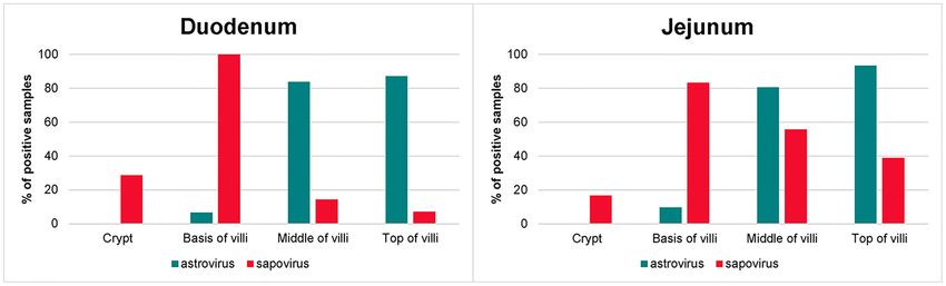
Birch et al.     3

Table 1. VLS of astro- and sapovirus detected by in situ hybridization.                       PWD. Furthermore, we have demonstrated coinfection with
                                                                                              these viruses in the same tissues by use of the RNAscope in
                                        Median         q1    q3      Min.        Max.         situ hybridization duplex assay. Astrovirus was most frequently
                                                                                              detected (92% of the animals) compared with sapovirus, which
All samples
                                                                                              was observed in 50% of the study group. These findings cor-
Astrovirus (n = 36)
                                                                                              respond well with other studies in which these two viruses
Duodenum                                    4           2     5         0           6
                                                                                              have been found to increase the risk of PWD (Englund et al.
Jejunum                                     5           2     6         0           6
Colon                                       0           0     2         0           5
                                                                                              2002; Birch et al. 2018b). The load of both astro- and sapovirus
Sapovirus (n = 36)                                                                            was significantly higher in the small intestine (duodenum and

                                                                                                                                                                       Downloaded from https://academic.oup.com/femsmicrobes/article/doi/10.1093/femsmc/xtab005/6228835 by guest on 09 October 2021
Duodenum                                    0           0     5         0           6         jejunum) than in the colon, which indicates that potentially
Jejunum                                     2           0     5         0           6         harmful effects of the viruses are also predominantly located to
Colon                                       0           0     0         0           2         the small intestine. However, the astrovirus infection extended
Virus positive samples                                                                        to enterocytes of the colon in some cases, and here the crypts
Astrovirus (n = 33)                                                                           were deeper, which suggests that these colon sections might
Duodenum                                    4           3     5         0           6         have been collected more proximally compared with the rest of
Jejunum                                     5           3     6         0           6         the samples. This is in line with other studies regarding astro-
Colon                                       0           0     2         0           5         virus infections in humans (Sebire et al. 2004), calfs (Woode
Sapovirus (n = 18)                                                                            et al. 1984), turkeys (Behling-Kelly et al. 2002; Nighot et al. 2010)
Duodenum                                    5           3     6         0           6         and lambs (Snodgrass et al. 1979) in which astroviruses have
Jejunum                                     6           5     6         3           6         been located to the small intestine in juvenile individuals. We
Colon                                       0           0     0         0           2         observed striking differences in the localization between astro-
                                                                                              virus and sapovirus, especially in samples with coinfection with
VLS was generated by adding VIL and VPE/VIL, where VIL was infected villi [‘0’
                                                                                              the two viruses, which suggests that these two viruses have
(0%), ‘1’ (
4 FEMS Microbes, 2021, Vol. 2
Downloaded from https://academic.oup.com/femsmicrobes/article/doi/10.1093/femsmc/xtab005/6228835 by guest on 09 October 2021
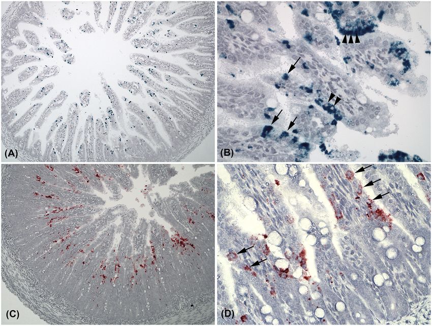
Figure 1. Photomicrographs of in situ hybridization targeting astro- and sapovirus in intestinal samples from mink kits with PWD. (A) Transverse section of a jejunum
with astrovirus (green) infection (×10). (B) Astrovirus infection at the tips of villi in jejunum. Note that single enterocytes contain very high loads of virus next to not
infected neighbour cells (arrows), and that replication is located to the cytoplasm and not the nucleus (small arrowheads) (×40). (C) Jejunal transverse section with
sapovirus (red) infection (×10). (D) Higher magnification (×40) of sapovirus infection at the base of the villi in the duodenum. Replication of sapovirus is located to the
cytoplasms of infected enterocytes (arrows).
Tabel 2. Pairwise comparison of sapo- and astrovirus locations in the small intestine.
Pairwise comparison OR Lower CL (OR) Upper CL (OR) P-value
Duodenum
Crypt: sapo vs astro Ref. n.a. n.a. 1
Base-villi: sapo vs astro 10.8 4.8 24.2 0.14
Mid-villi: sapo vs astro 0.023 0.010 0.051Birch et al. 5
Downloaded from https://academic.oup.com/femsmicrobes/article/doi/10.1093/femsmc/xtab005/6228835 by guest on 09 October 2021
Figure 2. Photomicrographs of in situ hybridization targeting astro- and sapovirus in intestinal samples from mink kits with PWD. (A) Coinfection of both astro- (green)
and sapovirus (red) in a transverse section of jejunum. Notice the sharp distinction of the location of the two viruses (×20). (B) An astrovirus positive duodenal section.
PAS staining added to the duplex in situ hybridization assay before counterstaining. Note that astrovirus positive enterocytes are positioned apart from the goblet cells
containing mucin (red) (×20). (C) Jejunum, negative control for both green and red channels (×20). (D) A colon section negative for both astro- and sapovirus (×10).
Figure 3. Locations of astro- and sapovirus in the small intestinal sections.
distinct pattern, which suggests that these viruses by different SUPPLEMENTARY DATA
cell tropisms or predilection sites may have evolved a strategy
Supplementary data are available at FEMSMC online.
for co-existence.
ACKNOWLEDGEMENTS FUNDING
The authors would like to express their gratitude to laboratory This study was funded by Pelsdyrafgiftsfonden (https://www.
technicians Elisabeth Wairimu Petersen, Betina Gjedsted Ander- pelsdyrafgiftsfonden.dk/) (HEJ) and Dansk Pelsdyravlerforen-
sen and Christina Tirsdal Kjempff for their valuable technical ings Forskningsfond (https://www.kopenhagenfur.com) (TS).
support. Also thanks to Klaus Juel Hansen and Anne Sofie Boyum The funders had no role in study design, data collection and
Johansen for their practical help during material collection. analysis, decision to publish or preparation of the manuscript.6 FEMS Microbes, 2021, Vol. 2
Conflict of Interest. JMB and TS received salary from Kopenhagen plant recipients. J Pediatr Gastroenterol Nutr 2005;40:
Fur. This does not alter our adherence to FEMS’s policies on shar- 328–33.
ing data and materials. Mittelholzer C, Hedlund KO, Englund L et al. Molecular character-
ization of a novel astrovirus associated with disease in mink.
J Gen Virol 2003;84:3087–94.
REFERENCES
Møller SH, Chriél M. Health effects of the feeding strategies
Behling-Kelly E, Schultz-Cherry S, Koci M et al. Localization of in the pre-mating and gestation periods of mink. Scientifur
astrovirus in experimentally infected turkeys as determined 2000;24:37–41.
by in situ hybridization. Vet Pathol 2002;39:595–8. Møller SH. Management of health in mink. A HACCP plan for
Downloaded from https://academic.oup.com/femsmicrobes/article/doi/10.1093/femsmc/xtab005/6228835 by guest on 09 October 2021
Berman JJ. Group IV viruses: single-stranded (+)sense RNA. In: energy allowance during winter and gestation in order to
Bermann JJ (ed). Taxonomic Guide to Infectious Diseases. Else- control sticky kits. Scientifur 2004;28:50–7.
vier, 2012, 237–46. Myers RB, Fredenburgh JL, Grizzle WE. Carbohydrates. In: Ban-
Birch JM, Agger JF, Aalbæk B et al. Dam characteristics associated croft LT, Gamble M (ed). Theory and Practice of Histological Tech-
with pre-weaning diarrhea in mink (Neovison vison). Acta Vet niques, 6th edn. Philadelphia: Churchill Livingstone, Elsevier,
Scand 2018;60:73. 2008, 168–71.
Birch JM, Agger JF, Dahlin C et al. Risk factors associated with Nighot PK, Moeser A, Ali RA et al. Astrovirus infection induces
diarrhea in Danish commercial mink (Neovison vison) dur- sodium malabsorption and redistributes sodium hydrogen
ing the pre-weaning period. Acta Vet Scand 2017;59:43. exchanger expression. Virology 2010; 401:146–54.
Birch JM, Agger JF, Leijon M et al. Comparing the treatment effect Oka T, Lu Z, Phan T et al. Genetic characterization and clas-
of narrow spectrum antimicrobial, probiotic and fluid with sification of human and animal sapoviruses. PLoS One
amoxicillin in mink kits (Neovison vison) with pre-weaning 2016;11:e0156373.
diarrhea. Res Vet Sci 2019;125:121–9. Pietsch C, Liebert UG. Intrahost viral evolution during chronic
Birch JM, Ullman K, Struve T et al. Investigation of the viral and sapovirus infections. J Clin Virol 2019;113:1–7.
bacterial microbiota in intestinal samples from mink (Neovi- R Core Team. R: A language and environment for statistical com-
son vison) with pre-weaning diarrhea syndrome using next puting. R Foundation for Statistical Computing, Vienna, Aus-
generation sequencing. PLoS One 2018b;13:e0205890. tria. https://www.r-project.org/ (13 August 2020, date last
Bowman K, Rose J. Estradiol stimulates glycogen synthesis accessed).
whereas progesterone promotes glycogen catabolism in the Sebire NJ, Malone M, Shah N et al. Pathology of astrovirus associ-
uterus of the American mink (Neovison vison). Anim Sci J ated diarrhoea in a paediatric bone marrow transplant recip-
2017;88:45–54. ient. J Clin Pathol 2004;57:1001–3.
Chriél M. Lad minktæverne selv bestemme! (Let the mink Snodgrass DR, Angus KW, Gray EW et al. Pathogenesis of diar-
females decide themselves). Dansk Pelsdyravl 1997;60:196–8. rhoea caused by astrovirus infections in lambs. Arch Virol
Cortez V, Boyd DF, Crawford JC et al. Astrovirus infects actively 1979;60:217–26.
secreting goblet cells and alters the gut mucus barrier. Nat Spencer LT, Bancroft JD. Tissue Processing. In: Bancroft LT,
Commun 2020;11:2097. Gamble M (ed). Theory and Practice of Histological Techniques,
Englund L, Chriél M, Dietz HH et al. Astrovirus epidemiologi- 6th edn. Philadelphia: Churchill Livingstone, Elsevier, 2008,
cally linked to pre-weaning diarrhoea in mink. Vet Microbiol 83–92.
2002;85:1–11. Svansson V. Studie af en række virusbetingede infektioner hos mink
Guardabassi L, Schmidt KR, Petersen TS et al. Mustelidae are (Study of a number of virus-induced infections in mink). 1991.
natural hosts of Staphylococcus delphini group A. Vet Microbiol Ph.D. Thesis. University of Copenhagen.
2012;159:351–3. Vulfson L, Pedersen K, Chriél M et al. Assessment of the aero-
Guo M, Chang KO, Hardy ME et al. Molecular characterization of a bic faecal microflora in mink (Mustela vison Schreiber) with
porcine enteric calicivirus genetically related to sapporo-like emphasis on Escherichia coli and Staphylococcus intermedius.
human caliciviruses. J Virol 1999;73:9625–31. Vet Microbiol 2003;93:235–45.
Guo M, Evermann JF, Saif LJ. Detection and molecular characteri- Vulfson L, Pedersen K, Chriél M et al. Serogroups and antimicro-
zation of cultivable caliciviruses from clinically normal mink bial susceptibility among Escherichia coli isolated from farmed
and enteric caliciviruses associated with diarrhea in mink. mink (Mustela vison Schreiber) in Denmark. Vet Microbiol
Arch Virol 2001;146:479–93. 2001;79:143–53.
Guo M, Hayes J, Cho KO et al. Comparative pathogenesis of tis- Wang QH, Souza M, Funk JA et al. Prevalence of noroviruses and
sue culture-adapted and wild-type cowden porcine enteric sapoviruses in swine of various ages determined by reverse
calicivirus (PEC) in gnotobiotic pigs and induction of diar- transcription-PCR and microwell hybridization assays. J Clin
rhea by intravenous inoculation of wild-type PEC. J Virol Microbiol 2006;44:2057–62.
2001;75:9239–51. Woode GN, Pohlenz JF, Kelso Gourley NE et al. Astrovirus and
Hothorn T, Bretz F, Westfall P. Simultaneous inference in general Breda virus infections of dome cell epithelium of bovine
parametric models. Biom J 2008;50:346–63. ileum. J Clin Microbiol 1984;19:623–30.
Jørgensen M, Scheutz F, Strandbygaard B. Escherichia coli and Zhang X, Moore JN, Newsted JL et al. Sequencing and characteri-
virus isolated from “sticky kits.” Acta Vet Scand 1996;37:163–9. zation of mixed function monooxygenase genes CYP1A1 and
Kaufman SS, Chatterjee NK, Fuschino ME et al. Character- CYP1A2 of Mink (Mustela vison) to facilitate study of dioxin-
istics of human calicivirus enteritis in intestinal trans- like compounds. Toxicol Appl Pharmacol 2009;234:306–13.You can also read