What Constitutes a Compromised Aortic Neck in 2018?
←
→
Page content transcription
If your browser does not render page correctly, please read the page content below
E VA R

What Constitutes a
Compromised Aortic
Neck in 2018?
Anatomic risk factors associated with an increased risk of proximal seal failure with standard
EVAR in the era of device and technique advancement.

BY GRAEME M c FARLAND, MD, AND JASON T. LEE, MD

E
       ndovascular aneurysm repair (EVAR) is now               criteria. The aortic wall should be parallel when viewed
       clearly the preferred and predominant approach          in two-dimensional imaging and absent of reverse taper
       for treatment of infrarenal abdominal aortic            configuration. The suprarenal and infrarenal angulation
       aneurysms (AAAs), including those that rupture.
Despite advancements in endograft technology over
the last several years, approximately 40% of patients are
deemed unsuitable for standard EVAR based on ana-
tomic criteria, particularly if device-specific instructions
for use (IFU) are followed.1,2 A substantial number of
these patients are anatomically unsuitable due to proxi-
mal aortic neck anatomy. As the boundaries of anatom-
ic suitability are challenged with newer endografts and
as more interventions proceed outside the IFU, which
occurs up to 60% to 70% of the time in some series,3,4
the number of “unsuitable” candidates considerably
decreases. Although treating AAAs outside the IFU has
shown mixed results,3-6 surgeon comfort and experience
with more complex endovascular interventions, includ-
ing branched or fenestrated EVAR (FEVAR), snorkel or
chimney EVAR (ChEVAR), or anchoring devices, has
called the use of standard infrarenal endovascular devic-
es into question for these more challenging anatomies.
The basis for this question primarily stems from proxi-
mal aortic neck anatomy and which intervention is best
suited to obtain a durable proximal seal.
   Discussions of the compromised neck ultimately
derive from what is considered an ideal aortic neck,
which is typically related to the IFUs of commercially
available endografts. For most devices, the infrarenal         Figure 1. Severe infrarenal aortic angulation challenges the
neck must be a minimum of 10 to 15 mm in length                flexibility of the endograft and makes the desired circumfer-
and 17 to 32 mm in diameter based on current IFU               ential wall apposition for proximal seal difficult.

                                                                           VOL. 17, NO. 3 MARCH 2018 ENDOVASCULAR TODAY 57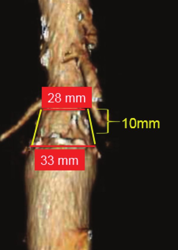
E VA R
is < 60° and the aortic wall should be void of significant
calcium or thrombus. Anything outside of these bound-
aries is considered treating outside the IFU, potentially
compromising the durability of the proximal seal.
Recently, concerns have been raised over the durabil-
ity of landing a standard device in a relatively large-
diameter neck despite still falling within the IFU criteria.
This article highlights the anatomic variables associated
with the compromised aortic neck and increased risk of
proximal seal failure with standard EVAR.
NECK DIAMETER
Current commercially available endografts have
proximal diameters ranging from 22 to 36 mm within
the IFU to treat aortic neck diameters of 18 to 32 mm.
Despite IFU parameters, including diameters up to
29 to 32 mm, recent studies have shown adverse out-
comes in treating AAAs with large neck diameters.
Although a few earlier studies demonstrated the fea-
sibility of this approach,7 several others have shown
that aortic neck diameters ≥ 28 mm are a risk factor
for proximal seal failure.4,8-11 Oliveira et al reported on
a multi-institutional study utilizing the Endurant endo-
graft (Medtronic), which demonstrated an increased
risk of type Ia endoleak and neck-related secondary
interventions in patients with infrarenal necks ≥ 30 mm
(odds ratio [OR], 3.8; 95% confidence interval [CI],
1.6–9.1).10 Similarly, our group recently explored our
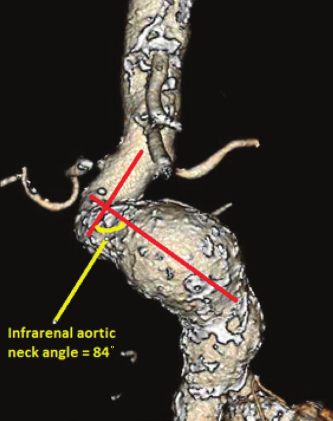
Figure 2. A reverse taper neck configuration with progressive
institutional data of all standard EVARs from 2000 to
dilatation within the proximal seal zone complicates accurate
2016 and noted a fourfold increase in failure of proxi-
graft oversizing and the long-term durability of standard EVAR.
mal fixation in patients treated with devices that had
large proximal diameters (34–36 mm).12 Further, on
multivariate analysis, we showed that a neck diameter est renal artery was 11%, as opposed to 3% to 5% at
≥ 29 mm was an independent risk factor for proximal the renal arteries and < 3% at the superior mesenteric
seal failure (OR, 2.5; 95% CI, 1.12–5.08). artery and celiac trunk, suggesting more durable endo-
A primary concern with the durability of standard vascular approaches should involve proximal fixation
infrarenal EVAR in patients with a dilated neck is into a healthier segment of perivisceral aorta for this
progressive aortic dilatation after EVAR. Our group subset of patients with dilated infrarenal necks.11
has also demonstrated a mean increase in aortic neck
diameter of 3.3 ± 0.6 mm at latest follow-up scans NECK LENGTH
in 86 patients, with a median radiologic follow-up of Most commercially available endografts recommend
21.9 months. No significant difference in neck dilation an aortic neck length of 10 to 15 mm for treatment
across devices was identified, but a positive correlation within the IFU. The concept of why a short aortic neck
between percent change in neck diameter and degree length leads to poorer outcomes is relatively simple:
of oversizing did exist (rs = 0.41; P < .001).13 This sug- the lesser the amount of seal zone, the more likely
gests that perhaps evolution in device fixation strategy that seal zone will fail. This has been described and
needs to be explored. Gargiulo and colleagues exam- demonstrated in multiple studies.14-16 Data from the
ined this concept further and not only demonstrated EUROSTAR registry indicate both increased risk of early
progressive neck dilatation in patients with wide aortic type Ia endoleak (OR, 4.46; 95% CI, 2.61–7.61) and late
necks (≥ 28 mm), but also demonstrated that the rate type Ia endoleaks (hazard ratio [HR], 2.13; 95% CI, 1.17–
of dilatation differed at distinct levels of the aorta. 4.60) in patients with aortic necks < 10 mm in length.15
The mean increase in diameter at the level of the low- A study by AbuRahma et al found similar results in
58 ENDOVASCULAR TODAY MARCH 2018 VOL. 17, NO. 3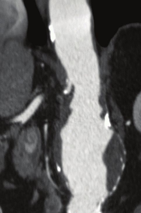
E VA R

   patients with neck lengths < 10 mm in both the early
                                                                 A                                B
   occurrence of type Ia endoleak (53% vs 12% in necks
   > 15 mm; P < .001) and late (3-year follow-up) freedom
   from type Ia endoleaks (53% vs 80%; P = .0263).14
      A more recent study by AbuRahma et al found that
   neck lengths < 10 mm had an OR of 4.26 (95% CI, 1.33–
   13.68) for type Ia endoleak.8 Further, Jordan and col-
   leagues, through the ANCHOR database, demonstrated
   that shorter neck lengths with a cut point of 17 mm
   were associated with a higher rate of type Ia endoleak
   (P = .017).16 Many reports on FEVAR and ChEVAR
   describe the concept of additional neck length gained
   that might lengthen an already compromised region of
   potential fixation.

   NECK ANGULATION                                              Figure 3. Aortic neck calcification signifies a diseased neck and
      Neck angulation includes both suprarenal and infra-       compromises active fixation of the endograft proximally (A).
   renal (Figure 1) aortic neck angulation measurements         Circumferential aortic neck thrombus has long been believed
   as standardized by van Keulen and colleagues in their        to be a risk factor for proximal seal failure; however, recent
   2010 publication.17 A cutoff of ≥ 60° is used to classify    data have called this into question, leading some to believe
   an aortic neck as angulated. The main concern when           thrombus to be protective against proximal seal failure (B).
   attempting to obtain a proximal seal within an angu-
   lated aortic neck is the ability for the device to contort
   enough to achieve circumferential wall apposition.           was a significant predictor for early type I endoleak
      Several studies have documented neck angulation           (OR, 5.25; P < .0001).8 In another study of patients
   as an independent risk factor for type Ia endoleak.6,8,18    with short aortic necks (< 15 mm), the reversed taper
   In the previously mentioned study by AbuRahma et al,         configuration was the most significant contributor to
   neck angulation > 60° had an OR of 2.81 (95% CI, 1.06–       proximal failure when compared to other associated
   7.47) for sac expansion and 3.28 (95% CI, 1.71–6.29) for     hostile neck characteristics.20
   early reintervention.6 Further data from the EUROSTAR
   registry determined that the risk of type Ia endoleak        NECK CALCIFICATION AND THROMBUS
   was higher in the perioperative period (OR, 2.17;               Neck calcification and neck thrombus have long
   95% CI, 1.20–3.91; P = .0105) than it was in the long        been considered risk factors for proximal seal failure in
   term (HR, 1.80; 95% CI, 1.25–2.58; P = .0016).19 Finally,    EVAR (Figures 3A and 3B). Despite this, there is no uni-
   Schanzer et al reported that in over 10,000 patients         versally agreed upon method to quantify the extent of
   undergoing EVAR between 1999 and 2008 with M2S               thrombus or calcification in the aortic neck. Kaladji and
   core laboratory analysis of both pre- and post-EVAR          colleagues previously studied predictive anatomic fac-
   anatomy, patients with neck angulation > 60° had an          tors for sac regression after EVAR by assigning a severity
   increased risk of aneurysm sac enlargement (HR, 1.96;        score to the aortic neck, AAA, and iliac arteries. On
   95% CI, 1.63–2.37; P < .0001).4                              multivariate analysis, they demonstrated that patients
                                                                with sac regression had a significantly lesser amount of
   NECK CONFIGURATION                                           calcification within the neck, and the lesser calcification
      Aortic necks with nonparallel walls risk compromis-       burden decreased the rate of type Ia endoleak.21
   ing the full 10 to 15 mm of seal required to stay within        Although neck calcification continues to result in
   the IFU. This primarily refers to a reverse taper or coni-   adverse outcomes in EVAR, recent studies have found
   cal configuration (Figure 2) that is often defined as a      neck thrombus to be less of a risk and more a protec-
   > 10% increase in diameter over a 5-mm increment             tive factor with regard to type Ia endoleak. Jordan et
   in the aortic neck. Again, the concern lies in decreas-      al found that the presence of aortic neck calcification
   ing the length of seal as the neck dilates, which in turn    was not a significant predictor for type Ia endoleak, and
   makes it more challenging to accurately oversize the         each degree of aortic neck thrombus decreased the risk
   selected endograft. In a 2011 study, AbuRahma et al          of type Ia endoleak by 1% (P = .001).16 Another study
   demonstrated that the reverse taper configuration            by Wyss et al similarly demonstrated neck thrombus to

60 ENDOVASCULAR TODAY MARCH 2018 VOL. 17, NO. 3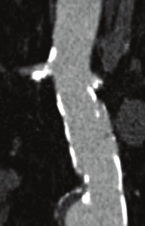
E VA R

have a protective effect against graft-related complica-                                                         5. Lee JT, Ullery BW, Zarins CK, et al. EVAR deployment in anatomically challenging necks outside the IFU. Eur J Vasc
                                                                                                                 Endovasc Surg. 2013;46:65-73.
tions (HR, 0.96; 95% CI, 0.92–0.99; P = .018), whereas                                                           6. AbuRahma AF, Yacoub M, Mousa AY, et al. Aortic neck anatomic features and predictors of outcomes in
aortic neck calcification was associated with a higher                                                           endovascular repair of abdominal aortic aneurysms following vs. not following instructions for use. J Am Coll Surg.
                                                                                                                 2016;222:579-589.
risk of graft-related complications (HR, 1.06; 95% CI,                                                           7. Zayed HA, Bell RE, Clough RE, et al. Results of endovascular repair of abdominal aortic aneurysms with an unfavor-
1.00–1.12; P = .044).22                                                                                          able proximal neck using large stent-grafts. Cardiovasc Intervent Radiol. 2009;32:1161-1164.
                                                                                                                 8. AbuRahma AF, Campbell JE, Mousa AY, et al. Clinical outcomes for hostile versus favorable aortic neck anatomy in
                                                                                                                 endovascular aortic aneurysm repair using modular devices. J Vasc Surg. 2011;54:13-21.
THE COMPROMISED NECK                                                                                             9. Chaikof EL, Fillinger MF, Matsumura JS, et al. Identifying and grading factors that modify the outcome of endovas-
   Each individual risk factor mentioned increases the                                                           cular aortic aneurysm repair. J Vasc Surg. 2002;35:1061-1066.
risk of proximal seal failure; however, the sum of two                                                           10. Oliveira NFG, Bastos Goncalves FM, Van Rijn MJ, et al. Standard endovascular aneurysm repair in patients with
                                                                                                                 wide infrarenal aneurysm necks is associated with increased risk of adverse events. J Vasc Surg. 2017;65:1608-1616.
or more of these findings may significantly increase the                                                         11. Gargiulo M, Gallitto E, Wattez H, et al. Outcomes of endovascular aneurysm repair performed in abdominal aortic
overall risk and truly define the compromised or hostile                                                         aneurysms with large infrarenal necks. J Vasc Surg. 2017;66:1065-1072.
neck. Few studies have attempted to quantify the added                                                           12. McFarland G, Tran K, Downey W, et al. Infrarenal EVAR with large device (34-36 mm) diameters are associated
                                                                                                                 with higher risk of proximal fixation failure. J Vasc Surg, In press.
detriment that each factor adds to the next. Kaladji and                                                         13. Kret MR, Tran K, Lee JT. Change in aortic neck diameter after endovascular aortic aneurysm repair. Ann Vasc Surg.
colleagues quantified an anatomic severity score and                                                             2017;43:115-120.
                                                                                                                 14. AbuRahma AF, Campbell J, Stone PA, et al. The correlation of aortic neck length to early and late outcomes in
noted that the higher the score, the lower the likelihood                                                        endovascular aneurysm repair patients. J Vasc Surg. 2009;50:738-748.
of aneurysmal regression over time.21 A meta-analysis of                                                         15. Leurs LJ, Kievit J, Dagnelie PC, et al. Influence of infrarenal neck length on outcome of endovascular abdominal
seven observational studies by Antoniou and colleagues                                                           aortic aneurysm repair. J Endovasc Ther. 2006;13:640-648.
                                                                                                                 16. Jordan WD Jr, Ouriel K, Mehta M, et al. Outcome-based anatomic criteria for defining the hostile aortic neck.
demonstrated that patients with the umbrella term                                                                J Vasc Surg. 2015;61:1383-1390.
hostile neck anatomy had a fourfold increased risk of devel-                                                     17. van Keulen JW, Moll FL, Tolenaar JL, et al. Validation of a new standardized method to measure proximal
oping a type Ia endoleak (OR, 4.563; 95% CI, 1.430–14.558)                                                       aneurysm neck angulation. J Vasc Surg. 2010;51:821-828.
                                                                                                                 18. Gallitto E, Gargiulo M, Freyrie A, et al. Results of standard suprarenal fixation endografts for abdominal aortic
and a ninefold increased risk of aneurysm-related mor-                                                           aneurysms with neck length ≤ 10 mm in high-risk patients unfit for open repair and fenestrated endograft. J Vasc
tality within 1 year of intervention (OR, 9.378; 95% CI,                                                         Surg. 2016;64:563-570.
1.595–55.137).23 Finally, a similar meta-analysis by                                                             19. Hobo R, Kievit J, Leurs LJ, Buth J; EUROSTAR Collaborators. Influence of severe infrarenal aortic neck angulation
                                                                                                                 on complications at the proximal neck following endovascular AAA repair: a EUROSTAR study. J Endovasc Ther.
Stather et al demonstrated that those with hostile neck                                                          2007;14:1-11.
anatomy had a significant increase in 30-day type Ia                                                             20. Pitoulias GA, Valdivia AR, Hahtapornsawan S, et al. Conical neck is strongly associated with proximal failure in
                                                                                                                 standard endovascular aneurysm repair. J Vasc Surg. 2017;66:1686-1695.
endoleak (OR, 2.92; 95% CI, 1.61–5.30; P < .001) and late                                                        21. Kaladji A, Cardon A, Abouliatim I, et al. Preoperative predictive factors of aneurysmal regression using the report-
type Ia endoleak (OR, 1.71; 95% CI, 1.31–2.23; P < .0001).24                                                     ing standards for endovascular aortic aneurysm repair. J Vasc Surg. 2012;55:1287-1295.
However, even with these warning signs, and particularly                                                         22. Wyss TR, Dick F, Brown LC, Greenhalgh RM. The influence of thrombus, calcification, angulation, and tortuosity of
                                                                                                                 attachment sites on the time to the first graft-related complication after endovascular aneurysm repair. J Vasc Surg.
with interventionalists wanting more access to complex                                                           2011;54:965-971.
devices, the treatment of the compromised neck is likely                                                         23. Antoniou GA, Georgiadis GS, Antoniou SA, et al. A meta-analysis of outcomes of endovascular abdominal aortic
going to continue to increase, and accurate reporting out-                                                       aneurysm repair in patients with hostile and friendly neck anatomy. J Vasc Surg. 2013;57:527-538.
                                                                                                                 24. Stather PW, Wild JB, Sayers RD, et al. Endovascular aortic aneurysm repair in patients with hostile neck anatomy.
comes are necessary to truly understand which strategy                                                           J Endovasc Ther. 2013;20:623-637.
will work in different scenarios.

CONCLUSION                                                                                                           Graeme McFarland, MD
  The compromised or hostile aortic neck has been                                                                    Clinical Fellow
shown to increase the risk of proximal seal failure in stan-                                                         Division of Vascular Surgery
dard EVAR. As endograft technology and surgeon com-                                                                  Stanford University Medical Center
fort with complex aortic repair progress, the use of stan-                                                           Stanford, California
dard EVAR in this particular patient population should                                                               Disclosures: None.
be considered only in those who are not candidates for a
more complex approach. Otherwise, in suitable patients,                                                              Jason T. Lee, MD
more complex endovascular interventions such as FEVAR                                                                Professor of Surgery
or ChEVAR versus a traditional open repair should likely                                                             Director of Endovascular Surgery
be the standard of care. n                                                                                           Program Director, Vascular Surgery Residency/
                                                                                                                     Fellowship
1. Wilderman M, Sanchez LA. Fenestrated grafts or debranching procedures for complex abdominal aortic aneu-          Division of Vascular Surgery
rysms. Perspect Vasc Surg Endovasc Ther. 2009;21:13-18.                                                              Stanford University Medical Center
2. Moise MA, Woo EY, Velazquez OC, et al. Barriers to endovascular aortic aneurysm repair: past experience and
implications for future device development. Vasc Endovasc Surg. 2006;40:197-203.                                     Stanford, California
3. Beckerman WE, Tadros RO, Faries PL, et al. No major difference in outcomes for endovascular aneurysm repair       jtlee@stanford.edu
stent grafts placed outside of instructions for use. J Vasc Surg. 2016;64:63-74.                                     Disclosures: None.
4. Schanzer A, Greenberg RK, Hevelone N, et al. Predictors of abdominal aortic aneurysm sac enlargement after
endovascular repair. Circulation. 2011;123:2848-2855.

                                                                                                                                         VOL. 17, NO. 3 MARCH 2018 ENDOVASCULAR TODAY 61
You can also read