2019 Report to the Fachbeirat Department of Physical Chemistry
←
→
Page content transcription
If your browser does not render page correctly, please read the page content below
Report to the Fachbeirat
Department of Physical Chemistry

2019
Fritz-Haber-Institut
der Max-Planck-Gesellschaft

 Department of Physical Chemistry
 Prof. Dr. Martin Wolf

 Director, Fritz Haber Institute of the MPG (since 2008)
 Professor, Freie Universität Berlin, Department of Physics (2000 – 2010)
 Habilitation, Fritz Haber Institute of the MPG with Prof. Gerhard Ertl (1998)
 Visiting Scientist, IBM Yorktown Heights & Columbia University
 with Prof. Tony Heinz (1993, 1999)
 Postdoc, University of Texas at Austin with Prof. Mike White (1991 – 1992)
 Dr. rer nat., Freie Universität Berlin (1991)

 Recent Development of the Department of Physical Chemistry

 General remarks
 Since early 2018 the Department of Physical Chemistry became fully operational in
 its new building, which provides a very high quality laboratory infrastructure and
 an excellent working environment with close interaction between all members of
 the department. In this stimulating atmosphere we have sharpened our research
 profile with a stronger focus on light-matter interaction and elementary processes.
 This development is supported by the implementation of new techniques and
 instrumentation in the various research groups headed by younger scientists. We
 have celebrated the opening of the new building in an inauguration ceremony on
 March, 21st 2018 including scientific lectures, speeches, lab tours and an opening
 party (see Fig. 1).

 Several changes have occurred among the group leaders of the department since
 the last meeting of the Fachbeirat:

 • Dr. Yujin Tong has started in January 2018 a new research group on “Non-linear
 Spectro-electrochemistry” to investigate electrochemical reactions and molecular
 species at electrified interfaces by vibrational sum-frequency generation spec-
 troscopy.

 • Dr. Matthias Koch has started in July 2018 a new research group entitled
 “Quantum Transport and Nanoelectronics“ and develops experiments for elec-
 tronic transport in nanostructures on a silicon platform combining top-down
 with bottom-up techniques in low temperature scanning microscopy.

 • Prof. Kramer Campen received an offer for a W3 professorship in physics at the
 University of Duisburg-Essen and has started on this position in July 2019. He will
 move his group to Duisburg early next year. Also Dr. Yujin Tong will take up a
 permanent scientist position in his group, but will continue in parallel with his
 research at the FHI and will pursue spectro-electrochemistry experiments at the
 FHI FEL exploiting the newly developed capabilities for MIR-Vis vibrational SFG
 spectroscopy.

 • In December 2018 Dr. Michael Zürch assumed a W2 position to set up a new
 independent Max-Planck-Research-Group (MPRG) for attosecond time-resolved

 2
Department of
 Physical Chemistry

 X-ray absorption and diffraction experiments on solids. Soon later he received
 an offer for an assistant professorship from the University of California at
 Berkeley and started on this tenure track position in July 2019. He will finish the
 construction of an experimental chamber at FHI during a phase-out period and
 will setup his experiments in the new labs at Berkeley in 2020.

• Most recently Dr. Julia Stähler received an offer for a W3 professorship at the
 Humboldt University Berlin and will likely relocate her activities to the HU Chem-
 istry Department in the next two years.

The Mechanical Workshops of the institute are associated with our department
and are now headed by Franck Kubitz. Their service and high quality output is well
received by all scientists. In the context of a necessary reconstruction of workshop
building, we have developed a concept for future reorganization, which will in-
corporate the activities of the crystal lab and will enable improved workflow and
machine infrastructure.

Figure 1: The inauguration ceremony for the new building of the Department of Physical Chemistry
took place on March, 21st 2018 (front page of the invitation).

Promotion of younger scientists
Guidance and promotion of younger scientists in their career development is an
important goal of our department and is implemented by several measures (e.g.
within the IMPRS graduate school, PhD student days and department workshops,
regular status discussions, the nomination for awards or invited talks). During the
last two years a remarkable number of younger scientists in the department re-
ceived academic offers, awards, distinguished research grants or fellowships. This
can be considered as an indicator demonstrating the success of the department
in the promotion of younger scientists:

Kramer Campen (ERC consolidator grant 2018, W3 professorship, University of
Duisburg-Essen, Dep. of Physics 2019), Sarah King (AvH fellowship 2018, assistant
professorship with tenure track, University of Chicago, Dep. of Chemistry 2018),
Julia Stähler (offer for W3 professorship, Humboldt University Berlin, Dep. of
Chemistry 2019), Michael Zürch (Max Planck Research Group (MPRG) 2018,
assistant professorship with tenure track, University of California at Berkeley,

 3
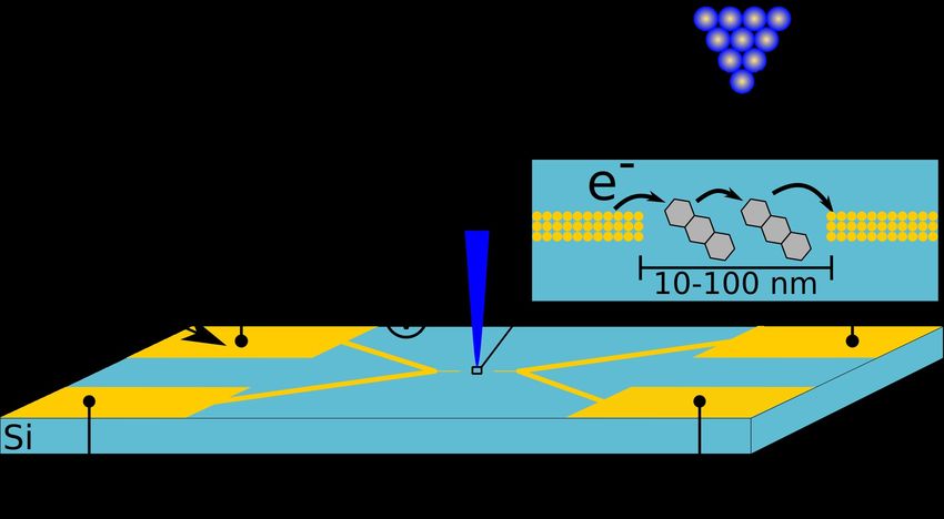
Fritz-Haber-Institut
der Max-Planck-Gesellschaft

 College of Chemistry), Helene Seiler (SNF postdoc mobility grant, Switzerland 2019),
 Samuel Beaulieu (Banting postdoctoral fellowship, Canada 2019), Chris Nicholson
 (Carl-Ramsauer-Award for excellent PhD thesis, Physical Society Berlin 2018),
 Daniela Zahn (Physics Study Award 2018, Physical Society Berlin 2018).

 An important career development for advanced postdocs and junior group lea-
 ders is gaining experience with grant applications and becoming project leader
 with grants from, e.g., German Research Foundation (DFG), European Research
 Council (ERC), or Japanese Science and Technology Agency (JST). In particular,
 establishing an independent junior research group with substantial own funding is
 one major achievement and career step for younger scientists. The department
 has been very successful in this regard as currently six research groups receive
 such independent funding and are hosted by the department: ERC Group of
 Kramer Campen, ERC Group of Ralph Ernstorfer, ERC Group of Tobias Kampfrath,
 DFG Emmy Noether Group of Laurenz Rettig, Max Planck Research Group of Julia
 Stähler, and Max Planck Research Group of Michael Zürch. Four of these group
 leaders have recently been appointed as professors (or received offers). In
 addition, several group leaders are project leaders in DFG funded collaborative
 research centers (Sfb 951, Sfb 1109, TRR 227), the DFG research unit FOR 1700 or
 have received individual research grants funded by DFG, JST, or from the European
 Commission.

 Research profile and structure
 Our research focusses on the dynamics of elementary processes at surfaces,
 interfaces and in solids aiming at a microscopic understanding of the coupling
 between various degrees of freedom (electrons, spins and phonons/vibrations).
 The goal is to obtain mechanistic insights at an atomistic or molecular level into
 various dynamic phenomena like ultrafast phase transitions, excited state dyna-
 mics or molecular processes and (electro-)chemical reactions at interfaces. Our
 strategy is to address these problems from several sides using complementary
 approaches, in particular by the development and application of various time- or
 spatial-resolved spectroscopic techniques dedicated to the specific physical ques-
 tions. Research in the department is performed by small teams with specific, com-
 plementary expertise, creating various synergies and exchange between the
 different groups.

 The current research topics of the department consist of three pillars: (1) Ultrafast
 dynamics of elementary processes and phase transitions in solids with specific
 focus on the coupling between electrons, phonons and spins. These processes are
 probed on their relevant time scales by time-resolved spectroscopy using ultrashort
 laser pulses from THz to XUV and ultrafast electron diffraction (UED). (2) A rather
 recent development is our new focus on localized excitations and transport
 phenomena at the nanoscale. Here we employ scanning probe microscopy to
 investigate plasmonic excitations and light confinement and inelastic scattering in
 STM junctions as well as electronic transport. The increasing emphasis on spatial-
 temporal phenomena combining ultrafast lasers with local scanning probe
 techniques is nicely complemented by ultrafast diffraction methods like UED. (3)
 The third pillar addresses molecular structure of adsorbates, electro-chemical
 reactions and dynamical processes at interfaces and in (molecular) liquids. These
 are probed by nonlinear optical techniques, in particular IR-vis vibrational sum

 4
Department of
 Physical Chemistry

frequency generation and nonlinear THz spectroscopy. Based on these pillars the
department currently supports the following groups:

Time-resolved Dynamics of Solids: Electrons, Phonons and Spins
 • Dynamics of Correlated Materials (Laurenz Rettig)
 • Structural & Electronic Surface Dynamics (Ralph Ernstorfer)
 • Lattice Dynamics (Alexander Paarmann)
 • Terahertz Physics (Tobias Kampfrath)

Localized Excitations and Transport
 • Nanoscale Surface Chemistry (Takashi Kumagai)
 • Ultrafast Scanning Probe Microscopy (Melanie Müller)
 • Quantum Transport & Nanoelectronics (Matthias Koch)

Molecular Processes and their Dynamics
 • Interfacial Molecular Spectroscopy (Kramer Campen)
 • Nonlinear Spectro-Electrochemistry (Yujin Tong)
 • THz Driven Molecular Dynamics (Mohsen Sajadi)

Max-Planck-Research Groups
 • Electron Dynamics (Julia Stähler)
 • Transient X-ray Spectroscopy & Diffraction (Michael Zürch)

This structure reflects a number of new research topics developed over the last
few years, which are often linked with the development of new techniques and in-
strumentation together with the inauguration of new research groups. This
includes: (1) Far-infrared nonlinear optical spectroscopy of solids and photonic
structures of phonon polaritons) at the FHI FEL and development of laser
synchronization at the FEL by Alexander Paarmann. (2) Spectro-electrochemistry of
electrochemical interfaces employing vibrational SFG and dedictated sample cells
by Kramer Campen and Yujin Tong (3) Generation of high field THz pulses to study
THz driven molecular dynamics in liquids and confined environment by Mohsen
Sajadi, (4) Light coupling into a custom designed low-temperature STM for atomic-
scale optical spectroscopy and studies of plasmon-mediated physical and
chemical processes in nanocavities by Takashi Kumagai. (5) Ultrafast THz scanning
probe microscopy developed by Melanie Müller and (6) Electronic transport and
scanning gate microscopy using nanoscale integrated circuits on silicon performed
by Matthias Koch. As several senior group leaders will move onto academic
positions in near future, the number of research groups will consolidate. However,
we will continue with our policy to create new groups headed by young scientists
with their specific profile as pursued in the past.

Furthermore, we also provide lab space for the new experimental setup of Hendrik
Bluhm (FHI Department of Inorganic Chemistry), who proposes challenging surface
science experiments to probe chemical reactions on liquid surfaces (like water)
using high pressure photoemission (HPXPS) and other characterization tools. We
are looking forward to collaborate with him and plan to add an interface-sensitive,
phase-resolved SFG spectroscopy setup to this experiment in a later stage.

 5
Fritz-Haber-Institut
der Max-Planck-Gesellschaft

 Research Highlights of the Department

 The following topics give a selection and comprehensive overview of research
 achievements as well as instrument developments in the department, obtained in
 the last two years. For more details see reports by the individual research groups:

 • Multidimensional time- and angle-resolved photoelectron spectroscopy has
 been developed combining a 500 kHz extreme ultraviolet (XUV) laser source with
 a time-of-flight momentum microscope. Together with novel multidimensional
 data analytics, this development enables the reconstruction of the equilibrium
 and non-equilibrium electronic band structure of solids with exceptional high
 sensitivity and paves the way for future experimental electronic structure
 benchmarking [Rev. Sci. Inst. 90, 023104 (2019)].

 • The sensitivity of the core level spectral function to excitonic excitations in the
 semiconductor WSe2 has been demonstrated using the free-electron laser
 FLASH at DESY in conjunction with a momentum microscope. Based on an
 analytical model for the screening of the core holes by the excited states, the
 dynamics of a Mott transition could be revealed.

 • Photo-induced chemical reactions and phase transitions are typically governed
 by dynamics on a Born-Oppenheimer potential energy surface, which depends
 on the transient electronic structure and its occupation. Using XUV time- and
 angle-resolved photoemission spectroscopy in conjunction with ab-initio mole-
 cular dynamics simulations, a detailed reaction pathway of a structural phase
 transition of In/Si(111) nanowires could be unraveled, revealing a close corres-
 pondence between changes of bonds in real space and the electronic band
 structure [Science 362, 821 (2018) & Phys. Rev. B 99, 155107 (2019)].

 • The microscopic origin of non-equilibrium properties of ZnO was resolved by the
 combination of several complementary time-resolved spectroscopies. Previous
 conflicting results originate from the competition of intrinsic and defect-related
 relaxation dynamics of charge carriers and excitons. This first comprehensive
 picture of quasiparticle dynamics in ZnO offers the possibility of making use of
 the materials' susceptibility to defects that can be exploited to manipulate re-
 laxation pathways on ultrafast timescales. [Structural Dynamics 6, 034501(2019)].

 • Yttrium iron garnet (YIG) is a model insulating ferrimagnet with interesting appli-
 cations in spintronics. The flow of energy and angular momentum between
 electron spins and ionic lattice has been directly probed after ultrafast resonant
 lattice excitation with intense THz pulses. On the 1-ps time scale, spins and
 phonons reach quasi-equilibrium in terms of energy through phonon-induced
 modulation of the exchange interaction. On the much slower, 100-ns scale, the
 excess of spin angular momentum is released to the crystal lattice, resulting in
 full equilibrium [Science Advances 4, eaar5164 (2018)].

 • Spin angular momentum can be also transported out of YIG by just heating an
 adjacent metal layer. The initial steps of this spin Seebeck effect was analyzed
 with
Department of
 Physical Chemistry

 on the same ~100 fs time scale on which the metal electrons thermalize and
 subsequently follows their temperature. This instantaneous response arises
 because the metal spins have a correlation time of only ~4 fs and deflect the
 ferromagnetic moments without inertia. In a first application, we used this
 principle to characterize spin-to-charge-current in various alloys [Nature
 Communications 9, 2899 (2018) & Nano Letters 18, 1064 (2018)].

• Only recently discovered antiferromagnetic metals such as CuMnAs are currently
 in the focus of magnetism research as their magnetic order parameter (the
 staggered magnetization or Neel vector L) can be rotated by 90° and back by
 simply applying an electrical current parallel to L. It has been demonstrated that
 such reversible switching can even be obtained by driving the current with an
 ultrashort terahertz electric-field pulse as stimulus [Science Advances 4, eaar3566
 (2018)].

• Femtosecond time-resolved resonant soft-X-ray diffraction is used to syste-
 matically investigate the ultrafast magnetization dynamics in various members of
 the rare-earth (RE) intermetallics family RERh2Si2. Demagnetization timescales
 varying by almost three orders or magnitude reveal a fundamental dependence
 of the angular momentum transfer rate on the strength of indirect RKKY
 exchange interaction in this prototypical class of antiferromagnets.

• Nanoscale heterostructures show rich structural dynamics in non-equilibrium
 states. Hot-electron induced atomic disordering and ultrafast nanoparticle
 motion has been revealed by femtosecond electron diffraction in Au nano-
 cluster-thin film heterostructures [ACS Nano 12, 7710 (2018) & Nanoscale
 Horizons 4, 1164 (2019)].

• Inelastic light or electron scattering contains information on the momentum
 distributions of fundamental excitations in a material. Employing femtosecond
 electron diffraction, the time-dependent inelastic (diffuse) scattering reveals the
 localization of transient phonon distributions in momentum space arising from
 electron-phonon coupling or photo-induced phase transitions. This new ap-
 proach has been successfully applied to different layered semiconductors and
 semimetals with thicknesses down to monolayers.

• In atomic-scale heterostructures, the macroscopic properties can be strongly
 influenced by the hetero-interfaces, due to the large interface-to-volume ratio.
 For optical phonons in Nitride semiconductor heterostructures, pronounced
 frequency shifts and the emergence of new interface modes have been
 demonstrated providing the possibility to modify the infrared dielectric function
 [ACS Nano 2019, 136, 6730].

• Strong coupling phenomena are ubiquitous in many areas of physics, providing
 hybrid modes with new physical properties. In ultrathin polar crystal films, the
 epsilon-near-zero phonon polariton hybridizes with the polar substrate
 polariton, resulting in a novel kind of hybrid polariton in the mid-infrared. [Nano
 Letters 2018, 18, 4285].

 7
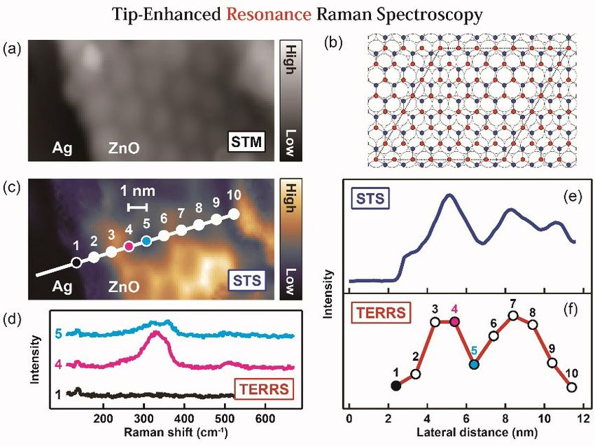
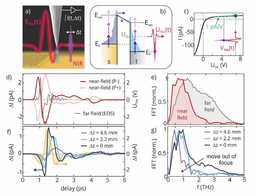
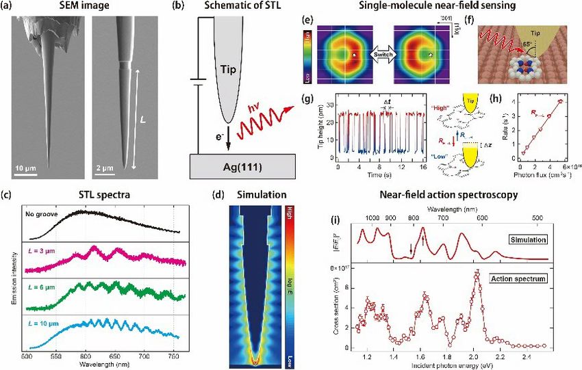
Fritz-Haber-Institut
der Max-Planck-Gesellschaft

 • Scanning probe microscope (SPM) pulling has the advantage that the structure
 of a molecule can be characterized before performing transport measurements.
 The influence of individual defects on the electromechanical properties of single
 graphene nanoribbons was obtained by combining SPM-Pulling experiments
 with atomistic simulations [Phys. Rev. Lett., 121, 047701 2018].

 • Plasmon-assisted resonant electron transfer under cw laser excitation has been
 demonstrated in nanoscale junctions of gold or silver tips and Ag(111). Resonant
 electron tunneling is induced from the tip to field emission resonances of
 Ag(111) through localized surface plasmon excitation in the STM junction [Phys.
 Rev. Lett. 121, 226802 (2018)].

 • Near-field-induced tautomerization in single porpycene molecules on Cu(111) is
 mediated by photogenerated carriers and the reaction cross section is signi-
 ficantly enhanced in the presence of a Au tip through localized surface plasmon
 excitation [Nano Lett. 18, 152 (2018)].

 • Using focused ion beam nanofabrication of gold tips, spectral features of a
 plasmonic STM junction can be manipulated. An exemplary Fabry−Pérot type
 resonator of surface plasmons is demonstrated by producing the tip with a
 single groove on its shaft and the spectral response is characterized by scanning
 tunneling luminescence [Nano Lett. 19, 3597 (2019)].

 • Tip-enhanced resonant Raman scattering (TERRS) was demonstrated on ultra-
 thin zinc oxide layers epitaxially grown on a Ag(111) surface. In this process both
 physical and chemical enhancement mechanisms support efficient TERRS with
 exceptional high (1-nm) spatial resolution arising from local variations of the
 electronic structure [Nano Lett. in press].

 • Multi-angle reflectance measurements on Germanium in the extreme ultraviolet
 are used to recover the complex-valued index of refraction in unprecedented
 resolution. Besides resolving formerly undetected transitions, experimental
 proof is provided that critical angle reflectivity is largely insensitive to the
 electronic structure [J. Opt. Soc. Am. B, 36, 1716 (2019)].

 • Sum frequency generation spectroscopy is interface specific within the electric
 dipole approximation for bulk phases with inversion symmetry. However many
 systems of interest may either have large quadrupole responses in the bulk or
 are non-inversion symmetric. A novel approach to quantitatively separate the
 two contributions from surface and bulk was demonstrated by taking advantage
 of the differing symmetries of bulk and surface contributions and performing
 high accuracy phase resolved SFG measurements. [J. Chem. Phys. 151, 064707
 (2019)].

 • Probing the mechanism of electro-catalytic reactions experimentally requires
 time domain characterization after rapid initiation of a reaction of interest. By
 quantifying photocurrents induced by fs optical pulse excitation we show the
 reactivity of Pt surfaces towards H2 generation requires consideration of both
 ultrafast charge transfer and more slowly evolving electrolyte structural
 dynamics. [ChemElectroChem., 6, 2675 2019].

 8
Department of
 Physical Chemistry

• The potential dependent Stark shift in vibrational line shapes of adsorbates in
 electrochemical systems is typically described as a linear function of applied bias
 where deviations from linearity are often attributed to changes in surface
 structure or electrochemistry. For (bi)sulfate on a Pt(111) electrode SFG spectro-
 scopy reveals such a nonlinear Stark shift. A microscopic model accounting for
 dipole-dipole coupling of adsorbates was developed showing that deviations
 from linearity are likely to occur in the absence of chemical or structural change.
 [Surface Science 678, 78–85 (2018)].

• Ion polarizability is important in understanding adsorption thermodynamics at
 liquid interfaces but has proven challenging to quantify. Using polarization re-
 solved, vibrationally resonant sum frequency spectroscopy the anisotropy of
 perchlorate’s polarizability at the air/water interface has been quantified, pro-
 viding constrains on the anion and solvation shell structure as function of
 surface coverage [Nature Communications 9, 1313 (2018)].

• Low-frequency collective molecular motions in the terahertz (THz) and sub-THz
 frequency range are decisive to the thermodynamic properties and chemical
 reactivity of liquids. It is demonstrated that the microscopic nature of the
 collective molecular dynamics of liquids, as complex as methanol, can be
 revealed by comparing their THz pulse and optical pulse induced Raman
 responses. [J. Phys. Chem. Lett. 9 1279-1283 (2018)].

Outlook

Aiming at spatial-temporal resolution at atomistic length and ultrafast time scales
we will enhance our activities in scanning probe microscopy coupled with local optical
excitation and spectroscopy by several measures (e.g. low temperature operation in
THz STM, fs-laser implementation to a LTSTM or significantly improved spectral re-
solution and sensitivity in TERS). Complementary to such local probes are diffrac-
tion techniques (like ultrafast electron diffraction). Here we plan to implement ad-
vanced detector technology to cover a much higher dynamic range for detection
of very weak diffuse scattering together with the intense Bragg peaks. We also
consider supporting a new junior research group in this area.

A further important direction will be the development of nonlinear vibrational spec-
troscopy and microscopy at the FHI FEL enabling investigations of (Chi(2) and Chi(3))
non-linear optical processes at wavelength not accessible with typical laboratory
sources. In addition we will further develop phase-sensitive SFG spectroscopy and
implement a new laser system. This will enable, e.g., spectroscopy of protons at
liquid interfaces or oxide formation at electrochemical interfaces.

Furthermore we will improve our infrastructure and capabilities for sample pre-
paration techniques for 2 D materials and heterostructures.

 9
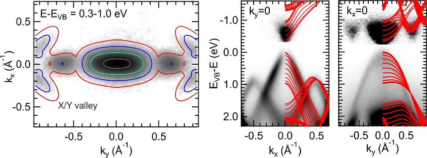
Fritz-Haber-Institut
der Max-Planck-Gesellschaft

 Emmy Noether Group Dynamics of Correlated Materials
 Dr. Laurenz Rettig

 Group Leader, Fritz-Haber-Institut der MPG, Berlin (since 2016)
 Postdoc, Paul Scherrer Institute, Switzerland (2013 - 2015)
 Postdoc, Universität Duisburg-Essen (2012 - 2013)
 Dr. rer. nat., Freie Universität Berlin (2012)
 Diploma in Physics, Freie Universität Berlin (2008)

 Dynamics of Correlated Materials Probed by trARPES and X-ray Diffraction

 The Emmy Noether group Dynamics of Correlated Materials focusses on under-
 standing the ultrafast dynamics of complex materials with many interacting de-
 grees of freedom after optical excitation, and on characterizing photo-induced
 phase transitions and out-of-equilibrium states using momentum-resolved tech-
 niques such as femtosecond time-resolved photoelectron spectroscopy (trARPES)
 or time-resolved (resonant) X-ray diffraction. Using a combination of such tech-
 niques, we aim at understanding the interactions between the various degrees of
 freedom inside such materials (e.g. electrons, spins or lattice), which in complex
 materials often lead to novel ground states with fascinating physical properties.

 Multidimensional photoelectron spectroscopy
 Many of the group’s activities are performed in close collaboration with the ERC
 group of Ralph Ernstorfer, with which we share our high-repetition rate XUV
 trARPES setup. A new electron detector, a momentum microscope (SPECS Metis),
 was installed during the last two years allowing simultaneous and very efficient
 detection of transient electronic band structures in the complete surface Brillouin
 zone (BZ) of most materials. This development of multidimensional photoelectron
 spectroscopy (MPES) enables a completely new approach towards the investi-
 gation of transient electronic band structures and their properties, as well as its
 dynamics, treating three (energy, kx, ky) or even four dimensions (including time) on
 the same footing (see also report of the Ernstorfer group).
 This approach is particularly useful for the investigation of anisotropic electronic

 Figure 1: Time-resolved ARPES data of black phosphorous. Left: Constant energy contour of the con-
 duction band states, showing the “hidden” X/Y valleys close to the BZ boundary. Right: Energy-
 momentum cuts along the armchair (kx) and the zigzag (ky) direction. Overlaid red lines are DFT
 calculations for different kz values.

 10
Department of
 Physical Chemistry

systems, such as e.g. the layered semiconductor black phosphorous (Fig. 1). This
material shows strong in-plane anisotropy associated with a glide-plane symmetry,
leading to a large polarization contrast in the photo-absorption cross-section.
Using MPES, we could identify a new “hidden” Y/X valley in the conduction band
dispersion off the high symmetry directions, which was not observed before, and
which plays an important role for the relaxation of excited carriers. Owing to the
full momentum and energy information obtained, we could carry out a detailed
analysis of the carrier relaxation between the different valleys, and for different
excitation conditions, and reveal a long memory effect of the initial carrier
distribution on the final state, governed by the glide plane symmetry in the system.

Photoinduced phase transitions
The spectroscopic access to the momentum-resolved unoccupied electronic band
structure provided by MPES is also particularly useful to investigate photoinduced
phase transitions (PIPTs) in complex materials, e.g. of changes in the topology of
the electronic structure or from an insulating ground state into a metallic excited
state. A key goal is to reveal the underlying reaction mechanisms and pathways. In
addition, by comparison of the experimental data to electronic band structure
calculations, their validity for predicting excited state band dispersion and poten-
tial deviations e.g. due to many-body phenomena can be assessed.

A fascinating system that we recently studied is the type-II Weyl semimetal Td-
MoTe2, where a strong sensitivity of the γ conduction band pockets on the
electron-electron interaction has been predicted by DFT calculations (Fig 2, c-d),
leading to a Lifshitz transition. Such a scenario can also happen in a photoexcited
semiconductor or semimetal, where the photoexcitation of a large number of free
carriers strongly enhances electronic screening and thereby suppresses electronic
correlations. Indeed, using MPES, we observe the emergence of additional pockets
on the Fermi surface after strong infrared photoexcitation (Fig. 2 b), supporting
this scenario. The strong correlation of the excited state carrier population and the
energy position of the γ pocket provide further evidence, supported by theory.

A model system for a structural phase transition, concomitant to an insulator-
metal transition, are In nanowires on a Si(111) surface. In this system, a detailed
comparison of the unoccupied electronic band structure to DFT and GW calcula-

Figure 2: Time-resolved Fermi surface spectroscopy of MoTe2. Fermi surface of MoTe2 before (a) and
 2
at 500 fs after (b) excitation with 1030 nm pump pulses (0.64 mJ/cm ). DFT+T calculated Fermi
surface with (c) U=3 eV and (d) U=0 eV. Calculations adapted from PRL 121,136401 (2018).

 11
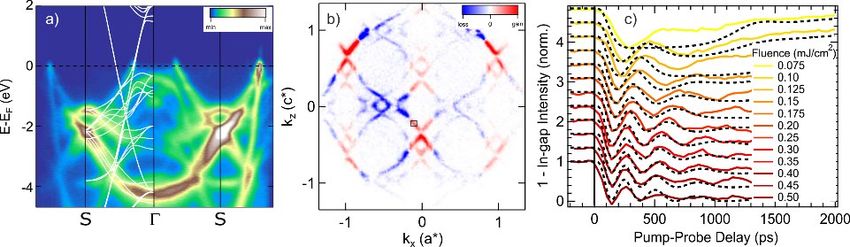
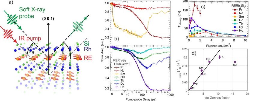
Fritz-Haber-Institut
der Max-Planck-Gesellschaft

 tions revealed a distinct momentum-dependent deviation pointing towards ex-
 citonic contributions in the low temperature phase. Furthermore, the structural
 PIPT has been probed by trARPES and a detailed reaction pathway with several
 distinct timescales of the transition in the electronic structure identified.
 Remarkably, the momentum-resolved dynamics is completely determined by the
 transient electronic temperature after excitation.

 A similar mechanism is present in the prototypical charge density wave (CDW)
 model system TbTe3, where large energy gaps open up in the energy dispersion
 due to a coupled structural and electronic instability. Using the momentum
 microscope, we are now able to investigate the transient electronic fingerprints of
 this transition in a wide energy and momentum range throughout the BZ,
 including the unoccupied band structure (Fig. 3a), which is well described by DFT
 band structure calculations. Upon optical excitation, the CDW distortion is transi-
 ently suppressed, as seen by a large transfer of spectral weight on the Fermi
 surface, and closing of the energy gaps (Fig. 3b). A detailed investigation of the
 fluence dependent dynamics of the spectral weight in the gapped regions yields
 access to the dynamics of the electronic order parameter of the CDW (Fig. 3c).
 Within a time-dependent Ginzburg-Landau model, we can extract the time and
 fluence dependent transient energy potential, which we find to be dominated by
 the evolution of the energy content in the electronic system. In conjunction with
 time-resolved structural dynamics of the CDW superlattice reflections measured
 by time-resolved X-ray diffraction, this model yields a detailed description of the
 dynamics of CDW suppression and recovery.

 Figure 3: Ultrafast CDW dynamics in TbTe3: (a) Band structure along the Γ-S high symmetry direction
 in a wide energy range, alongside with DFT band structure calculations (for YTe3). (b) Differential
 Fermi surface, at 250 fs after excitation with 800nm laser pulses. The transfer of spectral weight
 from the metallic (blue) to the gapped (red) Fermi surface regions indicates the transition into the
 high-temperature state. (c) The coherent dynamics of the spectral weight inside the gap (black box
 in (b)) is well described by a modified time-dependent Ginzburg-Landau model.

 Ultrafast dynamics of antiferromagnetic systems
 The technique of time-resolved resonant soft X-ray diffraction is sensitive to the
 dynamics of magnetic ordering, in particular, of antiferromagnetic (AFM) systems.
 We have performed a series of experiments at the FemtospeX slicing source of
 BESSY II to investigate the ultrafast demagnetization dynamics of prototypical rare-
 earth (RE) intermetallic antiferromagnets, RERh2Si2. In this system, the rare-earth
 ions are enclosed in a fixed crystal lattice of Rh and Si atoms (Fig. 4a), and
 replacing the RE ions changes the electronic and magnetic properties relatively
 little. Therefore, this system is ideally suited to study the influence of the indirect

 12
Department of
 Physical Chemistry

RKKY exchange interaction on the demagnetization dynamics in an antiferro-
magnetic system. The transient diffraction intensity of the (001) magnetic reflec-
tion shows a drastically different behavior for different rare-earth ions in the
system, ranging from a sub-picosecond demagnetization in SmRh2Si2 up to several
100 ps in PrRh2Si2 (Fig. 4b). In addition, we find a pronounced critical slow-down of
the dynamics upon approaching a critical fluence in all materials (Fig. 4c). This
strongly varying demagnetization behavior can be well understood by considering
the transfer rate of angular momentum, and its dependence on the strength of
the RKKY interaction, which we parametrize by the de Gennes factor (g-1)2 J(J+1).
Thereby, our systematic study of AFM demagnetization dynamics in this system
reveals the indirect exchange interaction as the fundamental process responsible
for angular momentum transfer. In addition, in GdRh2Si2, we could identify a co-
herent in-plane rotation of the whole AFM structure after excitation, due to a
transient change of the magnetocrystalline anisotropy.

Figure 4: Time-resolved resonant x-ray diffraction in RERh2Si2. (a) RE atoms are embedded in a fixed
Rh/Si matrix, and always order in a similar AFM structure. Soft x-rays tuned to the RE M4/5 ab-
sorption edges are used to detect the out-of-plane (001) magnetic diffraction peak. (b) Normalized
magnetic diffraction signal for various RERh2Si2 compounds. Lines are biexponential fits. (c)
Extracted slow time constants as a function of excitation fluence. (d) Derived angular momentum
transfer rate as a function of the de Gennes factor.

References

M. Puppin, Y. Deng, C. W. Nicholson, J. Feldl, N. B. M. Schröter, H. Vita,
P. S. Kirchmann, C. Monney, L. Rettig, M. Wolf, and R. Ernstorfer, Time- and angle-
resolved photoemission spectroscopy of solids in the extreme ultraviolet at 500
kHz repetition rate, Rev. Sci. Instr. 90, 023104 (2019).

C. W. Nicholson, A. Lücke, W. G. Schmidt, M. Puppin, L. Rettig, R. Ernstorfer, and
M. Wolf, Beyond the molecular movie: Dynamics of bands and bonds during a
photoinduced phase transition, Science 362, 821-825 (2018).

C. W. Nicholson, M. Puppin, A. Lücke, U. Gerstmann, M. Krenz, W. G. Schmidt,
L. Rettig, R. Ernstorfer, and M. Wolf, Excited-state band mapping and momentum-
resolved ultrafast population dynamics in In/Si(111) nanowires investigated with
XUV-based time- and angle-resolved photoemission spectroscopy,
Phys. Rev. B 99, 155107 (2019).

 13
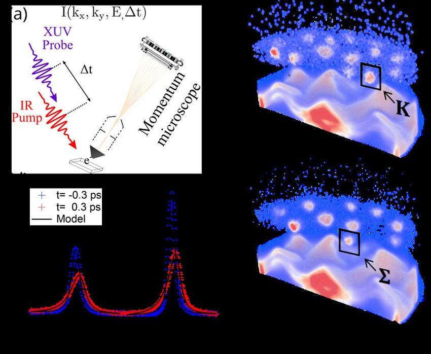
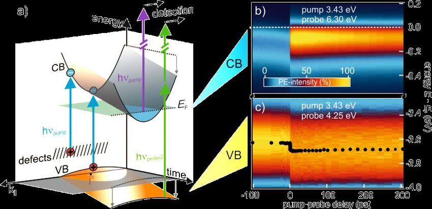
Fritz-Haber-Institut
der Max-Planck-Gesellschaft

 ERC Group Structural & Electronic Surface Dynamics
 Dr. Ralph Ernstorfer

 Group Leader, Department of Physical Chemistry (since 2017)
 ERC CoG project FLATLAND (since 2016)
 Max Planck Research Group, Department of Physical Chemistry (2010-2017)
 Scientist, TU Munich and MPI for Quantum Optics (2007-2010)
 Postdoctoral Fellow, University of Toronto (2005-2007)
 Dr. rer. nat., Freie Universität Berlin (2004)

 Ultrafast Dynamics of Electrons and Lattice in Nanoscale Matter

 The Structural & Electronic Surface Dynamics Group aims for a microscopic under-
 standing of fundamental interactions in nanoscale condensed matter, ultimately
 on the level of individual quantum states. To achieve this, we develop and employ
 experimental techniques providing momentum-resolved information on transient
 states of the electronic structure and the atomic structure. This methodology is
 applied to a broad spectrum of material systems ranging from thin-film (semi-)
 metals and semiconductors to nanoscale heterostructures and nanocrystals. We
 closely collaborate with the Dynamics of Correlated Materials Group headed by
 Laurenz Rettig.

 Development of multidimensional photoemission spectroscopy
 In recent years, we developed time- and angle-resolved photoemission spectro-
 scopy (trARPES) based on a femtosecond extreme ultraviolet (XUV) laser source,
 which provides access to the non-equilibrium electronic structure in the entire
 Brillouin zone of crystalline materials. We extended this approach by adding a
 momentum microscope detector, which enables four-dimensional trARPES as
 depicted in Fig. 1(a). The high repetition rate (500 kHz) of the XUV source provides
 enough statistics for mapping the weak and transient excited state signal in
 addition to the occupied states in the material’s valence band. This is illustrated in
 panels b and c, showing two snapshots from a 3D movie of the formation and
 evolution of excitons in WSe2, a prototypical layered semiconductor. Additional
 examples of use of this approach can be found in the report of the Dynamics of
 Correlated Materials Group.

 The combination of a high-repetition rate light source and an every-electron de-
 tection scheme generates large data volumes. Making such data accessible to a
 broader community, for instance for benchmarking electronic structure calcula-
 tions, represents a new challenge for the photoemission community. Within the
 BiGmax Research Network, we develop unsupervised learning algorithms for
 extracting experimental band structure from multidimensional data sets. We
 share this development with the community by developing a general multi-
 dimensional photoemission data analytics toolbox (https://github.com/mpes-kit).

 14
Department of
 Physical Chemistry

Figure 1: Multidimensional photoemission spectroscopy. (a) Illustration of the trARPES scheme with
a momentum microscope spectrometer, which resolves the photoelectrons in energy, both parallel
momentum dimensions and (pump-probe) time. Panels (b,c) show snapshots of the electronic
structure of WSe2 during the formation of an A exciton in the K valleys of the Brillouin zone (b) and
after scattering of the bright A exciton to optically-dark excitons (c). The electronic contribution to
the dark-exciton wave function is localized in momentum space near the Σ valleys of the conduction
band. (d) Time-resolved spectral function of the W 4f core levels before (blue) and 300 fs after
optical excitation of the A exciton resonance. The solid lines show fits with a time-dependent
Doniach-Sunjic model, which reveals a Mott transition through the temporal evolution of the core-
valence interaction.

Complementary to the study of ultrafast dynamics of valence electrons with
trARPES, we investigated the response of core levels to excitations of the valence
states in WSe2. Using a momentum microscope and the free-electron laser FLASH
at DESY, we probe the spectral function of W 4f core levels during the formation
and evolution of excitons, see Fig. 1(d). The evolution of the spectral function is
well reproduced by an analytic model, which extends the Doniach-Sunjic line
shape model to excited semiconductors. The time-dependence of the spectral
function is governed by the core-valence Coulomb interaction, which differs largely
between excitonic and single-particle excitations of the valence electrons. Core-
level photo-emission spectroscopy proves to be a sensitive tool for the
investigation of electronic phase transitions like, as shown here, a Mott transition
in a photo-excited semiconductor.

Elastic and inelastic femtosecond electron diffraction
Time-resolved diffraction techniques have primarily been employed for retrieving
structural dynamics from elastic scattering signals, which reveal the net effect of all

 15
Fritz-Haber-Institut
der Max-Planck-Gesellschaft

 phonon modes on the atomic motion. Only recently, the analysis of inelastic
 scattering signal has been established as technique for obtaining momentum-
 resolved information on transient phonon populations. We employ this approach
 to study electron-phonon and phonon-phonon interactions as well as phase
 transitions in strongly coupled electron-lattice systems like charge density wave
 materials and certain topologically nontrivial semimetals. Fig. 2 shows exemplary
 data of the anisotropic semiconductor black phosphorous. The diffraction
 difference between excited and ground states shows the momentum distribution
 of the inelastic scattering, which reflects the transient phonon distribution in
 momentum space. After 300 ps, the pattern reflects a hot, yet thermal phonon
 distribution, see panel (b). On the sub-100 ps timescale, however, the inelastic
 scattering reveals nonthermal phonon distributions and highly anisotropic phonon
 population dynamics, as shown for small-momentum phonons in the armchair
 (red) and zigzag (black) crystalline directions, panel (c).

 Figure 2: Femtosecond electron diffraction of a black phosphorous thin film. (a) Raw diffraction
 pattern showing the anisotropic structure in the armchair-zigzag plane. (b) Diffraction difference
 observed 300 ps after optical excitation of the electronic structure. Red signal: increase of star-
 shaped inelastic scattering, which reflects the transient phonon population at this time. The
 anisotropy of the phonon dynamics is retrieved from the momentum-dependent scattering signal,
 e.g., along the armchair (red dots) and zigzag (black dots) directions in the vicinity of elastic
 scattering peaks (see inset). Panel (c) shows the pronounced differences in the phonon dynamics
 along these high-symmetry directions (color code relates to the inset in (b)). These are explained by
 the anisotropy of the electron-phonon scattering phase space, which we independently measured
 with complementary trARPES measurements.

 Ultrafast diffraction and microscopy of nanoscale materials
 The large scattering cross section of nonrelativistic electrons allows for the investi-
 gation of structural dynamics in nanocrystals. We investigated size-selected magic-
 number Au clusters composed of ~920 atoms, which were soft-landed on different
 thin film substrates like graphene, see Fig. 3(a, b). The time-resolved diffraction
 patterns of these nanoscale heterostructures revealed the ultrafast flow of energy
 within each constituent as well as across the boundary and a hot-electron-assisted
 surface-melting mechanism. In the case of Au clusters on graphene, the dynamics
 of ultrafast restricted rotations, i.e., librations, could be extracted from the
 diffraction data. Molecular dynamics simulations reveal efficient coupling between
 flexural phonons of graphene and rotational motion of the cluster as quasi-
 impulsive driving mechanism of the librations.

 16
Department of
 Physical Chemistry

 Figure 3: (a) Illustration of
 librational motion of Au-932
 clusters on graphene. (b) The
 diffraction pattern of this 0D-2D
 heterostructure shows separa-
 ted graphene and Au peaks,
 which allows retrieving ultrafast
 energy flow in the hetero-
 structure. Deviations from
 Debye-Waller dynamics in the
 diffraction data could be identi-
 fied as rotational motion of the
 entire nanocrystals. (c) Electron
 transmission image of free-
 standing graphene recorded
 with 220 eV electrons. A crack in
 the graphene causes single-slit
 scattering and single-electron
 interference (top right and
 lineout profile).

Complementary to the momentum-space technique trARPES, we also aim to
observe ultrafast dynamics in real space with femtosecond electron point-pro-
jection microscopy. This technique is sensitive to the electric potential in the
vicinity of nanostructures; time-resolved experiments hence reveal changes in the
electric potential and allow the reconstruction of the underlying motion of
charges. To enhance the sensitivity of the technique towards the limit of a single
elementary charge, we aim to realize femtosecond electron holography. Im-
provements of the mechanical stability of the microscope and optimization of the
photoemission process from nanotips in terms of source coherence have recently
allowed us to record first images with clear signatures of electron interference, see
Fig. 3.

References

M. Puppin, Y. Deng, C. W. Nicholson, J. Feldl, N. B. M. Schroeter, H. Vita,
P. S. Kirchmann, C. Monney, L. Rettig, M. Wolf, and R. Ernstorfer, Time- and angle-
resolved photoemission spectroscopy of solids in the extreme ultraviolet at
500 kHz repetition rate, Rev. Sci. Inst. 90, 023104 (2019).

T. Vasileiadis, E. N. Skountzos, D. Foster, S. P. Coleman, D. Zahn, F. Krecinic,
V. G. Mavrantzas, R. E. Palmer, and R. Ernstorfer, Ultrafast rotational motions of
supported nanoclusters probed by electron diffraction, Nanoscale Horizons 4,
1164 (2019).

T. Vasileiadis, L. Waldecker, D. Foster, A. Da Silva, R. Bertoni, R. E. Palmer, and
R. Ernstorfer, Ultrafast Heat Flow in Heterostructures of Au Nanoclusters on Thin
Films: Atomic Disorder Induced by Hot Electrons, ACS Nano 12, 7710 (2018).

 17
Fritz-Haber-Institut
der Max-Planck-Gesellschaft

 Lattice Dynamics Group
 Dr. Alexander Paarmann

 Group Leader, Department of Physical Chemistry (since 2014)
 Postdoctoral Fellow, FHI, Department of Physical Chemistry (2010-2013)
 Ph.D., University of Toronto (2010)
 Diploma, TU Berlin (2005)

 Nonlinear Phonon Spectroscopy using Infrared Free-Electron Lasers

 The Lattice Dynamics group uses the FHI infrared Free-Electron Laser (FEL) to
 study optical phonons in polar dielectric crystals, and the associated resonances in
 the nonlinear-optical response of materials in the mid- to far-infrared spectral
 region. Specifically, we are interested in phonon polaritons in hetero- and nano-
 structures, where we use the structure to control the resonances while the
 polaritonic field confinement leads to enhancement of the nonlinear-optical
 signals. For these studies, we develop novel experimental approaches and instru-
 mentation, as for instance FEL-based far-infrared surface-vibrational spectroscopy
 and microscopy. These new methods will, for instance, also be applied to in-
 vestigations of transient structural motifs in electrochemical systems and domain
 structure in correlated oxide heterostructures in the future.

 Phonon polaritons in polar dielectric heterostructures
 Phonon polaritons – phonon-light coupled quasiparticles - arise in thin films or at
 surfaces of polar dielectric crystals due to the infrared-active optical phonons.
 They are supported in the negative permittivity region between transverse and
 longitudinal optical phonon resonances - the so-called Reststrahlen region. The
 long lifetimes of these excitations offer unique advantages for infrared nano-
 photonics over lossy, short-lived plasmon polaritons. However, a major drawback
 arises from the limited, material-specific spectral range over which phonon polari-
 tons are supported, as well as their limited tunability. The Lattice Dynamics group
 follows various approaches to lift these restrictions by means of planar hetero-
 structures, where the polariton modes can be tuned either via polariton-polariton
 coupling between the different layers, or even by phonon hybridization at the
 material interfaces in atomic-scale heterostructures.

 For ultrathin polar dielectric films of 10-100 nm thickness, the phonon polariton
 dispersion is strongly modified, resulting in a unique mode that naturally resides at
 the zero-crossing of the dielectric function (epsilon-near-zero, ENZ). This ENZ con-
 dition has many attractive properties otherwise only accessible through carefully
 designed metamaterials, such as infinite phase velocity, scatter-free propagation,
 and drastically enhanced nonlinear-optical effects. When brought onto a polar
 dielectric substrate, the ENZ polariton in the thin film can hybridize with the
 substrate polariton, to generate strongly coupled modes with unique properties of
 both surface and ENZ polaritons.

 In contrast, a (non-polar) dielectric ultrathin film placed on a polar crystal surface
 results in a compression of the substrate polariton dispersion. This is particularly

 18
Department of
 Physical Chemistry

interesting when using phase change materials like GST as the dielectric, where
this compression can be modulated, see Fig. 1. Additionally, the perpendicularly
polarized waveguide mode was found to, due to the polar crystal substrate, have
very similar (polariton-like) properties, making the GST/polar crystal hetero-
structure an extremely attractive platform for infrared nanophotonics.

Figure 1: Tuning of phonon polariton resonances: (a) Schematic of Otto-type prism coupling ex-
periment that allows mapping of the polariton dispersion in heterostructures. (b) For ultrathin layers
of the phase-change material GST, switching of the of the GST phase results in tremendous tuning
of the GST-compressed surface polariton (p-pol, blue/green) and the newly discovered polariton-like
waveguided mode (s-pol, red/orange).

For heterostructures with ultrathin film thicknesses approaching atomic length
scales, the modified chemical bonding at the layer interfaces additionally leads to
hybridization of the optical phonons, and thereby modulation of the phonon
polaritons. Simultaneously, due to strong anisotropy (in-plane vs. out-of-plane
modes) of the heterostructure, this new material now also supports so-called
hyperbolic polaritons – volume-confined modes with unbound momenta. As such,
atomic-scale heterostructures establish a new way to design materials specifically
for phonon-polariton-based nanophotonics.

Far-Infrared sum-frequency generation spectroscopy and microscopy
Infrared-visible sum-frequency generation (SFG) spectroscopy is a powerful tool to
reveal the structure and symmetry of interfaces. This technique is well established
in the mid-infrared range using table-top laser sources providing access to high-
frequency vibrations. This has led to many major discoveries, for instance regar-
ding the surface structure of liquid and solid water, or the transient interface
structure in electrochemical systems. Using the FHI FEL, the Lattice Dynamics
group is now pushing SFG spectroscopy into the far-infrared range, opening up a
whole new field of exciting possibilities, owing to the vibrational and electronic
excitations in that spectral range which are currently inaccessible. This applies for
instance - but is by far not limited to - most optical phonon modes in the large
class of metal oxides. Therefore, far-infrared SFG spectroscopy could be employed
to study many important problems in, for instance, aqueous oxide chemistry or
strongly correlated oxide heterostructures.

Many interface problems in physics, chemistry, and even biology involve significant
spatial heterogeneity. Therefore, spatially resolved information about the interface

 19
Fritz-Haber-Institut
der Max-Planck-Gesellschaft

 structure and symmetry can tremendously enhance the understanding of under-
 lying mechanisms. Notably, SFG is perfectly suited for infrared microscopy,
 whereby the spatial resolution is limited only by the wavelength of the visible SFG
 photons, i.e., well below the diffraction limit for infrared radiation. Motivated by
 these possibilities, we have started to implement FEL-based far-infrared SFG
 spectroscopy and microscopy.

 Figure 2: Timing tool for the FHI FEL: (a) Concept of balanced optical cross correlation (BOC) used for
 online monitoring of the relative arrival time of visible and FEL pulses. (b) BOC signal when scanning
 the delay time т between FEL and visible laser. (c) The timing distribution function yields a timing
 jitter of ~ 100 fs.

 To enable far-infrared SFG using the FHI FEL as the powerful far-infrared light
 source, a time-synchronized visible laser system is required. The Lattice Dynamics
 group has, over the last 4 years, established the respective instrumentation. A
 radio-frequency timing infrastructure at the FEL facility was installed, which was
 demonstrated to allow synchronization of a low-power fiber laser to the FEL pulses
 with ~100 fs precision (see Fig. 2). We also developed a new concept for and
 implemented a timing tool for online-monitoring of the relative timing between the
 two lasers, a crucial advantage of the current approach over earlier attempts at
 other FEL facilities.

 Using the synchronized low-power visible laser for a proof-of-concept experiment,
 SFG spectroscopy and microscopy measurements were performed, see Fig. 3.
 Here we took advantage of the polaritonic field confinement in phonon polariton
 sub-diffractional resonators that leads to dramatic enhancement of the nonlinear
 signal, resulting in detectable signal levels despite the low visible laser power

 Figure 3: (a) Concept of scanning probe SFG microscopy of nanophotonic structures made of (b)
 Silicon Carbide nanopillars. Experimental resonant (c) and non-resonant (d) SFG images. (e) Concept
 for FEL-based wide-field SFG microscopy to be implemented

 20
Department of
 Physical Chemistry

available. This also allowed for first far-infrared SFG microscopy experiments, here
done with spatially scanning tightly focused beams.

To harvest the full potential of far-infrared SFG spectroscopy and microscopy, the
department applied for and was recently granted financial support by the Max-
Planck-Society to acquire a new high-power visible laser operating in burst mode,
synchronized with the FEL. This new laser will be installed in early 2020, and our
group will implement FEL-based SFG spectroscopy and microscopy as an ex-
perimental end station at the FHI FEL. Notably, the concept also includes to
establish wide-field SFG microscopy. Once operational, this SFG end station will be
used for many different experiments, ranging from infrared nanophotonics to
quantum materials and electrochemistry.

References

N. C. Passler, C. R. Gubbin , T. G., Folland, I. Razdolski, D.S. Katzer, D. F. Storm,
M. Wolf, S. De Liberato, J. D. Caldwell, and A. Paarmann, A. Strong Coupling of
Epsilon-Near-Zero Phonon Polaritons in Polar Dielectric Heterostructures, Nano
Lett., 18, 4285–4292 (2018).

D. C. Ratchford,* C.J. Winta,*, I. Chatzakis, C. T. Ellis, N. C. Passler, J. Winterstein,
P. Dev, I. Razdolski, J. R. Matson, J. R. Nolen, J. G. Tischler, I. Vurgaftman, M. B. Katz,
N. Nepal, M. T. Hardy, J. A. Hachtel, J.-C. Idrobo, T. L. Reinecke, A. J. Giles,
D. S. Katzer, N. D. Bassim, R. M. Stroud, M. Wolf, A. Paarmann*, and J. D. Caldwell*,
Controlling the Infrared Dielectric Function through Atomic-Scale
Heterostructures, ACS Nano 136, 6730-6741 (2019).
(*: equal contributions)

R. Kiessling, W. B. Colson, S. Gewinner, W. Schöllkopf, M. Wolf, and A. Paarmann,
Femtosecond single-shot timing and direct observation of subpulse formation in
an infrared free-electron laser, Phys. Rev. Accel. Beams 21, 080702 (2018).

 21
Fritz-Haber-Institut
der Max-Planck-Gesellschaft

 ERC Group Terahertz Physics
 Prof. Dr. Tobias Kampfrath

 Full professor, Freie Universität Berlin (since 2016)
 Group Leader, FHI, Department of Physical Chemistry (since 2010)
 Postdoctoral Fellow, AMOLF Amsterdam/The Netherlands, University of Konstanz
 and Freie Universität Berlin (2006-2010)
 Ph.D., Freie Universität Berlin (2010)
 Diploma, Universities of Erlangen and Göttingen (2001)

 Ultrafast Terahertz Spectroscopy of Complex Materials

 The terahertz (THz) frequency range is attracting increasing interest for both
 applied and fundamental reasons. On one hand, bit rates in current information
 technology may soon approach the THz range. Therefore, it is warranted to study
 the behavior of materials at THz frequencies. This goal is, on the other hand, also
 highly interesting from a scientific viewpoint because its low photon energy
 (4.1 meV at 1 THz) makes THz radiation an excellent probe of many elementary
 excitations of solids, for instance lattice vibrations (phonons), electronic intraband
 transport, excitons and spin waves (magnons).

 The Terahertz Physics Group makes use of ultrashort THz and optical laser pulses

 • To gain insight into the interplay of low-energy excitations in complex materials.
 An example is the elusive interaction of lattice and electron spins in magnetically
 ordered solids.
 • To push various physical effects so far studied at low frequencies into the THz
 range. Currently, the group has a strong focus on fundamental spintronic effects
 such as the only recently discovered spin Hall effect and the spin-type Seebeck
 effects. These studies are conducted in the framework of the PI’s ERC
 Consolidator Grant project TERAMAG.
 • To develop new spectroscopic tools which permit, for example, control over
 elementary motions such as lattice vibrations and the detection of ultrafast spin
 currents (“ultrafast spin amperemeter”), with interface sensitivity.

 Recent examples of these activities are illustrated below.

 Ultrafast spin currents in magnetic bilayers
 A fundamental operation of spintronic devices is the transport of spin information,
 for example from a magnetic layer into an adjacent nonmagnetic metal (see
 Fig. 1a). Remarkably, such spin current js can be triggered by simply heating the
 metallic layer, even if the magnetic layer is an insulator such as yttrium iron garnet
 (YIG).

 To reveal the possibly ultrafast formation of this spin Seebeck effect in the model
 bilayer system YIG|Pt, we made use of a femtosecond laser pulse to instan-
 taneously heat the Pt film (Fig. 1a). Note that any spin current jc flowing from YIG to
 Pt is converted into a transient transverse charge current jc by the inverse spin Hall

 22
You can also read