AML: WHO CLASSIFICATION, BIOLOGY AND PROGNOSIS - DIMITRI BREEMS, MD, PHD INTERNIST-HEMATOLOOG ZIEKENHUIS NETWERK ANTWERPEN
←
→
Page content transcription
If your browser does not render page correctly, please read the page content below
AML: WHO classification,
biology and prognosis

Dimitri Breems, MD, PhD
Internist-Hematoloog
Ziekenhuis Netwerk Antwerpen
Acute myeloid leukemia
 Clonal expansion of undifferentiated myeloid precursors
 Impaired hematopoiesis and bone marrow failure
 Heterogeneous response to treatment and prognosis

              Löwenberg et al, NEJM, 2011
Acute myeloid leukemia
                Prognosis according age

Age                Complete     Overall survival
                   remission
18 - 60 years       80%         40% at 5 years

>60 years           65%         28% at 2 years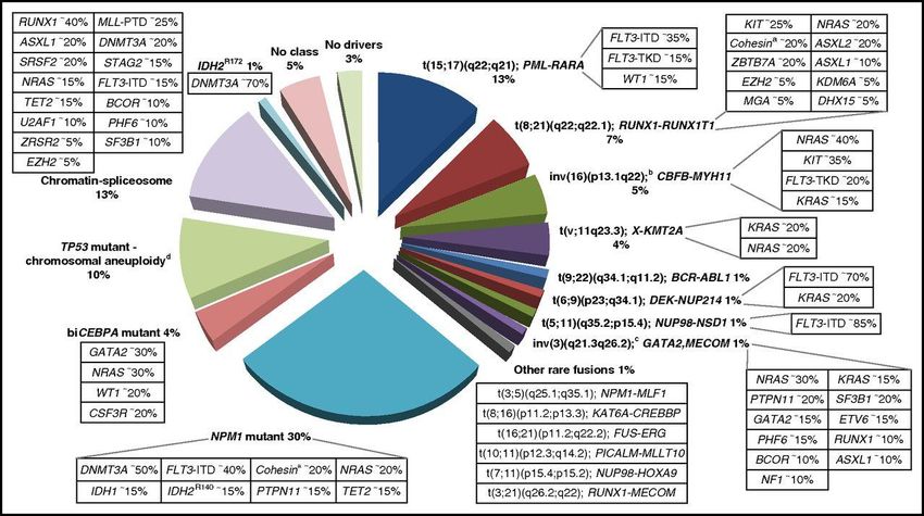
FAB classification of AML

     Bennett et al et al, BJH, 1976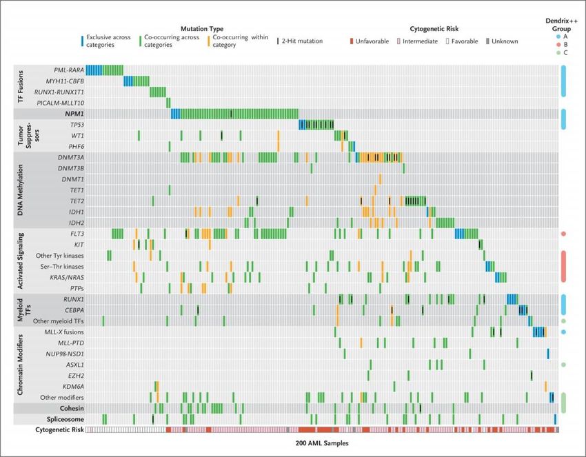
Modern diagnosis of AML
Cytogenetic distribution of AML

                                            7

     Based on Grimwade et al, Blood 1998;
     Grimwade et al, Blood, 2001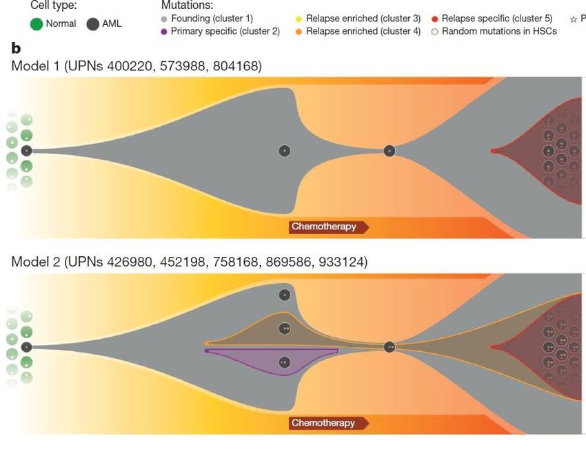
Impact of specific genetic
aberrations on survival in AML

                                    8

       Grimwade et al, Blood 2010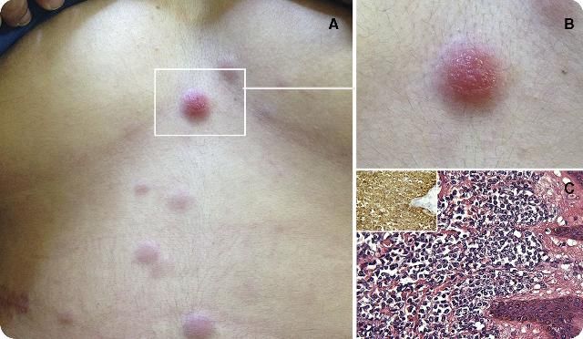
Impact of karyotype complexity on survival for
  AML patients not belonging to favourable
                 subgroups

                                             9

                Grimwade et al, Blood 2010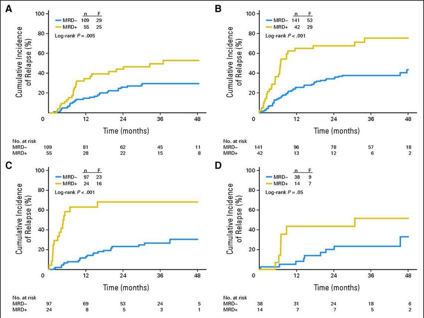
Overall survival in AML patients categorized into
  favourable, intermediate, adverse and very
         adverse cytogenetic risk groups
                        100
 Overall survival (%)

                        75                                 inv(16), t(8;21)
                                                                                          66%

                        50                                normal karyotype, -X, -Y
                                                                                           41%
                                                          other abnormal karyotype

                        25                                                                 26%

                                                          monosomal karyotype*             4%
                                  p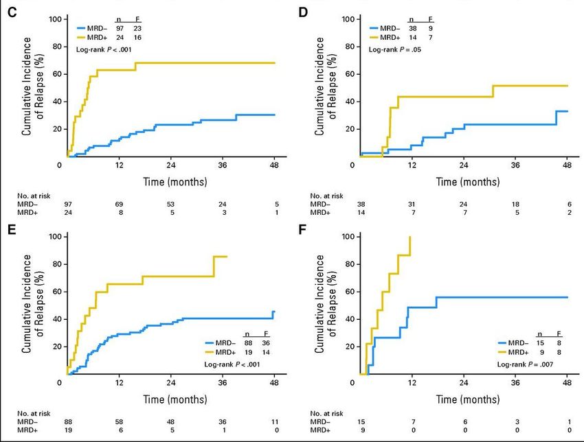
Prognostic value of cytogenetics
in acute myeloid leukemia
Cytogenetic analysis of 1975 patients, 18-60 years
Karyotype Number of Four-year overall
patients (%) survival, % (SE)
Normal, -X, -Y 1001 (51) 41 (2)
inv(16)/t(16;16) 120 (6) 70 (4)
t(8;21) 134 (7) 63 (4)
Abnormal, no 535 (27) 26 (2)
monosomal karyotype
Monosomal karyotype 184 (9) 4 (1)
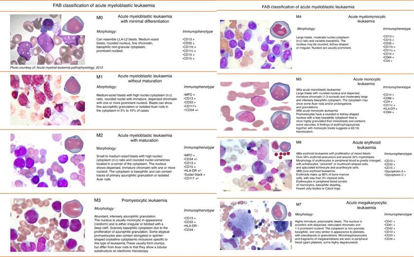
Breems et al. J Clin Oncol 2008Mutational complexity of AML Patel JP et al. N Engl J Med 2012;366:1079-1089
Organization of mutations into categories of related genes

The Cancer Genome Atlas Research Network. N Engl J Med 2013;368:2059-
2074
Comprehensive mutational profiling for risk
  stratification and clinical management of AML.

Patel JP et al. N Engl J Med 2012;366:1079-1089
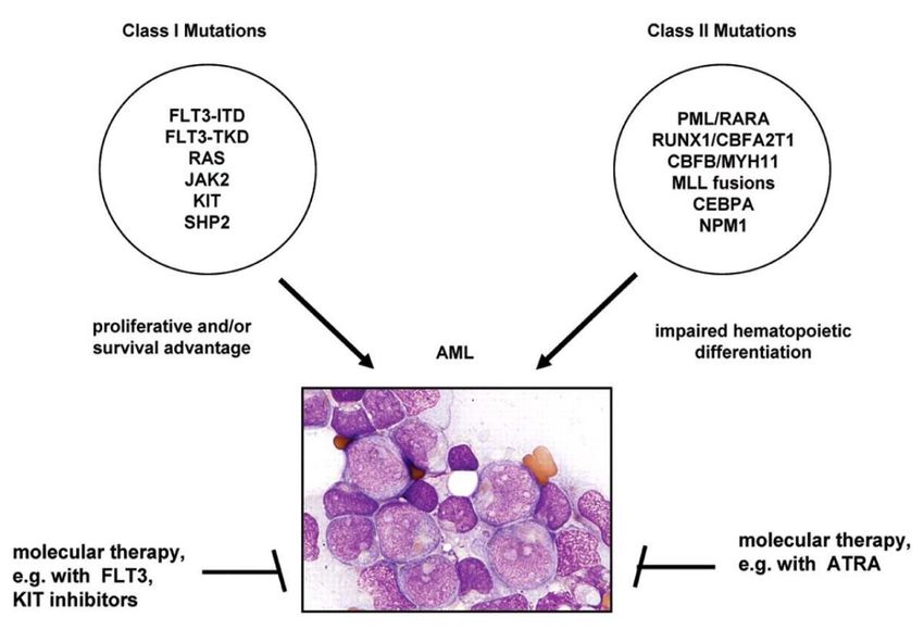
Two cooperating classes of mutations in AML

Adapted from Speck & Gilliland, Nat Rev Cancer. 2002
Evolution of mutations in AML

Welch et al, Cell, 2012
Patterns of relapse in AML

Ding et al, Nature, 2012
WHO classification

Swerdlow et al, Revised 4th Edition, 2017
Contents
Chapter 7: Myeloid neoplasma with germline predisposition

Chapter 8: Acute myeloid leukemia and related precursor
neoplasms

Chapter 9: Blastic plasmacytoid dendritic neoplasm

Chapter 10: Acute leukemias of ambiguous lineage
      Mixed phenotype acute leukemia (MPAL)
Principles WHO classification Integration of all available information Definition, ICD-O Code, Synonyms Epidemiology Clinical features Microscopy Immunophenotype Genetic profile Prognosis and predictive factors
Tests/procedures
For a patient with AML

Tests to establish the diagnosis                Additional tests/procedures at diagnosis (cont'd)

  Complete blood count and differential count     Analysis of comorbidities
  Bone marrow aspirate                            Biochemistry, coagulation tests, urine analysis**
  Bone marrow trephine biopsy*                    Serum pregnancy test††

  Immunophenotyping                               Information on oocyte and sperm cryopreservation‡‡

                                                  Eligibility assessment for allogeneic HCT (including
Genetic analyses
                                                HLA typing)a
  Cytogenetics†                                   Hepatitis A, B, C; HIV-1 testing
                                                  Chest radiograph, 12-lead electrocardiogram, and
  Screening for gene mutations including‡
                                                echocardiography or MUGA (on indication)
     NPM1, CEBPA, RUNX1, FLT3, TP53, ASXL1        Lumbar punctureb
  Screening for gene rearrangements§              Biobankingc

    PML-RARA, CBFB-MYH11, RUNX1-RUNX1T1, BCR-
                                                Sensitive assessment of response by RT-qPCR or MFCd
ABL1, other fusion genes (if available)

                                                  RT-qPCRe,f for NPM1 mutation, CBFB-MYH11, RUNX1-
Additional tests/procedures at diagnosis
                                                RUNX1T1, BCR-ABL1, other fusion genes (if available)d

  Demographics and medical history||              MFCf,g
  Detailed family history¶
  Patient bleeding history#
                                                                     Blood, 2017, Döhner et al.
  Performance status (ECOG/WHO score)
Markers for the diagnosis of AML and MPAL
Expression of cell-surface and cytoplasmic markers
Diagnosis of AML*
Precursors† CD34, CD117, CD33, CD13, HLA-DR
Granulocytic markers‡ CD65, cytoplasmic MPO
Monocytic markers§ CD14, CD36, CD64
Megakaryocytic markers|| CD41 (glycoprotein IIb/IIIa), CD61 (glycoprotein IIIa)
Erythroid markers CD235a (glycophorin A), CD36
Diagnosis of MPAL¶
MPO (flow cytometry, immunohistochemistry, or
cytochemistry) or monocytic differentiation (at least 2 of the
Myeloid lineage
following: nonspecific esterase cytochemistry, CD11c, CD14,
CD64, lysozyme)
Strong# cytoplasmic CD3 (with antibodies to CD3 ε chain) or
T-lineage
surface CD3
Strong# CD19 with at least 1 of the following strongly
expressed: cytoplasmic CD79a, cCD22, or CD10 or weak CD19
B-lineage**
with at least 2 of the following strongly expressed: CD79a,
cCD22, or CD10
Blood, 2017, Döhner et al.8: Acute myeloid leukemia and related precursor neoplasms AML with recurrent genetic abnormalities AML with myelodysplasia-related changes Therapy-related myeloid neoplasms AML not otherwise specified Myeloid sarcoma Myeloid proliferations associated with Down syndrome
AML with recurrent genetic
abnormalities
 AML with t(8;21)(q22;q22); RUNX1-RUNX1T1
 AML with inv(16)(p13.1;1q22) or t(16;16)(p13.1;q22); CBFB-MYH11
 Acut promyelocytic leukemia with PML-RARA     FAB M3
 AML with t(9;11)(p21.3;q23.3); KMT2A-MLLT3
 AML with t(6;9)(p23;q34.1); DEK-NUP214
 AML with inv(3)(q21.3q26.2) or t(3;3)(q21.3;q26.2); GATA2, MECOM
 (=EVI1)
 AML (megakaryoblastic) with t(1;22)(p13.3;q13.1); RBM15-MKL1
 AML with BCR-ABL1
 AML with with gene mutations
   AML with mutated NPM1
   AML with biallelic mutation of CEBPA
   AML with mutated RUNX1
AML with recurrent genetic
abnormalities favorable prognosis
 AML with t(8;21)(q22;q22); RUNX1-RUNX1T1
 AML with inv(16)(p13.1;1q22) or t(16;16)(p13.1;q22); CBFB-MYH11
 Acut promyelocytic leukemia with PML-RARA     FAB M3
 AML with t(9;11)(p21.3;q23.3); KMT2A-MLLT3
 AML with t(6;9)(p23;q34.1); DEK-NUP214
 AML with inv(3)(q21.3q26.2) or t(3;3)(q21.3;q26.2); GATA2, MECOM
 (=EVI1)
 AML (megakaryoblastic) with t(1;22)(p13.3;q13.1); RBM15-MKL1
 AML with BCR-ABL1
 AML with with gene mutations
   AML with mutated NPM1
   AML with biallelic mutation of CEBPA
   AML with mutated RUNX1
AML with recurrent genetic
abnormalities adverse prognosis
 AML with t(8;21)(q22;q22); RUNX1-RUNX1T1
 AML with inv(16)(p13.1;1q22) or t(16;16)(p13.1;q22); CBFB-MYH11
 Acut promyelocytic leukemia with PML-RARA     FAB M3
 AML with t(9;11)(p21.3;q23.3); KMT2A-MLLT3
 AML with t(6;9)(p23;q34.1); DEK-NUP214
 AML with inv(3)(q21.3q26.2) or t(3;3)(q21.3;q26.2); GATA2, MECOM
 (=EVI1)
 AML (megakaryoblastic) with t(1;22)(p13.3;q13.1); RBM15-MKL1
 AML with BCR-ABL1
 AML with with gene mutations
   AML with mutated NPM1
   AML with biallelic mutation of CEBPA
   AML with mutated RUNX1
2017 ELN risk genetic stratification
Risk category*                                 Genetic abnormality
                 t(8;21)(q22;q22.1); RUNX1-RUNX1T1
                 inv(16)(p13.1q22) or t(16;16)(p13.1;q22); CBFB-MYH11
Favorable
                 Mutated NPM1 without FLT3-ITD or with FLT3-ITDlow†
                 Biallelic mutated CEBPA
                 Mutated NPM1 and FLT3-ITDhigh†
                 Wild-type NPM1 without FLT3-ITD or with FLT3-ITDlow† (without adverse-risk
Intermediate     genetic lesions)
                 t(9;11)(p21.3;q23.3); MLLT3-KMT2A‡
                 Cytogenetic abnormalities not classified as favorable or adverse
                 t(6;9)(p23;q34.1); DEK-NUP214
                 t(v;11q23.3); KMT2A rearranged
                 t(9;22)(q34.1;q11.2); BCR-ABL1
                 inv(3)(q21.3q26.2) or t(3;3)(q21.3;q26.2); GATA2,MECOM(EVI1)

Adverse          −5 or del(5q); −7; −17/abn(17p)
                 Complex karyotype,§ monosomal karyotype||
                 Wild-type NPM1 and FLT3-ITDhigh†
                 Mutated RUNX1¶
                 Mutated ASXL1¶
                 Mutated TP53#                                 Blood, 2017, Döhner et al.
AML with myelodysplasia-related
changes
≥ 20% blasts in PB or BM
AND one of the following:
History of MDS or MDS/MPN
Myelodysplasia-related cytogenetic abnormality
Complex karyotype: 3 or more chromosomal abnormalities
Unbalanced abnormalities: -7, del(7q), -5, del(5q), i(17q), t(17q), -13, del(13q),
del(11q), del(12p), t(12p) or idic(X)(q13)
Balanced abnormalities: t(11;16)(q23.3;p13.3), t(3;21)(q26.2;q22.1),
t(1;3)(p36.3;q21.2), t(2;11)(p21;q23.3), t(5;12)(q32;p13.2), t(5;7)(q32;q11.2),
t(5;17)(q32;p13.2), t(5;10)(q32;q21.2) or t(3;5)(q25.3;q35.1)
Multilineage dysplasia: dysplasia in ≥50% of cells in ≥2 myeloid lineages
AND absence of both prior cytotoxic therapy for unrelated
disease and aforementioned recurring genetic abnormalitiesTherapy-related myeloid neoplasms t-AML, t-MDS or t-MDS/MPN Excluded: progression from MPN or evolution of primary MDS or MDS/MPN to AML (secondary AML) Cytotoxic agents implicated in therapy-related myeloid neoplasms Alkylating agents Ionizing radiation therapy Topoisomerase II inhibitors Others
AML not other specified

 AML with minimal differentiation            FAB M0
  MPO negative, CD13+, CD117+, CD33+ (60%)
 AML without maturation                      FAB M1
  >90% blasts of NEC
 AML with maturation                         FAB M2
 Acute myelomonocytic leukemia               FAB M4
 Acute monoblastic/monocytic leukemia FAB M5a/b
 Acute erythroid leukemia                    FAB M6
 Acute megakaryoblastic leukemia             FAB M7
 Acute basophilic leukemia
 Acute panmyelosis with myelofibrosis
Myeloid sarcoma

Tumor mass consisting of myeloid blasts with or without
maturation
Occurring in other anatomical site than bone marrow
Not: Infiltration of any site of the body by myeloid blasts in a
patient with AML
Localization, any site, most frequent:
  Skin, lymph nodes, GI tract, bone, soft tissue, testes
Molecular classes of AML and concurrent gene
      mutations in adult patients ≤65 years

Döhner et al. Blood 2017
Genomic classification and prognosis in AML
11 discrete genetic subsets of AML on the basis of the
expression and coexpression of particular mutations
Papaemmanuil et al, N Engl J Med, Volume 374(23):2209-2221, June 9, 2016Molecular subclassification and
overall survival
• 11 discrete genetic subsets of AML on the basis of the expression and
coexpression of particular mutations.
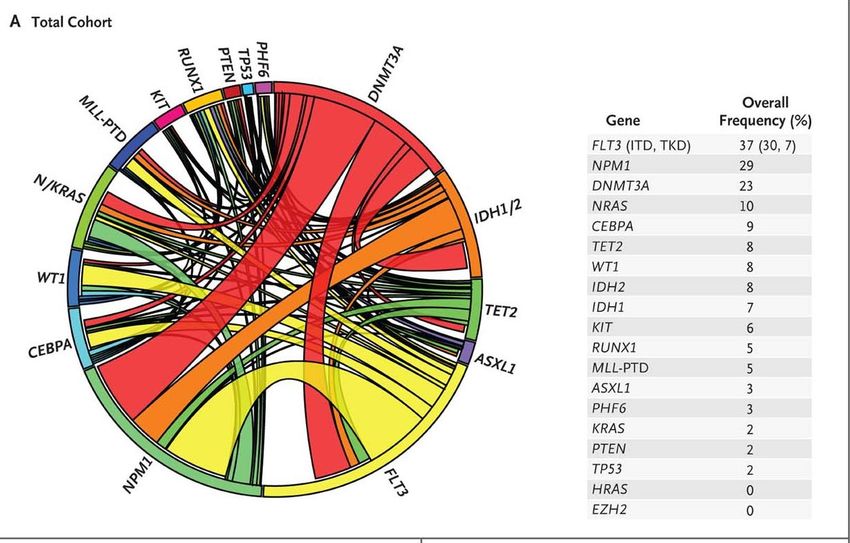
Papaemmanuil et al, N Engl J Med, Volume 374(23):2209-2221, June 9, 2016Proposed genomic classification of AML Papaemmanuil et al, N Engl J Med, Volume 374(23):2209-2221, June 9, 2016
Genomic classification and prognosis
              in AML
    • The driver landscape in AML reveals distinct molecular
      subgroups that reflect discrete paths in the evolution of
      AML, informing disease classification and prognostic
      stratification.
    • Prospective studies may elucidate distinct approaches to
      their management.

Papaemmanuil et al, N Engl J Med, Volume 374(23):2209-2221, June 9, 2016
Prognostic value of minimal residual disease
detection in AML with flowcytometry
517 AML patients, 18-60 years
85% of all AMLs:
Leukemia-associated phenotype by immunoflow cytometry
is determined at diagnosis
Minimal residual disease assessment in complete
remission:
After chemotherapy induction cycle 1
After chemotherapy cycle 2
After consolidation treatment
Terwijn et al. J Clin Oncol 2013Relapse incidence by minimal residual disease
A: After chemotherapy induction cycle 1
B: After chemotherapy cycle 2
C: After consolidation treatmentRelapse incidence by minimal residual disease After chemotherapy cycle 2 D: Good risk C: Intermediate risk F: Poor risk
Literature AML
 Diagnosis and management of AML in adult: 2017 ELN
 recommendations from an international expert panel. Döhner
 H et al. Blood. 2017;129(4):424-447.
You can also read