An exonic insertion in the NAGLU gene causing Mucopolysaccharidosis iiiB in Schipperke dogs - Nature
←
→
Page content transcription
If your browser does not render page correctly, please read the page content below
www.nature.com/scientificreports

            OPEN           An exonic insertion in the NAGLU
                           gene causing Mucopolysaccharidosis
                           IIIB in Schipperke dogs
                           Karthik Raj1, N. Matthew Ellinwood1,2 & Urs Giger1*
                           Mucopolysaccharidosis (MPS) IIIB (Sanfilippo syndrome B; OMIM 252920), is a lysosomal
                           storage disease with progressive neurological signs caused by deficient activity of alpha-N-
                           acetylglucosaminidase (NAGLU, EC 3.2.1.50). Herein we report the causative variant in the NAGLU
                           gene in Schipperke dogs and a genotyping survey in the breed. All six exons and adjacent regions of
                           the NAGLU gene were sequenced from six healthy appearing and three affected Schipperkes. DNA
                           fragment length and TaqMan assays were used to genotype privately owned Schipperkes. A single
                           variant was found in exon 6 of MPS IIIB affected Schipperkes: an insertion consisting of a 40–70 bp
                           poly-A and an 11 bp duplication of the exonic region preceding the poly-A (XM_548088.6:c.2110_21
                           11ins[A(40_70);2100_2110]) is predicted to insert a stretch of 13 or more lysines followed by either
                           an in-frame insertion of a repeat of the four amino acids preceding the lysines, or a frameshift. The
                           clinically affected Schipperkes were homozygous for this insertion, and the sequenced healthy dogs
                           were either heterozygous or homozygous for the wild-type allele. From 2003–2019, 3219 Schipperkes
                           were genotyped. Of these, 1.5% were homozygous for this insertion and found to be clinically affected,
                           and 23.6% were heterozygous for the insertion and were clinically healthy, the remaining 74.9% were
                           homozygous for the wild-type and were also clinically healthy. The number of dogs homozygous and
                           heterozygous for the insertion declined rapidly after the initial years of genotyping, documenting the
                           benefit of a DNA screening program in a breed with a small gene pool. In conclusion, a causative NAGLU
                           variant in Schipperke dogs with MPS IIIB was identified and was found at high frequency in the breed.
                           Through genotyping and informed breeding practices, the prevalence of canine MPS IIIB has been
                           drastically reduced in the Schipperke population worldwide.

                           The mucopolysaccharidoses (MPS) are a group of hereditary lysosomal storage disorders in which specific gly-
                           cosaminoglycans accumulate in lysosomes due to various enzyme deficiencies. There are up to 12 individual MPS
                           types described in humans1 and animals2 with all but MPS II showing autosomal recessive inheritance. Genetic
                           variants in dogs have been identified in the genes associated with MPS I3, IIIA4,5, VI6,7, and VII8,9. Clinical man-
                           ifestations of MPS in dogs are most commonly ocular and musculoskeletal2. In contrast, MPS III, also known as
                           Sanfilippo Syndrome, causes a progressive and primarily neurological disease4,5,10. At the cellular level it is charac-
                           terized by primary lysosomal accumulation of heparan sulfate and secondary lysosomal storage of gangliosides1.
                               Mucopolysaccharidosis IIIB (also known as Sanfilippo syndrome B) is caused by variants in the
                           alpha-N-acetylglucosaminidase (NAGLU) gene and has been previously characterized at the clinical and molecu-
                           lar level in humans, emus11, cattle12, knockout mice13, and transgenic swine14,15. Furthermore, MPS IIIB has been
                           clinicopathologically reported in Schipperke dogs (OMIA 001342-9615)10. At approximately two years of age,
                           affected Schipperkes develop a slowly progressive ataxia leading to humane euthanasia before six years of age10.
                           This study documents the causative NAGLU gene variant in Schipperke dogs with MPS IIIB and also demon-
                           strates the high initial frequency of this allele in the breed, and its reduction after inauguration of genotype
                           screening for this variant allele in the breed.

                           1
                            Section of Medical Genetics (PennGen), School of Veterinary Medicine, University of Pennsylvania, Philadelphia,
                           PA, 19104-6010, USA. 2Present address: College of Agriculture and Life Sciences, Iowa State University, Ames, Iowa,
                           USA. *email: giger@upenn.edu

Scientific Reports |   (2020) 10:3170 | https://doi.org/10.1038/s41598-020-60121-3                                                              1
www.nature.com/scientificreports/ www.nature.com/scientificreports
Exon length Amplicon Annealing
Exon # (bp) Forward Primer Reverse Primer Length (bp) Temperature (°C)
1 424 ATGTGAAAGCTCTCCAGGTACA CGATGTCACCGTTTCCATTCTTC 925 66
2 148 GTGAGTCCTGGAGTGAAACAGT TAGCGTTTCTAGTGAGGTGCTG 485 65
147 (Exon 3)
3–4 TTGCAACAAAGCTGACCCATTAG GCTGCCATTTGCTAAGACTGTG 798 67
86 (Exon 4)
5 257 CACTGCTCCATCTAGGACTCTG AGTGCTTGGTCAACTGTCAAGG 682 67
6a GACAACACTGCCCTAGAGATCC CCTCGCCTCCACATAGTACAAG 953 67
1450
6b ATGGTTACCACTGTCTGGTACA AAACGTATTGGGAGAGGATTCCC 1043 65
Genotyping GCATTCCCTTCCAACAGCACCAGT GCCCACAAGGAGCCAGCCACCAAT 169 68
Table 1. Primers used for amplifying and Sanger sequencing exonic and flanking regions of canine NAGLU
gene and for genotyping the causative insertion and wild-type alleles in Schipperkes. Primer pairs 6a and 6b
were designed to amplify and sequence exon 6 to make it across this relatively long exon.
Methods
Ethylenediaminetetraacetic acid (EDTA) anticoagulated blood and cheek swab/brush samples were sent for the
diagnosis of MPS IIIB or genotyping to PennGen Laboratories at the University of Pennsylvania, Philadelphia,
Pennsylvania USA from clinically affected and healthy-appearing Schipperke dogs. Affected status was based on
the reported onset of severe and progressive ataxia with onset at two to three years of age based on communica-
tion from owners or referring veterinarians. The studies were approved by the Institutional Animal Care and Use
Committee (IACUC) of the University of Pennsylvania.
Briefly, DNA was extracted from EDTA blood and cheek swab/brush samples using the QIAamp DNA Blood
Mini Kit (Qiagen, Hilden, Germany). The six exons of the NAGLU gene were sequenced from nine Schipperkes
including six healthy appearing and three clinically affected with MPS IIIB. Primers were designed, based on the
published canine reference sequences (CanFam3.1 and XM_548088.6) to cover all six exons and at least 90 bp of
intronic sequence adjacent to the exons (Table 1). The PCR-amplified products (amplified using KOD Xtreme Hot
Start DNA Polymerase, EMD Millipore Corp., Billerica, MA, USA) were submitted for direct Sanger sequenc-
ing at the University of Pennsylvania’s Sequencing Core Facility. The DNA sequences were then aligned to dog
reference sequences (CanFam3.1 and XM_548088.6) to find genetic variants in the exons and splice-sites of the
NAGLU gene.
Sanger sequencing was unable to accurately determine the exact sequence of the long homopolymer poly-A
insertion in the affected dogs (data not shown). Consequently, the insert sizes were estimated based upon either
gel separation or Sanger sequencing. In addition, for genotyping, a specific primer pair (Table 1) was designed
to amplify the region surrounding the exon 6 insertion, with subsequent PCR products subjected to fragment
length analysis using electrophoresis on a 6% polyacrylamide gel. The fragments’ size, either consistent with
the mutant and/or wild-type alleles, was the basis for genotyping. To control for any potential allelic dropout
issues, samples were separately tested twice. A TaqMan genotyping assay was subsequently developed: briefly,
a VIC dye-labelled probe (TGACAAGAATGCCTTCCAGCT) was designed to anneal to the insertion site of
wild-type allele and a FAM dye-labelled probe (CAAGAATGCCTTCCAAAAA) to a unique sequence of the
variant allele with the insertion and the primers used for the amplification are CTGGGTGCCGAAGATAAAGGT
and CCCTTCCAACAGCACCAGTT. Genotyping data for MPS IIIB in the Schipperke pet population based on
samples submitted to the PennGen Laboratories from 2003 to 2019 was gathered and analyzed.
Results
When aligning Sanger sequencing data of the exonic and surrounding intronic regions of the NAGLU gene from
clinically healthy Schipperkes and those Schipperkes affected with MPS IIIB to the canine reference sequences
(CanFam3.1 and XM_548088.6), only one single variant was discovered. The sixth and last exon of the NAGLU
gene contains an insertion (XM_548088.6:c.2110_2111ins[A(40_70);2100_2110]) comprised of a homopolymer
of A residues (poly-A) and an 11 bp duplication of the sequence directly upstream of the poly-A. Three clinically
affected dogs were found to be homozygous for this insertion, four clinically healthy dogs were heterozygous,
and two clinically healthy dogs were homozygous for the wild-type allele. The poly-A region was at least 40 bp in
length, however the exact length of the poly-A in the insert could not be determined from the Sanger sequencing
data as the sequencing read quality decreased, presumably due to variation between the two allele insert sizes,
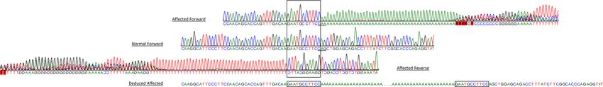
and/or due to “slippage” of the polymerases during amplification or sequencing (Fig. 1).
The primer pair designed to be used for genotyping by gel electrophoresis amplified a wild-type fragment pre-
dicted to be 169 bp and a longer fragment for the variant allele with the insertion (Fig. 2). The fragments with the
insertion were at least 50 bp longer than the wild-type fragment, but the insert lengths varied markedly between
individual Schipperkes, ranging between 50–80 bp.
From 2003 to 2019, a total of 3,219 Schipperkes were genotyped at PennGen Laboratories. Of the total number
genotyped 2,411 (74.9%) Schipperkes were homozygous for the wild-type allele, 760 (23.6%) were heterozygous,
and 48 (1.5%) were homozygous for the insertion (Table 2 and Fig. 3). All Schipperkes homozygous for the vari-
ant had or developed clinical signs of MPS IIIB unless lost to follow up before reaching the age of onset of clinical
signs (≥2 years). As this was a genotyping survey, there was no means to follow cases closely. Of the 48 animals
tested as homozygous for the mutation, 54.2% (n = 26) were of an age where they were definitively displaying
clinical signs of MPS IIIB. The remaining animals were younger than the extreme limit for onset of signs (3 years
Scientific Reports | (2020) 10:3170 | https://doi.org/10.1038/s41598-020-60121-3 2www.nature.com/scientificreports/ www.nature.com/scientificreports
Figure 1. Sanger sequencing chromatograms of a region in exon 6 of NAGLU gene from a Schipperke with
MPS IIIB (forward and reverse) compared to the normal canine sequence (forward). The affected reverse
sequence is flipped horizontally to align to the normal. The sequence traces show the poly-A/poly-T and loss of
sequence quality, while trying to make it past the poly-A/poly-T insertion. An 11 bp region (boxed) is seen in
both the forward and reverse sequences before the poly-A and poly-T respectively, demonstrating a duplication.
The 704th codon in the normal forward (CAG) and in the affected forward (CAA) at the junction of the
insertion are underlined, it codes for glutamine in both (a synonymous variant). The affected sequence flanking
the insertion deduced from the forward and reverse sequences is shown at the bottom of the figure.
Figure 2. Polyacrylamide gel (6%) electrophoresis of the amplified fragments to detect the NAGLU insertion in
Schipperkes with and without MPS IIIB. M is 100 bp marker, 1–4 heterozygotes, 5–7 are homozygous dogs for
the insertion, 8 and 9 homozygous wild-type/normal, and B blank/negative control. Notable is the considerable
variation in insert size in different individuals. This is a cropped gel image.
of age). Of these dogs (n = 22), none were subsequently reported as not developing disease, but may well have
been euthanized before the expected age of disease onset.
The number of Schipperke samples submitted for genotyping rapidly and drastically declined from the first
years of screening. Similarly, the number of samples from Schipperkes that were genotyped as homo- and hete-
rozygous for the insertion declined. However, while in absolute numbers, the mutant allele numbers decreased
strikingly, the frequency of the allele in submitted samples did not decrease per year, and there have even been
recent carriers and (rarely) affected dogs identified (Table 2 and Fig. 3). Screening of Schipperkes from North
America, Europe, Australasia, and Russia revealed carrier dogs in all these regions, indicating the worldwide
distribution of the mutant allele (data not shown).
Discussion
The canine NAGLU gene is on chromosome 9 and comprised of six exons (XM_548088.6) with exon 6 being by
far the longest (1450 bp). It codes for the lysosomal acid hydrolase alpha-N-acetylglucosaminidase (EC 3.2.1.50)
which consists of 747 amino acids (XP_548088.2), including the signal sequence. The exonic NAGLU sequence
from all nine dogs sequenced was identical to the reference genomic sequence except for the disease-associated
variant. The protein sequence shows close homology to human (86% identity) and other mammalian (76–90%)
sequences which is expected for a housekeeping gene (https://www.ncbi.nlm.nih.gov/homologene/222).
Schipperkes with MPS IIIB have a NAGLU insertion near the end of exon 6, which contains a poly-A insertion
followed by a duplication of the preceding 11 bp of wild-type sequence. The sequence of this 11 base pair repeat
in the native context is flanked by AA (two adenines) at the 5′ end and A (one adenine) at the 3′ end. Since the
insertion is a poly-A sequence, and the molecular mechanism of the sequence repetition is not known, it could
actually have been a 14 bp repeat of the native sequence. We have chosen the conservative assessment that the A
residues at both ends of the insertion were part of an exogenous poly-A insert. The insertion is predicted to result
in the addition of many lysines after the 704th amino acid (a glutamine that results from a synonymous variant
caused by the insertion) in the NAGLU protein with three potential consequences past the stretch of inserted
lysines depending on the actual length of the poly-A insertion: (1) It stays in-frame with an insertion of a repeat of
the four native amino acids preceding the lysines (asparagine, alanine, phenylalanine, glutamine), or (2) it causes
a frameshift with an early stop-codon, or (3) frameshift with the lack of a stop-codon. In any case, the exonic
insertion is predicted to disrupt the C-terminal end of the enzyme in affected dogs. We had shown the lack of
NAGLU enzyme activity and lysosomal storage in affected dogs, but neither immunoblotting nor gene expression
studies were performed to further confirm the disruptive nature of this genetic variant.
A review of the human NAGLU gene sequence in ClinVar (https://www.ncbi.nlm.nih.gov/clinvar) accessed
on (October 14, 2019) contains 181 variants. Of the 77 that are labelled as pathogenic or likely pathogenic, 42 are
in exon 6. However, only one (c.2116C>T, p.Gln706Ter) is near the location of the insertion seen in MPS IIIB
Scientific Reports | (2020) 10:3170 | https://doi.org/10.1038/s41598-020-60121-3 3www.nature.com/scientificreports/ www.nature.com/scientificreports
Gender Year Tested
All
Genotype Dogs Female Male NA* 2003 2004 2005 2006 2007 2008 2009 2010 2011 2012 2013 2014 2015 2016 2017 2018 2019
Affected 48 12 28 8 22 11 0 0 1 1 1 2 1 1 4 1 2 0 0 1 0
Carrier 760 391 343 26 234 86 60 72 34 49 22 39 17 45 20 26 17 17 8 7 7
Normal 2411 1290 1092 29 841 200 142 228 153 111 78 77 64 93 81 67 65 42 64 53 52
Total 3219 1693 1463 63 1097 297 202 300 188 161 101 118 82 139 105 94 84 59 72 61 59
Table 2. Genotyping results of Schipperkes for the exonic insertion in NAGLU gene from 2003–2019. *NA is
not available.
1000
100
Number of dogs (log)
10
1
0.1
2003 '04 '05 '06 '07 '08 '09 '10 '11 '12 '13 '14 '15 '16 '17 '18 '19
Years tested
Affected Carrier Normal
Figure 3. Survey of Schipperkes for the exonic insertion in NAGLU gene from 2003–2019. Note this is a semi-
log chart.
Schipperkes. The c.2116C>T variant was reported in a 6 year old female child with severe degenerative neurop-
athy due to MPS IIIB16.
Interestingly, there are several disease-causing poly-A insertions known in dogs that have the same pattern
of a poly-A flanked by a duplicated/repeated native sequence at both ends17–21. Such inserts with characteristic
repeats may likely be the result of a target primed reverse transcription mechanism22. Some are also known to
exhibit varied length of their poly-A, for example, the FXI variant in the Kerry Blue Terriers with Factor XI defi-
ciency17. In cattle and emus with MPS IIIB, the disease-causing NAGLU variants are a missense (c.1354G>A,
p.Glu452Lys) and frameshift deletion (c.1098_1099delGG), respectively, and both are also located in exon 6.
Occasionally when genotyping heterozygotes by fragment length, the amplification preferentially produced
the smaller wild-type amplicon and failed to amplify the larger fragment, resulting in allelic dropout in hete-
rozygotes. This did not appear to be a factor in the homozygous affected dogs. This preferential amplification in
rare circumstances could have led to misidentification of heterozygotes as homozygotes for the wild-type allele.
A cause was not identified and analyzing the samples in separate assays eliminated the allelic dropout issue. The
TaqMan genotyping assay clearly discriminated all three genotypes, and this technique was not affected by any
apparent allelic dropout artifact.
Based upon the devastating progressive clinical course of canine MPS IIIB in Schipperkes, breeders, owners,
and veterinary clinicians were eager to genotype their dogs and patients. And while this represents a biased
population within the breed, a striking number of homozygous and heterozygous dogs for the mutant allele were
identified. The survey of NAGLU variant genotyping results in Schipperkes shows characteristic dynamics for a
canine breed with a small gene pool. According to the UK Kennel Club only ≤51 (https://www.thekennelclub.
org.uk/media/129029/10yrstatsutility.pdf) puppies were registered each year, from 2009 to 2018. Before the dis-
ease was discovered at the turn of the century and the molecular basis was established, no clinical screening test
for MPS IIIB carrier dogs was available, and the prevalence of the mutant allele was not known. Following the
initiation of genotyping in 2003, about one third of 3,219 Schipperkes whose genotypes are presented herein were
tested in the first year of screening and another one third during the following five years. The last third of dogs
were genotyped during next 11 years withwww.nature.com/scientificreports/ www.nature.com/scientificreports
and/or close inbreeding affecting the Hardy-Weinberg equilibrium. Indeed a potential founder individual up
to eight generations deep was found in the pedigree, with six separate lines of descent, which was further com-
pounded by multiple lines of decent from two intermediate animals10. While a specific popular sire/dam was not
identified through testing, the mutant allele was widespread in the breeding population worldwide.
Our genotyping was in complete concordance with phenotype, except for those dogs too young (www.nature.com/scientificreports/ www.nature.com/scientificreports
20. Turba, M. E., Loechel, R., Rombolà, E., Gandini, G. & Gentilini, F. Evidence of a genomic insertion in intron 2 of SOD1 causing
allelic drop-out during routine diagnostic testing for canine degenerative myelopathy. Animal Genetics 48, 365–368 (2017).
21. Lit, L., Belanger, J. M., Boehm, D., Lybarger, N. & Oberbauer, A. M. Differences in Behavior and Activity Associated with a Poly(A)
Expansion in the Dopamine Transporter in Belgian Malinois. PloS one 8, e82948 (2013).
22. Ostertag, E. M. & Kazazian, H. H. Jr. Biology of mammalian L1 retrotransposons. Annual Review of Genetics 35, 501–538 (2001).
23. Fieten, H. et al. The Menkes and Wilson disease genes counteract in copper toxicosis in Labrador retrievers: a new canine model for
copper-metabolism disorders. Disease models & mechanisms 9, 25–38 (2016).
24. Kijas, J. M. H. et al. A Missense Mutation in the β-2 Integrin Gene (ITGB2) Causes Canine Leukocyte Adhesion Deficiency.
Genomics 61, 101–107 (1999).
25. Bhalerao, D. P., Rajpurohit, Y., Vite, C. H. & Giger, U. Detection of a genetic mutation for myotonia congenita among Miniature
Schnauzers and identification of a common carrier ancestor. American journal of veterinary research 63, 1443–1447 (2002).
Acknowledgements
The studies were in part supported by grants from the National MPS Society, the AKC Canine Health Foundation,
and NIH grants (RR002512 and OD010939). We thank Mr. Adam Seng for genotyping dogs. We also wish to
acknowledge the many Schipperke owners and breeders and the Schipperke breed associations for encouraging
the screening of their dogs. We acknowledge Mark E. Haskins and Paula S. Henthorn for the original funding and
helpful discussions.
Author contributions
K.R., M.E. and U.G. designed and did most of the studies. K.R. and U.G. wrote most of the manuscript. All
authors reviewed and revised the manuscript.
Competing interests
K.R., M.E. and U.G. are/were members of the not-for-profit PennGen Laboratories, which offer metabolic and
DNA testing for mucopolysaccharidosis and other hereditary diseases and inborn errors of metabolism.
Additional information
Supplementary information is available for this paper at https://doi.org/10.1038/s41598-020-60121-3.
Correspondence and requests for materials should be addressed to U.G.
Reprints and permissions information is available at www.nature.com/reprints.
Publisher’s note Springer Nature remains neutral with regard to jurisdictional claims in published maps and
institutional affiliations.
Open Access This article is licensed under a Creative Commons Attribution 4.0 International
License, which permits use, sharing, adaptation, distribution and reproduction in any medium or
format, as long as you give appropriate credit to the original author(s) and the source, provide a link to the Cre-
ative Commons license, and indicate if changes were made. The images or other third party material in this
article are included in the article’s Creative Commons license, unless indicated otherwise in a credit line to the
material. If material is not included in the article’s Creative Commons license and your intended use is not per-
mitted by statutory regulation or exceeds the permitted use, you will need to obtain permission directly from the
copyright holder. To view a copy of this license, visit http://creativecommons.org/licenses/by/4.0/.
© The Author(s) 2020
Scientific Reports | (2020) 10:3170 | https://doi.org/10.1038/s41598-020-60121-3 6You can also read