Mapping Immunodominant Antibody Epitopes of Abrin - MDPI
←
→
Page content transcription
If your browser does not render page correctly, please read the page content below
antibodies
Article
Mapping Immunodominant Antibody Epitopes
of Abrin
Ron Alcalay 1 , Reut Falach 1 , Yoav Gal 1 , Anita Sapoznikov 1 , Tamar Sabo 1 , Chanoch Kronman 1
and Ohad Mazor 2, *
 1    Department of Biochemistry and Molecular Genetics, Israel Institute for Biological Research,
      Ness-Ziona 76100, Israel; rona@iibr.gov.il (R.A.); reutf@iibr.gov.il (R.F.); yoavg@iibr.gov.il (Y.G.);
      anitas@iibr.gov.il (A.S.); tamars@iibr.gov.il (T.S.); chanochk@iibr.gov.il (C.K.)
 2    Department of Infectious Diseases, Israel Institute for Biological Research, Ness-Ziona 76100, Israel
 *    Correspondence: ohadm@iibr.gov.il; Tel.: +972-8-9385862; Fax: +972-8-9381544

 Received: 23 February 2020; Accepted: 22 April 2020; Published: 27 April 2020

 Abstract: Abrin, a toxin isolated from the seeds of Abrus precatorius (jequirity pea) is considered a
 biological threat agent by the Center for Disease Control and Prevention. To date, there is no effective
 postexposure treatment for abrin poisoning, and efforts are being made to develop an efficient vaccine
 and measures for postexposure therapy. Epitope mapping is widely applied as an efficient tool
 for discovering the antigenic moieties of toxins, thus providing invaluable information needed for
 the development of vaccines and therapies. Aiming to identify the immunodominant epitopes
 of abrin, several neutralizing antiabrin polyclonal antibodies were screened using a set of 15-mer
 peptides spanning the amino acid sequence of either the A or B subunits of abrin. Analysis of the
 antibody-binding pattern revealed 11 linear epitopes for the A subunit and 14 epitopes for the B subunit
 that are located on the surface of the toxin and thus accessible for antibody interactions. Moreover,
 the spatial location of several of these epitopes suggests they may block the galactose-binding pockets
 or the catalytic domain, thus neutralizing the toxin. These findings provide useful information and
 suggest a possible strategy for the development and design of an improved abrin-based vaccine and
 therapeutic antibodies.

 Keywords: abrin; toxin; polyclonal antibodies; epitope mapping

1. Introduction
     Abrin, a toxin isolated from the seeds of Abrus precatorius (jequirity pea), belongs to the family
of Type 2 ribosome inactivating glycoproteins (RIP) [1]. As such, abrin consists of two subunits,
the enzymatic A-chain (ATA) that depurinates a specific adenine residue of the 28S ribosomal RNA of
the 60S subunit, thereby arresting protein synthesis; and the B-chain (ATB), a lectin that binds galactose
residues at the cell surface, thereby mediating toxin internalization into the cells [2,3]. Owing to its
high toxicity, relative ease of purification, and accessibility, abrin is considered a biological threat agent
by the Center for Disease Control and Prevention (CDC). Indeed, over the past decade, terrorist plots
involving the use of abrin as a biological threat agent were uncovered prior to their execution [4].
     To date, there is no effective postexposure treatment for abrin poisoning, and efforts are made to
develop an efficient vaccine and measures for postexposure therapy [5]. Interestingly, abrin and ricin
toxin share a marked homology in their sequence (42% for their A-chain and 59% for the B-chain) [6],
and there is well-established body of knowledge on the immunodominant epitopes of ricin. However,
there is no known cross-reactivity between antibodies elicited against ricin and abrin [7], and hence no
shared neutralizing epitopes, reinforcing the need to map abrin epitopes. To date, only two neutralizing
epitopes of antiabrin monoclonal antibodies were identified, both located on the surface of ATA [8,9].

Antibodies 2020, 9, 11; doi:10.3390/antib9020011                                   www.mdpi.com/journal/antibodies
Antibodies 2020, 9, 11                                                                              2 of 9

     Epitope mapping of the polyclonal antibodies in the sera of immunized animals is widely applied
as an efficient tool for discovering antigenic moieties of pathogens, and thus provides invaluable
information needed for the development of vaccines and therapies [10]. The aim of this work was
to provide, for the first time, epitope-mapping analysis of several antiabrin polyclonal antibody
preparations in order to identify the immunodominant epitopes on both subunits of the toxin.

2. Materials and Methods

2.1. Antibodies
     Purified abrin was essentially prepared from Abrus precatorius seeds as described previously [9,11].
The immunization protocol of Serum R1 was described earlier [12], and that of Serum R3 (pooled from
several immunized rabbits) was detailed by Sabo et al. [13]. Serum R2 was derived from a pool of
rabbits that were immunized by three injections of alum-adsorbed abrin (4 µg per animal). M1 was
derived from mice (CD-1 females) previously immunized by three injections of alum-adsorbed abrin
(4 µg per animal), and the antibody-contained ascites fluid was collected and pooled.

2.2. ELISA Titer Determination
     Determination of antiabrin antibody titers was performed as described before [12]. In short,
maxisorp 96-well plates (Nunc, Sigma-Aldrich, St. Louis, MO, USA) were coated overnight with
2 µg/mL of abrin in 50 mM pH 9.6 carbonate–bicarbonate buffer, washed, and blocked with PBST
buffer (0.05% Tween 20, 2% BSA in PBS). Antibodies were added and incubated in threefold dilutions
for one hour; the plates were then washed with PBST and incubated with the reporting antibody
(AP-conjugated-goat antirabbit or antimouse), and developed with substrate (p-nitrophenyl phosphate).

2.3. In Vitro Abrin-Neutralization Assay
     Determination of antibody-neutralization potency was performed as described before [12]. In short,
Ub-FL cells (a kind gift from Professor Piwnica-Worms University of Texas, MD Anderson Cancer
Center, Austin, TX, USA) were seeded in 96-well plate (1.5 × 10⁴ cells/well) and incubated over night
at 37 ◦ C. Cell-culture medium was removed, and abrin (7 ng/mL) was added with serial dilutions of
antiabrin antibodies. Twenty-four hours later, cell-culture medium was replenished with fresh medium
containing proteasome inhibitor MG132 (Sigma, C2211 1 µM) for another hour. Cells were lysed by the
addition of 50 µL lysis buffer (Promega, E1941), and residual luciferase activity was determined.

2.4. Epitope Mapping
     A set of 15 amino acid long peptides (Figure S1), overlapping one another by 10 residues and
spanning the sequence of either the A or the B subunits of abrin were produced by JPT Peptide
Technologies (Berlin, Germany). Each peptide was biotinylated at the N-terminus and modified by
glycine amide at the C-terminus. Lyophilized peptides were reconstituted using 100% DMSO and
further diluted in PBST. Maxisorp 96-well microtiter plates were coated overnight with 5 µg/mL
streptavidin, washed and blocked as described above. Peptides (5 µg/mL in PBST) were then added
for 20 min, the plates were washed, and antiabrin antibodies diluted in PBST were added for 1 hour
of incubation. Plates were then washed with PBST and incubated with the detecting antibody (AP
conjugated goat antirabbit or antimouse) and developed with substrate (p-nitrophenyl phosphate).

3. Results

3.1. Characterization of Polyclonal Antiabrin Antibodies
     As an initial step, we analyzed the binding properties of several polyclonal antiabrin-antibody
preparations. These included ascitic fluid derived from mice immunized with purified abrin adsorbed
on alum hydroxide (M1) and hyperimmune serum from rabbits immunized with abrin with Freund’s
Antibodies 2020, 9, 11                                                                                                             3 of 9

adjuvant (R1), abrin adsorbed on alum hydroxide (R2), or abrin adsorbed on alum hydroxide, followed
by Freund’s incomplete adjuvant (R3).
     To this end, binding of the different preparations to the toxin was assessed by ELISA, and the
half-dilution values (Dil50 ) [13] were determined. Although all four preparations were found to bind
abrin with high affinity (Dil50 of ~10,000 and above), the binding values of R1 and R3 were significantly
higher than those of R2 and M1 (Table 1). Next, we in vitro determined the neutralizing potency of each
preparation and assessed their ability to prevent abrin from arresting luciferase synthesis. Residual
intracellular luciferase levels were measured, and the maximal dilution that allowed neutralization of
50% of abrin activity (ED50 ) was determined (Table 1). Overall, there was a positive correlation between
the binding properties and the neutralization potencies of the tested preparations, where antibodies
that exhibited high binding also possessed high PD50 values.

                                         Table 1. Characteristics of antiabrin antibodies.

                             Serum          Binding (DIL50 ) a      Neutralization (ED50 ) b         B:N
                               R1                110,000                     22,600                  4.9
                               R2                27,000                       8800                   3.1
                               R3                153,400                     112,300                 1.4
                               M1                 9000                         960                   9.4
      a   Half-dilution values of sera in ELISA towards abrin; b serum dilutions that neutralize 50% of abrin activity in vitro.

     Interestingly, the proportion of the neutralizing antibodies in overall antiabrin antibodies
(expressed as the ratio between binding and neutralization; B:N) in each preparation varied by
up to sevenfold (1.4 to 9.4). Different vaccination strategies using the homologous toxin ricin elicited
antibodies directed against the sugar moieties of the toxin to different degrees [14]. While antisugar
antibodies increased the overall binding titer toward the toxin, they did not contribute to toxin
neutralization. It may, therefore, follow that differences between B:N ratios reflect differences in the
fraction of antisugar antibodies in various antiabrin preparations, an issue that we intend to assess in
the future. Taken together, these results indicate that antiabrin preparations represent diverse sets of
antibodies and are therefore suitable for fingerprinting the immunodominant epitopes of abrin.

3.2. Immunodominant Epitopes of Abrin Subunit A
     To characterize the polyclonal antibody response toward abrin, a set of 15-mer biotinylated
peptides were prepared spanning the amino acid sequence of either the A or the B subunits of abrin,
each peptide overlapping with the previous peptide by 10 amino acids, thus resulting in a set of 49
and 52 peptides for ATA and ATB, respectively (full sequences listed in Figure S1). The four antiabrin
antibody preparations were first reacted with the ATA set of peptides and the binding to each peptide
was determined. Since the peptides overlap each other, the epitope was considered positive only if it
appeared in at least two successive peptides. The reactivity of Serum R1 toward ATA revealed the
most diverse epitope recognition (Figure 1) that could be assigned to 11 sequences (Table 2).

                                           Table 2. ATA immunodominant epitopes.

                              Epitope No.           Epitope Sequence             ATA Residue Number
                                     1                   EDRPI                              1–5
                                     2                 KQFIEALR                            18–25
                                     3                   IPVLP                             36–40
                                     4                  TNAYV                              71–75
                                     5                   GTQSY                             81–85
                                     6                 DYLFTGT                            96–102
                                     7                  GLQALT                           130–135
                                     8                QPDAAMISLE                         186–195
                                     9                  QESVQD                           206–211
                                    10                   PVIVD                           226–230
                                    11                  CNPPN                            247–251
Antibodies 2020, 9, 11                                                                                           4 of 9
  Antibodies 2020, 9, x FOR PEER REVIEW                                                                   4 of 10

      Figure 1. Binding of antiabrin sera to enzymatic A-chain (ATA) peptide array. Set of 15-mer biotinylated
        Figure 1. Binding of antiabrin sera to enzymatic A-chain (ATA) peptide array. Set of 15-mer
      peptides spanning amino acid sequence of A subunit of abrin were immobilized on microtiter plates
        biotinylated peptides spanning amino acid sequence of A subunit of abrin were immobilized on
      and incubated with antiabrin antibodies R1 (black), R2 (red), R3 (purple), or M1 (green). Plates were
        microtiter plates and incubated with antiabrin antibodies R1 (black), R2 (red), R3 (purple), or M1
      then washed, AP-conjugated secondary antibody was added, and antibody binding in each well
        (green). Plates were then washed, AP-conjugated secondary antibody was added, and antibody
      was determined.
        binding in each well was determined.
      The three-dimensional structure of ATA is classically divided into three folding domains: Domain 1
                                    Table 2. ATA immunodominant epitopes.
spans Residues 1–109, Domain 2 spans Residues 110–197, and Domain 3 spans Residues 198–251 [6].
                        Epitope
According to this division,       No. Epitope
                                Domains           Sequence
                                           1–3 contain          ATA
                                                         6, 2, and 3 Residue  Number epitopes, respectively
                                                                     of the identified
(Figure 2A).                   1               EDRPI                     1–5
      While Serum R3 exhibited 2             KQFIEALR
                                     the highest                        18–25
                                                  titer and neutralization   potency, it seems that it did not
                               3               IPVLP                    36–40
interact with any of the linear ATA epitopes (Figure 1). From the overall peptide-binding pattern,
a response toward five epitopes4      could beTNAYV
                                                deduced, all of which71–75
                                                                         are shared with R1 (epitopes 1–3, 8,
                               5               GTQSY                    81–85
and 9). There was a significant response of Serum R2 with Peptide 44 that might suggest that there
is another epitope located 6within that sequence.
                                              DYLFTGT However, since    96–102
                                                                           this serum did not recognize the
                               7              GLQALT                   130–135
adjacent peptides that largely overlapped in the sequence, we could not relate the high response to a
                               8          QPDAAMISLE                   186–195
novel epitope. In contrast to these findings, the murine antiabrin antibodies reacted with only one
                               9              QESVQD                   206–211
major epitope (overlapping epitope 3) and Serum R3 did not react with any of the ATA peptides. These
                              10               PVIVD                   226–230
results may suggest that the ATA epitopes of these sera are mainly directed against nonlinear epitopes.
                              11               CNPPN                   247–251
      The location of the 11 ATA epitopes within the crystal structure of abrin is shown in Figure 2B.
As3.3.
    expected  from antibody
       Immunodominant          epitopes,
                         Epitopes of Abrinall 11 epitopes
                                           Subunit  A      are located on the solvent-exposed surface of the
toxin. In the majority of the cases, the exposed residues represent the full amino acid sequence of the
        The three-dimensional structure of ATA is classically divided into three folding domains:
predicted   epitope. However, in some cases (i.e., Epitopes 3 and 10), only part of the assigned target
   Domain 1 spans Residues 1–109, Domain 2 spans Residues 110–197, and Domain 3 spans Residues
epitope is located on the surface of the toxin, suggesting that, for these epitopes, amino acid residues
   198–251 [6]. According to this division, Domains 1–3 contain 6, 2, and 3 of the identified epitopes,
that are in direct contact with the antibody are restricted, while other residues that are seemingly
   respectively (Figure 2A).
inaccessible are mainly responsible for maintaining the epitope 3D structure.
      The toxicity of abrin stems from its catalytic activity that causes irreversible depurination of a
specific adenine nucleotide within the 28S rRNA, thereby leading to the cessation of cellular-protein
synthesis and eventually to cell death. This catalytic activity is mediated at the active site cleft within
the A chain that consists of five residues (Y74, Y113, E164, R167, and W198) [6]. Though these residues
map to noncontiguous sites within the linear sequence of abrin (Figure 2A), they cluster together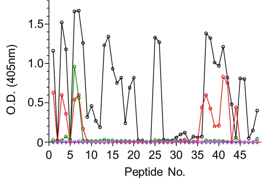
Antibodies 2020, 9, 11                                                                                                      5 of 9

to form the active site region (Figure 2B). It was, therefore, of interest to determine whether any of
the mapped epitopes are located in the vicinity of the active site. Indeed, active-site Residue Y74 is
part of Epitope 4, and that this epitope resides at the surface of the active site. It is thus tempting to
assume that the antibody binding to this epitope blocks the active site, thereby directly neutralizing
the catalytic activity of abrin. This notion may be supported by the study by Bagaria et al. [15] that
mapped the epitope of an antiabrin monoclonal neutralizing antibody, D6F10. This antibody binds to
Residues T112, G114, and R118 that are located also at the surface of the active site, contrapositioned to
Epitope   4. 2020, 9, x FOR PEER REVIEW
     Antibodies                                                                                   5 of 10

      Figure 2. Immunodominant epitopes on ATA. (A) Linear depiction of ATA subunits folding domain
      (1–3)Figure  2. Immunodominant
             and amino      acid residuesepitopes
                                            (markedon ATA.   (A) Linearcomprising
                                                       by asterisks)     depiction of ATA    subunits
                                                                                        catalytic       foldingLocation
                                                                                                   domain.      domain of
           (1–3) and amino acid residues (marked by asterisks) comprising catalytic domain. Location of
      immunodominant epitopes (1–11) marked as shaded boxes, whereas shading tones represent number
           immunodominant epitopes (1–11) marked as shaded boxes, whereas shading tones represent number
      of sera that reacted with each epitope (pale to darkest for 1 to 3 sera, respectively). (B) Crystal structure
           of sera that reacted with each epitope (pale to darkest for 1 to 3 sera, respectively). (B) Crystal structure
      of abrin (PDB 1abr; ATA in pale blue and enzymatic B-chain (ATB) in light brown). Immunodominant
           of abrin (PDB 1abr; ATA in pale blue and enzymatic B-chain (ATB) in light brown). Immunodominant
      epitopes color-coded and numbered; catalytic domain in orange.
           epitopes color-coded and numbered; catalytic domain in orange.

       As mentioned     earlier, very little is known about the targets of antiabrin-neutralizing antibodies; in
           While Serum R3 exhibited the highest titer and neutralization potency, it seems that it did not
fact,interact
      only two   such
               with  anyepitopes,   bothATA
                          of the linear    located   on ATA,
                                                epitopes        have
                                                           (Figure  1). been
                                                                         Fromdescribed
                                                                                the overallso   far—epitopes of
                                                                                             peptide-binding        antibodies
                                                                                                                pattern,  a
D6F10response toward five epitopes could be deduced, all of which are shared with R1 (epitopes 1–3, 8,that
         (discussed    above)   and   A7C4    ([9]. By   using   a set  of  toxin  mutants,    the  authors   concluded
Residues
     and 9).T82,  G83,
              There  wasH85,   D103, and
                          a significant      H105 of
                                         response    areSerum
                                                          crucial
                                                                R2for
                                                                    withthePeptide
                                                                             binding44 of this
                                                                                        that     antibody.
                                                                                             might   suggestHere,    we found
                                                                                                              that there is
that another
      these residues
               epitopeare   members
                         located        of that
                                   within   two sequence.
                                                  of the identified
                                                               However, immunodominant
                                                                             since this serumepitopes,     Epitopes 5the
                                                                                                  did not recognize      and 6,
     adjacent peptides
respectively.             that largelyinoverlapped
                Not surprisingly,         the foldedinformthe sequence,
                                                               of the toxin,we could
                                                                                these not
                                                                                       tworelate   the high
                                                                                             epitopes   are response
                                                                                                             adjacent to
                                                                                                                       to aeach
     novel  epitope.  In  contrast  to these   findings,   the murine     antiabrin  antibodies   reacted
other (Figure 2B), and they are positioned distal to the active site; however, to induce cell death, ATA   with  only  one
needsmajor   epitopewith
        to interact   (overlapping    epitope
                           other proteins     en3)route
                                                    and to
                                                         Serum    R3 did not(as
                                                             the cytoplasm       react
                                                                                   waswith
                                                                                         shownany in
                                                                                                   of detail
                                                                                                      the ATA    peptides.
                                                                                                             for ricin subunit
     These   results may   suggest  that  the ATA    epitopes   of these   sera are mainly   directed   against
A [15]. It is, therefore, possible that binding antibodies to Epitope(s) 5 and/or 6 may interfere with one      nonlinear
     epitopes.
or more abrin:protein interactions required for ATA cytotoxic performance.
          The location of the 11 ATA epitopes within the crystal structure of abrin is shown in Figure 2B.
     As expected from antibody epitopes, all 11 epitopes are located on the solvent-exposed surface of the
     toxin. In the majority of the cases, the exposed residues represent the full amino acid sequence of the
     predicted epitope. However, in some cases (i.e., Epitopes 3 and 10), only part of the assigned target
     epitope is located on the surface of the toxin, suggesting that, for these epitopes, amino acid residues
     that are in direct contact with the antibody are restricted, while other residues that are seemingly
     inaccessible are mainly responsible for maintaining the epitope 3D structure.
          The toxicity of abrin stems from its catalytic activity that causes irreversible depurination of a
     specific adenine nucleotide within the 28S rRNA, thereby leading to the cessation of cellular-protein
     synthesis and eventually to cell death. This catalytic activity is mediated at the active site cleft within
Antibodies 2020, 9, 11 6 of 9
3.3. Immunodominant Epitopes of Abrin Subunit B
Using the same strategy described above, the four antiabrin antibody preparations were allowed
to interact with peptides spanning the amino acid sequence of the abrin B subunit (ATB). In this case,
all four preparations interacted with the peptides (Figure 3), and 15 binding epitopes were identified
overall (Table 3). Sera R1 and R3 exhibited diverse recognition with 13 shared epitopes (1–8, 10–13,
and 15), whereas Serum R3 also interacted with Epitopes 9 and 14. Unlike the lack of interactions
between Serum R2 and ATA, this serum was found to interact with two ATB epitopes, 7 and 9.
The murine-derived antiabrin antibodies (M1) interacted with Epitopes 7, 12, and 15. The observation
that all sera interacted with ATB epitopes, while only a limited number of these sera interacted with
ATAAntibodies
epitopes, may
2020, imply
9, x FOR PEERthat ATB is more immunogenic than ATA.
REVIEW 7 of 10
Figure 3. Binding of antiabrin sera to ATB peptide array. Set of 15-mer biotinylated peptides spanning
Figure 3. Binding of antiabrin sera to ATB peptide array. Set of 15-mer biotinylated peptides spanning
amino acidacid
amino sequence
sequenceofofBB subunit
subunit ofofabrin
abrin were
were immobilized
immobilized on microtiter
on microtiter plates
plates and and incubated
incubated with
withantiabrin
antiabrinantibodies
antibodies R1 (black), R2 (red), R3 (purple), or M1 (green). Plates were
R1 (black), R2 (red), R3 (purple), or M1 (green). Plates were then washed,thenAP-
washed,
AP-conjugated secondaryantibody
conjugated secondary antibodywaswas added,
added, andand antibody
antibody binding
binding in eachinwell
each well
was was determined.
determined.
Table3.3.ATB
Table ATB immunodominant epitopes.
immunodominant epitopes.
Epitope No. No. Epitope
Epitope Epitope Sequence
Sequence ATB Residue
ATB Residue NumberNumber
1 1 VRIGG
VRIGG 16–20 16–20
2 2 VDVYD
VDVYD 26–30 26–30
3 3 NGYHNG
NGYHNG 31–36 31–36
4 4 DRLEE
DRLEE 46–50 46–50
5 5 WTLKSDK
WTLKSDK 54–60 54–60
6 6 YAPGSYV
YAPGSYV 74–80 74–80
7 7 IWDNGT
IWDNGT 97–10297–102
8 8 MGGTLTV
MGGTLTV 119–125
119–125
9 QGWRTGN 134–140
9 QGWRTGN 134–140
10 VTSIS 146–150
10 VTSIS 146–150
11 QAQGSNVWMAD 158–168
12 11 QAQGSNVWMAD
DGSI 158–168
183–186
13 12 DGSI
WVKFNDGSI 183–186
221–229
14 13 WVKFNDGSI
KGSDPSLKQ 221–229
241–249
15 14 KGSDPSLKQ
QIWLTLF 241–249
261–267
15 QIWLTLF 261–267Antibodies 2020, 9, 11 7 of 9
ATB comprises two homologous globular domains [6], each containing a galactose-binding pocket.
These domains can be further divided into four subdomains (Figure 4A), where a hydrophobic core is
formed by Subdomains α, β, and γ, while Subdomain λ connects the two globular domains. Overall,
mapped epitopes are distributed over the entire length of ATB, with Subdomain 1β being slightly more
populated with9,interacting
Antibodies 2020, epitopes when compared to the 3 subdomains.
x FOR PEER REVIEW 8 of 10
Figure
Figure 4. 4. Immunodominantepitopes
Immunodominant epitopes on
on ATB.
ATB. (A)
(A) Linear
Linear depiction
depictionofoftwo
twoATB
ATBsubunit
subunithomologous
homologous
globular domains and amino acid residues comprising galactose-binding pockets
globular domains and amino acid residues comprising galactose-binding pockets of each of each domain
domain
(marked by asterisks). Location of immunodominant epitopes (1–15) marked as shaded boxes, boxes,
(marked by asterisks). Location of immunodominant epitopes (1–15) marked as shaded whereas
whereas shading tones represent number of sera that reacted with each epitope (pale to darkest for 1
shading tones represent number of sera that reacted with each epitope (pale to darkest for 1 to 4 sera,
to 4 sera, respectively). (B) Crystal structure of abrin (PDB 1abr; ATA in pale blue and ATB in light
respectively). (B) Crystal structure of abrin (PDB 1abr; ATA in pale blue and ATB in light brown).
brown). Immunodominant epitopes color-coded and numbered; galactose-binding pockets in green.
Immunodominant epitopes color-coded and numbered; galactose-binding pockets in green.
As the main activity of ATB is to bind galactose moieties located on the cell surface and thus
Visualization of the 15 epitopes on the crystal structure of abrin revealed that all but one are located on
mediate toxin uptake, it was of interest to examine whether any of the identified ATB epitopes play
the surface of the toxin, securing their accessibility to antibody binding (Figure 4B). Epitope 12, however,
a role in abrin neutralization by blocking its ability to bind galactose. ATB contains two potential
is buried deep within the molecule, thus raising the question about its role as an antibody epitope.
galactose-binding sites, N51 and N260, for Subdomains 1 and 2, respectively [6]. In addition, on the
A possible explanation may rely on the fact that the sequence of this epitope (DGSI) also appears as a
basis of structure similarities to ricin, two residues (in each subdomain) were assumed to be involved
part of Epitope 13 (WVKFNDGSI) that is located at the surface of the toxin. It is thus possible that the
in hydrogen bonding to the sugar (D27 and W42 for Domain 1; D239 and W253 for Domain 2; Figure
antibodies
4). Indeed,that
theinteracted with thepocket
galactose-binding peptides encompassing
of Domain 1 seemed Epitope 12 were originally
to be populated by severalraised
of the against
ATB
Epitope 13.
epitopes. First, sugar-binding Residue D27 is a part of Epitope 2, and Epitopes 4 and 8 surround the
As thepocket.
binding main activity of ATB is
As for Domain 2, to bind galactose
it appears moieties
that Epitope 14 islocated
in closeonproximity
the cell surface and thus
to the second
mediate toxin uptake, it was of interest to examine whether any of the identified
galactose-binding pocket, and can thus also be regarded as a putative neutralizing epitope. Although ATB epitopes play
a role in abrin
the main neutralization
function of the ATBby blocking
is to bind theits ability
cell to bind
membrane, galactose.
it is ATB contains
highly possible two
that it also haspotential
a role
galactose-binding sites, N51(mainly
in intracellular trafficking and N260, in thefor Subdomains
early endosomes) 1 and
where 2, respectively
it may interact[6]. In other
with addition, on the
proteins.
basis of structure
Therefore, othersimilarities to ricin,
epitopes, though two residues
located (inthe
distally to each subdomain) were
galactose-binding assumed
pockets, might toalso
be involved
play a
role in antibody-mediated
in hydrogen bonding to the sugarabrin (D27
neutralization.
and W42 for Domain 1; D239 and W253 for Domain 2; Figure 4).
Indeed, the galactose-binding pocket of Domain 1 seemed to be populated by several of the ATB
4. Conclusions
epitopes. First, sugar-binding Residue D27 is a part of Epitope 2, and Epitopes 4 and 8 surround
the binding pocket. As for
We characterized forDomain
the first2, time
it appears that Epitope
the polyclonal 14 is inresponse
antibody close proximity
towards to the second
abrin, and
galactose-binding pocket, and can thus also be regarded as a putative neutralizing
identified the immunodominant epitopes of each of the toxin’s subunits. By screening with antibodies epitope. Although
thederived
main function
from two ofanimal
the ATB is to bind
species the and
(rabbits cell membrane, it is highly
mice) by different possible
vaccination that it also
strategies, we has a role in
increased
the possibility
intracellular to identify
trafficking a wide
(mainly in coverage
the earlyofendosomes)
epitopes. It where
is possible,
it may however,
interactthat
with these
otherantibody
proteins.
preparations also target nonlinear epitopes that cannot be identified by current method applied in
this study. As abrin is considered an imminent biothreat agent, there is an ongoing effort to develop
effective countermeasures to this toxin. Epitope mapping of the polyclonal antibodies in the sera of
immunized animals enhances our knowledge regarding the antigenic moieties of abrin and provides
important information for the development of such countermeasures. Indeed, two of the identified
ATA immunodominant epitopes were previously shown to be the target of neutralizing monoclonalAntibodies 2020, 9, 11 8 of 9
Therefore, other epitopes, though located distally to the galactose-binding pockets, might also play a
role in antibody-mediated abrin neutralization.
4. Conclusions
We characterized for the first time the polyclonal antibody response towards abrin, and identified
the immunodominant epitopes of each of the toxin’s subunits. By screening with antibodies derived
from two animal species (rabbits and mice) by different vaccination strategies, we increased the
possibility to identify a wide coverage of epitopes. It is possible, however, that these antibody
preparations also target nonlinear epitopes that cannot be identified by current method applied in
this study. As abrin is considered an imminent biothreat agent, there is an ongoing effort to develop
effective countermeasures to this toxin. Epitope mapping of the polyclonal antibodies in the sera of
immunized animals enhances our knowledge regarding the antigenic moieties of abrin and provides
important information for the development of such countermeasures. Indeed, two of the identified
ATA immunodominant epitopes were previously shown to be the target of neutralizing monoclonal
antibodies. In the future, the neutralization potency of the novel epitopes identified in this work
(especially on ATB) will be evaluated. To conclude, the findings of this study provide useful information
as part of an overall strategy to design improved vaccines and countermeasures to abrin.
Supplementary Materials: The following are available online at http://www.mdpi.com/2073-4468/9/2/11/s1.
Figure S1: Sequences of 15-mer peptides of ATA and ATB used for binding screening assay.
Author Contributions: Conceptualization and supervision, O.M. and C.K.; methodology and analysis, R.A.,
R.F., Y.G., A.S., T.S., C.K., and O.M.; writing—original-draft preparation, O.M. and C.K.; writing—review and
editing, O.M., C.K., R.A., R.F., Y.G., A.S., and T.S.; All authors have read and agreed to the published version of
the manuscript.
Funding: This study was supported by the Israel Institute for Biological Research.
Conflicts of Interest: The authors declare no conflict of interest.
References
1. Olsnes, S. The history of ricin, abrin and related toxins. Toxicon 2004, 44, 361–370. [CrossRef] [PubMed]
2. Audi, J.; Belson, M.; Patel, M.; Schier, J.; Osterloh, J. Ricin poisoning: A comprehensive review. JAMA 2005,
294, 2342–2351. [CrossRef] [PubMed]
3. Endo, Y.; Mitsui, K.; Motizuki, M.; Tsurugi, K. The mechanism of action of ricin and related toxic lectins on
eukaryotic ribosomes. The site and the characteristics of the modification in 28 S ribosomal RNA caused by
the toxins. J. Biol. Chem. 1987, 262, 5908–5912. [PubMed]
4. Biological Terrorism in Indonesia. Available online: https://globalbiodefense.com/headlines/biological-
terrorism-in-indonesia/ (accessed on 20 November 2019).
5. Janik, E.; Ceremuga, M.; Saluk-Bijak, J.; Bijak, M. Biological Toxins as the Potential Tools for Bioterrorism.
Int. J. Mol. Sci. 2019, 20, 1181. [CrossRef] [PubMed]
6. Tahirov, T.H.; Lu, T.H.; Liaw, Y.C.; Chen, Y.L.; Lin, J.Y. Crystal structure of abrin-a at 2.14 A. J. Mol. Biol. 1995,
250, 354–367. [CrossRef] [PubMed]
7. Lebeda, F.J.; Olson, M.A. Prediction of a conserved, neutralizing epitope in ribosome-inactivating proteins.
Int. J. Biol. Macromol. 1999, 24, 19–26. [CrossRef]
8. Bansia, H.; Bagaria, S.; Karande, A.A.; Ramakumar, S. Structural basis for neutralization of cytotoxic abrin by
monoclonal antibody D6F10. FEBS J. 2019, 286, 1003–1029. [CrossRef] [PubMed]
9. Kumar, M.S.; Karande, A.A. A monoclonal antibody to an abrin chimera recognizing a unique epitope on
abrin A chain confers protection from abrin-induced lethality. Hum. Vaccin. Immunother. 2016, 12, 124–131.
[CrossRef] [PubMed]
10. Dudek, N.L.; Perlmutter, P.; Aguilar, M.I.; Croft, N.P.; Purcell, A.W. Epitope discovery and their use in
peptide based vaccines. Curr. Pharm. Des. 2010, 16, 3149–3157. [CrossRef] [PubMed]
11. Falach, R.; Sapoznikov, A.; Gal, Y.; Israeli, O.; Leitner, M.; Seliger, N.; Ehrlich, S.; Kronman, C.; Sabo, T.
Quantitative profiling of the in vivo enzymatic activity of ricin reveals disparate depurination of different
pulmonary cell types. Toxicol. Lett. 2016, 258, 11–19. [CrossRef] [PubMed]Antibodies 2020, 9, 11 9 of 9
12. Mechaly, A.; Alcalay, R.; Noy-Porat, T.; Epstein, E.; Gal, Y.; Mazor, O. Novel Phage Display-Derived
Anti-Abrin Antibodies Confer Post-Exposure Protection against Abrin Intoxication. Toxins 2018, 10, 80.
[CrossRef] [PubMed]
13. Sabo, T.; Kronman, C.; Mazor, O. Ricin-Holotoxin-Based Vaccines: Induction of Potent Ricin-Neutralizing
Antibodies. Methods Mol. Biol. 2016, 1403, 683–694. [CrossRef] [PubMed]
14. Noy-Porat, T.; Rosenfeld, R.; Ariel, N.; Epstein, E.; Alcalay, R.; Zvi, A.; Kronman, C.; Ordentlich, A.; Mazor, O.
Isolation of Anti-Ricin Protective Antibodies Exhibiting High Affinity from Immunized Non-Human Primates.
Toxins 2016, 8, 64. [CrossRef] [PubMed]
15. Bagaria, S.; Karande, A. Abrin and Immunoneutralization: A Review. Toxinology 2014, 1–21. [CrossRef]
© 2020 by the authors. Licensee MDPI, Basel, Switzerland. This article is an open access
article distributed under the terms and conditions of the Creative Commons Attribution
(CC BY) license (http://creativecommons.org/licenses/by/4.0/).You can also read