3D morphology of nematode encapsulation in snail shells, revealed by micro CT imaging - Nature
←
→
Page content transcription
If your browser does not render page correctly, please read the page content below
www.nature.com/scientificreports
OPEN 3D morphology of nematode
encapsulation in snail shells,
revealed by micro‑CT imaging
P. Falkingham & R. Rae*
Many parasites and hosts are embroiled in an on-going arms race that affects the evolution of each
participant. One such battle is between parasitic nematodes and terrestrial gastropods which have
co-evolved for 90–130 MY. Recently, snails have been shown to encase and kill invading nematodes
using their shell as a defence mechanism. However, there is remarkably little known about this process
in terms of understanding where, when and how nematodes are fixed within the shell. Also there has
never been any attempt to observe this process using methods other than light microscopy. Therefore,
we used micro CT scanning of a Cepaea nemoralis shell (a common host for nematodes) to 3D visualise
encased nematode parasites and quantify morphological parameters. By taking this approach future
studies could use micro CT scanning of fossil shells in conchology collections to understand nematode/
snail co-evolution.
The co-evolutionary arms race between host and parasite has resulted in rapid changes in the evolution of the
immune system1. Terrestrial gastropods (slugs and snails) are parasitised regularly by flies, protozoa, trema-
todes and v iruses2, but nematodes are the most prolific parasites with 108 species (representing four out of five
clades of the Nematoda) using terrestrial gastropods as definitive, intermediate and paratenic hosts3–5. This arms
race has been on going for 90–130 MY6. Examples include Caenorhabditis elegans Maupas which is thought to
use slugs and snails for t ransport7 and Angiostrongylus vasorum Baillet (the casual agent of cardio/pulmonary
disease in dogs) uses snails as intermediate hosts to facilitate transmission to m ammals8. In order to combat
parasites, terrestrial gastropods use Reactive Oxygen Species (ROS), antimicrobial peptides and lectins to kill
invading parasites9, but in general, their immune system is poorly researched10. Interestingly, recent studies
examining the susceptibility of snails to the commercially available biological control agent nematode parasite
Phasmarhabditis hermaphrodita Schneider (sold as Nemaslug)11, observed nematodes being trapped, encased
and killed by unknown cells fusing the animals to the inner part of the shell en masse12–16. The shell is made of
an outer proteinaceous periostracum of conchiolin and crystalline calcium carbonate sub-layers17 and is used
for shelter from extreme environmental conditions but this recent research posits the shell has been co-opted
ematodes14. Upon nematode infection, cells on the shell surface aggregate and adhere to the nematode
to kill n
cuticle and fuse it to the inner shell, often hundreds at a time. This was initially observed in infection experi-
ments with the giant African snail (Lissachatina fulica) Férussac12 and has subsequently been observed in live
Cepaea nemoralis L.13, Arianta arbustorum L.15 and in museum collections of Cornu aspersum Müller16 and
across many representatives of the Stylommatophora14, even in the vestigial shell of slugs18. By examining shells
in conchology collections nematodes over 500 years old have been observed14. This in vivo fossilisation process
could allow an unprecedented insight into spatial and temporal changes in co-evolutionary dynamics between
nematodes and snails. Also as nematode DNA can be extracted from preserved shells to aid identification to
species14,16 the molecular evolution of nematodes could be tracked over time. As nematodes are soft bodied
and do not fossilise19,20 this approach has huge potential however, the basic processes involved in encapsulation
used to kill nematodes are poorly understood. This is primarily as the inner aperture and whorl of a snail’s shell
is difficult to observe. Light microscopy has been used to view nematodes fixed and fused in shells (Fig. 1) but
there have been no other techniques used to investigate this further. Hence, new, non-destructive approaches
are needed. One such approach is micro computed tomography (micro CT scanning) that has been successfully
used understand the structure of a rachnids21 and a mmonites22. Thus, we had two main aims; first, to use micro
CT scanning to discover if nematodes can be viewed in the snails’ shell and second, whether any morphometric
data can be gleaned from such an approach.
School of Biological and Environmental Sciences, Liverpool John Moores University, Byrom St, Liverpool L33AF,
UK. *email: r.g.rae@ljmu.ac.uk
Scientific Reports | (2021) 11:2523 | https://doi.org/10.1038/s41598-021-82106-6 1
Vol.:(0123456789)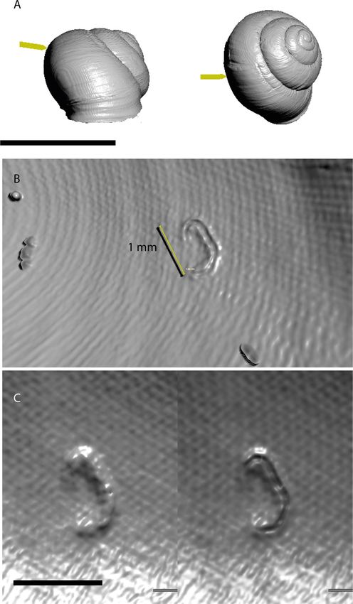
www.nature.com/scientificreports/
Figure 1. (A) Snails, such as C. nemoralis, regularly encase and kill nematodes in the inner whorl of their shell.
The nematodes can be seen by using light microscopy and are fused to the inner shell (B,C). Bars represent 1 cm
in (A) and 0.5 mm in (B,C).
Materials and methods
Observation and scanning of nematodes encased in snail shells. A collection of approximately
1–2 year old C. nemoralis shells collected from sand dunes in Formby, Sefton (n = 50) (Grid Reference: SD273075)
were examined for nematodes encased in the inner lip and whorl of the shells using light microscopy follow-
ing standard p rocedures12–16. One C. nemoralis shell had a prominent nematode fixed opposite the inner whorl
of the shell and was used for subsequent studies. The specimens were scanned using a SkyScan 1272, at voxel
size of 19.82 µm3. Voltage and Current were set to 50 kV and 200 µA respectively. CT data were analysed using
Dragonfly software, version 4 for Windows (http://www.theobjects.com/dragonfly/, Object Research Systems
(ORS) Inc, Montreal, Canada). Thresholding was used to isolate shell material in 3D and 2D views, and the
encapsulated nematode located. 3D views of the encapsulated nematode were rendered with a “hard gradient” to
illustrate the morphology of the feature. 2D slices detailing the internal morphology of the encapsulation were
produced with a rainbow colour map to indicate density (warmer colours indicate higher density). Tomographic
data are provided in supplemental data.
Results
Prior to scanning, one C. nemoralis shell was identified through light microscopy as exhibiting encapsulation
of an individual nematode located inside the dorsal portion of the shell (Fig. 2a; Supplementary Video S1).
The feature is C-shaped, curving and tapering at both ends. The encapsulation is ~ 1 mm long and 0.2 mm in
width, (Fig. 2b,c) and is raised 0.1 mm above the surrounding shell surface (Fig. 3). The outermost layer of the
encapsulation is extremely thin, only a few voxels in width. The interior of the encapsulation is approximately
80 µm in width, and has higher density than the surrounding air, but lower density than the shell around it. It is
clear there is a cavity produced when the nematode is covered in unknown cells (Fig. 3c). However, the lack of
resolution here makes it difficult to draw firm conclusions regarding the nature of the interior of the encapsula-
tion (see Fig. 3c).
Discussion
The number of shells found with nematodes present was surprisingly low in our study. This is unusual. The num-
ber of shells positive for nematode encapsulation as well as the number of nematodes found per shell has been
found to be high in field based studies. For example, from C. nemoralis collected from Merseyside, 4–60% of shells
had nematodes present ranging from 1 to 152 nematodes per s hell14. Similarly, 2–25% of C. hortensis shells from
north Scotland had from 1 to 51 nematodes p resent14. This high infection load is not restricted to snails from
the genus Cepaea. All shells of C. aspersum (n = 136) from an escargot farm in northern Ireland had nematodes
present in their shells with a mean of 31 ± 2 nematodes per s hell14,16. Snail shells hundreds of years old housed
in conchology collections have nematodes encased in their shells. For example, A. arbustorum from 1 90815, C.
aspersum and Helix pomatia L. from 1901 and 1 90416 respectively, as well as C. nemoralis from 1864 and even
Scientific Reports | (2021) 11:2523 | https://doi.org/10.1038/s41598-021-82106-6 2
Vol:.(1234567890)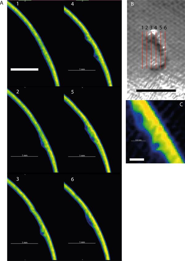
www.nature.com/scientificreports/

                                  Figure 2.  3D views of the nematode encapsulation. (A) Arrows indicate location of the encapsulation in
                                  anterior and dorsal views of the shell. (B) The encapsulation inside the shell. Fine sand grains are also adhered to
                                  the inside of the shell and are visible. (C) 3D view of the encapsulation at two slightly different thresholds—on
                                  the left, a broader threshold window showing the complete nature of the encapsulation, and right, a narrower
                                  thresholding window exposing the internal geometry of the encapsulation. Scale bar = 20 mm in (A), and 1 mm
                                  in (B,C).

Scientific Reports |   (2021) 11:2523 |                https://doi.org/10.1038/s41598-021-82106-6                                                   3

                                                                                                                                               Vol.:(0123456789)
www.nature.com/scientificreports/
Figure 3. (A) Six sequential slices through the encapsulation and surrounding shell. The complete
encapsulation, and tubular nature of the structure can be seen in slices 2–6. Slices are coloured according to
density—warmer colours indicate higher density. Slice locations are shown in the upper right (B). (C) Close up
slice shown as raw pixel data (non-interpolated). Scale bar in A + B = 1 mm, scale bar in C = 0.2 mm.
over 500 years old14 all had nematodes encased in their shells. Therefore, as nematode encapsulation is common
in many members of the S tylommatophora14 there is ample opportunity for studying the spatial and temporal
changes in nematode infection in many different species and locations using light microscopy and μCT scanning.
Previous attempts using standard light microscopy have been able to quantify nematode numbers fixed in
snail shells12–16 but 3D visualisation and measurements of individual animals have not been possible. Using
micro CT scanning we have been able to remedy this problem. The 1 mm long nematode fused to the inner shell
of C. nemoralis was covered with a thin layer of unknown snail cells leaving a clear cavity where the nematode
degrades. It was previously unknown whether snail cells would fill this void or if layers were produced on top of
the lesion during the encapsulation process. This is an interesting and important discovery for future research,
as this cavity will protect nematode DNA from agents responsible for degradation e.g. extreme temperatures and
water23. Molecular analyses of museum collections have yielded fascinating insights into the evolution of many
organism including h umans24, plants25 and even bacterial pathogens26. Perhaps this system could be no different.
Scientific Reports | (2021) 11:2523 | https://doi.org/10.1038/s41598-021-82106-6 4
Vol:.(1234567890)www.nature.com/scientificreports/
If shells are stored correctly in conchology collections this encapsulation process could allow molecular analysis
of nematodes over time using fossilised shells. In general, molecular approaches of preserved nematodes have
been restricted to genotyping of helminth eggs from coprolites and archaeological digs hundreds even thousands
of years old. For example, Ascaris eggs were extracted from coprolites from the Middle-Ages in B elgium27 and
Ascaris sp. and Trichuris sp. eggs have been identified from environmental samples from Viking age sediment
(dated 1018–1030 AD)28. This is due to helminth eggs being resistant to environmental stressors. From our
understanding there have been no molecular analyses (other than a few genes for genotyping) of preserved
nematodes at any other developmental stage as they do not fossilise. In contrast, adult stage nematodes encased
in C. nemoralis shells over 500 years old have been observed14. Examination of older shells is possible and is
highly likely to yield positive shells with evidence of nematode parasitism. One such group of snails to focus on
could be edible land snails (e.g. C. aspersum), reared by humans in the late Pleistocene and Holocene and are
often abundant in archeological deposits and hence m useums29. Analysis of these shells could potentially tell
us about the evolutionary history of nematodes infecting humans. For example, a common parasite of snails is
Angiostrongylus cantonensis Chen the causal agent of human eosinophilic meningoencephalitis w orldwide30.
Although our study using micro CT scanning was successful in providing information about nematode para-
sitism of snail shells there are a wealth of techniques to use in future studies including using higher resolution
scanners or microscopy including Transmission Election Microscopy (TEM). Scanning Electron Microscopy
(SEM) is another possibility though this would involve breaking shells to reveal the nematodes inside.
Providing these 3D observations opens up the opportunity of examining the fossil record for nematode-
snail relationships, and exploring the evolution of this defence mechanism. The three dimensional nature of
encapsulations like this makes it a very real possibility encapsulated nematodes might be observed in fossil snail
specimens via µCT imaging. Armed with this search image, fossil snail collections stretching back hundreds of
millions of years may hold important information on when this capability evolved, and how it might be spread
across the snail phylogeny.
Received: 30 April 2020; Accepted: 25 November 2020
References
1. Frank, S. A. Immunology and Evolution of Infectious Diseases (Princeton, Princeton University Press, 2002).
2. Barker, G. M. Natural Enemies of Terrestrial Molluscs (CABI Publishing, Wallingford, 2004).
3. Grewal, P. S., Grewal, S. K., Tan, L. & Adams, B. J. Parasitism of molluscs by nematodes: types of associations and evolutionary
trends. J. Nematol. 35, 146–156 (2003).
4. Blaxter, M. L. et al. A molecular evolutionary framework for the phylum Nematoda. Nature 392, 71–75 (1998).
5. Pieterse, A., Malan, A. P. & Ross, J. L. Nematodes that associate with terrestrial molluscs as definitive hosts, including Phasmarhab-
ditis hermaphrodita (Rhabditida: Rhabditidae) and its development as a biological molluscicide. J. Helminthol. 91, 517–527 (2017).
6. Tillier, S., Masselot, M. & Tillier, A. Phylogenic relationships of the pulmonate gastropods from rRNA sequences, and tempo and
age of the Stylommatophoran radiation. In Origin and Evolutionary Radiation of the Mollusca (ed. Taylor, J.D.) 267–284 (Oxford,
Oxford University Press, 1996).
7. Félix, M-A. & Braendle, C. The natural history of Caenorhabditis elegans. Curr. Biol. 20, R965-R969 (2010).
8. Bolt, G., Monrad, J., Koch, J. & Jensen, A. L. Canine angiostrongylosis: a review. Vet. Rec. 135, 447–452 (1994).
9. Loker E.S. Gastropod immunobiology in Invertebrate Immunity (ed. Soderhall, K.) 17–43 (Springer, 2010).
10. South, A. Terrestrial Slugs: Biology, Ecology and Control (Chapman & Hall, London, 1992).
11. Wilson, M. J., Glen, D. M. & George, S. K. The rhabditid nematode Phasmarhabditis hermaphrodita as a potential biological control
agent for slugs. Biocontrol Sci. Technol. 3, 503–511 (1993).
12. Williams, A. J. & Rae, R. Susceptibility of the Giant African Snail (Achatina fulica) exposed to the gastropod parasitic nematode
Phasmarhabditis hermaphrodita. J. Invertebr. Pathol. 127, 122–126 (2015).
13. Williams, A. & Rae, R. Cepaea nemoralis uses its shell as a defence mechanism to trap and kill parasitic nematodes. J. Mollus. Stud.
12, 1–2 (2016).
14. Rae, R. The gastropod shell has been co-opted to kill parasitic nematodes. Sci. Rep. 7, 4745. https://doi.org/10.1038/s41598-017-
04695-5 (2017).
15. Rae, R., 2018. Shell encapsulation of parasitic nematodes by Arianta arbustorum (Linnaeus, 1758) in the laboratory and in field
collections. J. Molluscan Stud. 84, 92–95 (2018).
16. Cowlishaw, R. M., Andrus, P. & Rae, R. An investigation into nematodes encapsulated in shells of wild, farmed and museum
specimens of Cornu aspersum and Helix pomatia. J. Conchol. 43, 1–8 (2020).
17. Lowenstam, H. A. & Weiner, S. On Biomineralization (Oxford University Press, Oxford, 1989).
18. Rae, R. G., Robertson, J. F. & Wilson, M. J. Susceptibility and immune response of Deroceras reticulatum, Milax gagates and Limax
pseudoflavus exposed to the slug parasitic nematode Phasmarhabditis hermaphrodita. J. Invertebr. Pathol. 97, 61–69 (2008).
19. Littlewood, D. T. J. & Donovan, S. K. Fossil parasites: a case of identity. Geol. Today. 19, 136–142 (2003).
20. Poinar, G. O. Jr. The geological record of parasitic nematode evolution. Adv. Parasitol. 90, 53–92 (2015).
21. Garwood, R., Dunlop, J.A. & Sutton, M.D. High-fidelity X-ray micro-tomography reconstruction of siderite-hosted Carboniferous
arachnids. Biol. Lett. 5, 6 https://doi.org/10.1098/rsbl.2009.0464 (2009).
22. Inoue, S. & Kondo, S. Structure pattern formation in ammonites and the unknown rear mantle structure. Sci. Rep. 6, 33689; https
://doi.org/10.1038/srep33689 (2016).
23. Shapiro, B. Ancient DNA. In Princeton Guide to Evolution (ed. Losos, J.) 475–481 (Princeton, Princeton University Press, 2013).
24. Slon, V. et al. The genome of the offspring of a Neanderthal mother and a Denisovan father. Nature 561, 113–116 (2018).
25. Swarts, K. et al. Genomic estimation of complex traits reveals ancient maize adaptation to temperate North America. Science 357,
512–515 (2017).
26. Spyrou, M. A. et al. Analysis of 3800-year-old Yersinia pestis genomes suggests Bronze Age origin for bubonic plague. Nat. Com-
mun. 9, 2234. https://doi.org/10.1038/s41467-018-04550-9 (2018).
27. Loreille, O., Roumat, E., Verneau, O., Bouchet, F. & Hänni, C. Ancient DNA from Ascaris: extraction amplification and sequences
from eggs collected from coprolites. Int. J. Parasitol. 31, 1101–1106 (2001).
28. Søe, M. J., Nejsum, P., Fredensborg, B. L. & Kapel, C. M. O. DNA typing of ancient parasite eggs from environmental samples
identifies human and animal worm infections in Viking-age settlement. J. Parasitol. 101, 57–63 (2015).
Scientific Reports | (2021) 11:2523 | https://doi.org/10.1038/s41598-021-82106-6 5
Vol.:(0123456789)www.nature.com/scientificreports/
29. Lubell, D. Prehistoric edible land snails in the cicum-Mediterranean: the archaeological evidence. In Petits Animaux et Societes
Humaines. Du Complement Alimentaire Aux Resources Utiliaires. XXIVe rencontres internationals d’archeologie et d’histoire d’Antibes
(eds. Brugal, J-J & Dess, J.) 77–98 (Editions APDCA, 2004).
30. Eamsobhana, P. Eosinophilic meningitis caused by Angiostrongylus cantonenses – a neglected disease with escalating importance.
Trop. Biomed. 31, 569–578 (2014).
Acknowledgements
We are grateful to LJMU for use of equipment and facilities.
Author contributions
R.R. and P.F. conceived the experiment and wrote the manuscript. P.F. carried out experiment and conducted
analysis.
Competing interests
The authors declare no competing interests.
Additional information
Supplementary Information The online version contains supplementary material available at https://doi.
org/10.1038/s41598-021-82106-6.
Correspondence and requests for materials should be addressed to R.R.
Reprints and permissions information is available at www.nature.com/reprints.
Publisher’s note Springer Nature remains neutral with regard to jurisdictional claims in published maps and
institutional affiliations.
Open Access This article is licensed under a Creative Commons Attribution 4.0 International
License, which permits use, sharing, adaptation, distribution and reproduction in any medium or
format, as long as you give appropriate credit to the original author(s) and the source, provide a link to the
Creative Commons licence, and indicate if changes were made. The images or other third party material in this
article are included in the article’s Creative Commons licence, unless indicated otherwise in a credit line to the
material. If material is not included in the article’s Creative Commons licence and your intended use is not
permitted by statutory regulation or exceeds the permitted use, you will need to obtain permission directly from
the copyright holder. To view a copy of this licence, visit http://creativecommons.org/licenses/by/4.0/.
© The Author(s) 2021
Scientific Reports | (2021) 11:2523 | https://doi.org/10.1038/s41598-021-82106-6 6
Vol:.(1234567890)You can also read