Timing of hospital admission for stillbirth delivery on maternal and obstetric outcome: a retrospective cohort study
←
→
Page content transcription
If your browser does not render page correctly, please read the page content below
www.nature.com/scientificreports
OPEN Timing of hospital admission
for stillbirth delivery on maternal
and obstetric outcome:
a retrospective cohort study
1*
Dana Anaïs Muin , Anke Scharrer2, Alex Farr1, Herbert Kiss1 & Helmuth Haslacher3
The effect of timing of hospital admission for stillbirth delivery following late intrauterine fetal death
(IUFD) has not yet been described. By this study, we aimed to gain an understanding of the impact of
“immediate” (i.e., on the same day of IUFD diagnosis) versus “delayed” hospital admission (i.e., on
the subsequent day or two days after IUFD diagnosis) on maternal and delivery outcome parameters.
This retrospective cohort study comprised all women who suffered a singleton IUFD ≥ 21 gestational
weeks and delivered the stillborn at our tertiary referral center between 2003 and 2019. We excluded
all terminations of pregnancy and women presenting with acute symptoms on the day of IUFD
diagnosis. In total, 183 women were included of whom 69.4% (n = 127) were immediately admitted
and 30.6% (n = 56) had delayed admission. Median gestational age of IUFD was 30+3 (21+0–41+3) weeks.
Whilst women with early signs of labor were more frequently admitted immediately (87.5%; 14/16),
neither maternal demographic and obstetric parameters, nor day of the week or presenting symptoms
influenced the timing of hospital admission. 77.6% (142/183) of women after IUFD diagnosis delivered
within the first 3 days after admission. Women after immediate admission equally often delivered
on admission day and the day after (26.0%; 33/127 each), women after delayed admission most
commonly delivered the day after admission (39.3%; 22/56). Stillbirth delivery on the day of diagnosis
was more common upon immediate admission (p = 0.006), especially in early gestational weeks
(p = 0.003) and with small fetal weight (p < 0.001), requiring less induction of labor. No significant
difference regarding delivery mode, labor duration, use of intrapartum analgesia, need for episiotomy
and risk of perineal injury was observed between the groups. Also rate of intrapartum hemorrhage
was independent of admission timing, although immediately admitted women experienced greater
median blood loss after vaginal delivery. Maternal laboratory parameters (hemoglobin, thrombocytes
and CRP) were independent of admission timing, except for higher levels of leucocytes, neutrophils
and lymphocytes in immediately admitted women. Our study shows no clinical superiority of
immediate hospital admission for stillbirth delivery. Under stable medical circumstances, it, therefore,
seems feasible to allow the woman delayed admission for labor and delivery.
Late intrauterine fetal death (IUFD) occurs with a prevalence of 2.68 per 1000 live births in Austria1. The diag-
nosis is usually made by real-time ultrasonography upon absent fetal heartbeats and umbilical blood flow2.
Learning the diagnosis of fetal death is immensely distressing and devastating to the affected w oman3,4. Unless
in case of an acute event, such as uterine rupture or placental abruption, current practice guidelines advise that
the woman can be discharged home and readmitted at a later stage for induction of labor (IOL), if she remains
clinically stable and well2,5.
In a recent national survey, we found that 41.3% of Austrian maternity units let the woman decide upon the
timing of admission for IOL after fetal death, whereas 17.4% institutions usually admit the woman straight a way6.
The remaining maternity units usually admit the woman on the following day or later. Institutions, which admit
the woman immediately, were more likely to perform maternal tests to rule out bleeding and clotting disorders.
It has been empirically thought that prolonged uterine retention of the demised fetus increased the maternal
1
Division of Feto‑Maternal Medicine, Department of Obstetrics and Gynecology, Medical University of Vienna,
Waehringer Guertel 18‑20, 1090 Vienna, Austria. 2Clinical Institute for Pathology, Medical University of Vienna,
Vienna, Austria. 3Department of Laboratory Medicine, Medical University of Vienna, Vienna, Austria. *email:
dana.muin@meduniwien.ac.at
Scientific Reports | (2021) 11:18854 | https://doi.org/10.1038/s41598-021-98229-9 1
Vol.:(0123456789)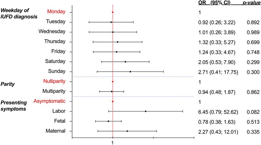
www.nature.com/scientificreports/
Figure 1. Flowchart on the selection of the study population following fetal death at the Department of
Obstetrics and Gynecology, Medical University of Vienna, Austria, between 2003 and 2019.
risk for disseminated intravascular coagulopathy (DIC) due to toxic releases during the maceration process7–9.
In this respect, we previously found no association between higher fetal maceration grade and increased risk
for maternal DIC10.
The paucity of data on the timing of hospital admission for labor induction following IUFD and obstetric
outcome, prompted us to design this study to better understand its impact on maternal care and safety. The lack
of a clinical standard on the timing of admission following IUFD allowed us to critically evaluate the outcome
after immediate versus delayed hospital admission for stillbirth delivery, whilst we hypothesized that the latter
was associated with greater maternal and obstetric complications.
Methods
Study design and subjects. We conducted a retrospective cohort study on women who suffered singleton
IUFD ≥ 21+0weeks of gestation and subsequently delivered the stillborn between January 2003 and December
2019 at the Department of Obstetrics and Gynecology at the Medical University of Vienna, Austria. Women
were excluded in case of termination of pregnancy, and if they had presented to our department with acute
symptoms needing immediate admission and prompt delivery (e.g., acute bleeding, signs of placental abruption,
uterine rupture, eclampsia, coagulation disorders, severe maternal or fetal trauma). Cases with missing data
regarding the timing of admission were excluded (Fig. 1).
Data collection. Perinatal, maternal and fetal data were retrieved from the electronic medical database
ViewPoint Version 5.6.28.56 (General Electric Company, Solingen, Germany). Variables regarding presenting
symptoms, date of diagnosis and date of admission were manually retrieved from the electronic medical records
for each case.
Maternal laboratory test results were extracted from the Laboratory Information and Management System
MOLIS (vision4health, Bochum, Germany) at the Department of Laboratory Medicine at the Medical University
of Vienna, Austria.
Upon admission, all women after IUFD routinely received laboratory tests including assessment of their
complete blood count [neutrophils, lymphocytes (Lymphos), monocytes (Monos), eosinophils (Eos), basophils
(Baso), erythrocytes (Ery), hemoglobin (Hb), leucocytes (Lc), thrombocytes (Tc)], coagulation parameters and
C-reactive protein (CRP).
Biochemical analyses were conducted at the Department of Laboratory Medicine, Medical University of
Vienna, Austria (ISO9001-certification; ISO15189-accreditation). Complete blood count and CRP were
Scientific Reports | (2021) 11:18854 | https://doi.org/10.1038/s41598-021-98229-9 2
Vol:.(1234567890)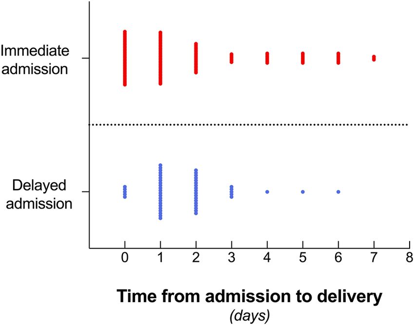
www.nature.com/scientificreports/
quantified on Sysmex hematology analyzers (Sysmex Europe GmbH, Norderstedt, Germany). Results below
the limit of detection (LOD) were estimated by their assumed expected value: x = 0+LOD2 and values above the
highest point of the standard curve (HQS) were fixed at xh = HQS + 1. At our institution, normal ranges are as
follows: Lc 4.0–10.0 G/L, CRP < 0.5 mg/dl, Hb 12.0–16.0 g/dl, Tc 150–350 G/L, Neutrophils (absolute) 2.0–7.5
G/L, Neutrophils (relative) 50.0–75.0%, Lymphos (relative) 25.0–40.0%, Monos (relative) 0.0–12.0%.
All data were transferred into a database, checked for accuracy and consistency and made anonymous prior
to analyses.
Definitions. At our institution, clinically stable women are offered to freely decide on the timing of their
hospital admission after stillbirth. For this retrospective study, women were included into the “immediate admis-
sion” group, if they had decided to be admitted to the hospital on the same day of IUFD diagnosis. Women of
“delayed admission" had opted for admission on the subsequent day or 2 days after the diagnosis of fetal death.
Symptoms at diagnosis were grouped into fetal (reduced fetal movements), maternal (bleeding, cardiovascular
symptoms, discomfort/pain, pain accompanied by bleeding), early signs of labor (bulging membranes, rupture
of membranes, contractions) and none (e.g., a woman attending routine antenatal check-up).
Maternal age was defined as age in years at the time of delivery. Ethnicity was self-reported by the woman.
Gravidity was defined as the number of the current pregnancy. Parity was defined as the number of previous
deliveries. Maternal weight (in kg) was obtained at antenatal booking or admission and Body-Mass-Index (BMI)
was grouped into six categories: underweight (≤ 18.5 kg/m2), normal weight (18.6–24.9 kg/m2), pre-obesity
(25–29.9 kg/m2), obesity class I (30–34.9 kg/m2), obesity class II (35–39.9 kg/m2) and obesity class III (≥ 40 kg/
m2). Smoking was defined as current smoker or non-smoker during pregnancy.
Indications for lower segment caesarean sections (LSCS) were categorized according to the Robson classifica-
tion based on parity, previous LSCS, LSCS before labor or after failed IOL11.
Intrapartum obstetric hemorrhage was defined as an estimated blood loss ≥ 500 ml during vaginal birth
or ≥ 1000 ml during LSCS12.
As per Austrian law, each fetus undergoes a standard post-mortem examination including autopsy (and occa-
sionally fetal imaging). Fetal autopsies were conducted in all cases according to standard operating procedures
at the Clinical Institute for Pathology, Medical University of Vienna, Austria. Upon receipt of all post-mortem
reports (including genetic testing, placental histology, and maternal tests results), we followed the algorithm as
proposed by Reddy et al. to conclude the cause of fetal d eath13. Cause of fetal death was defined as the “initial,
demonstrable pathophysiological entity initiating the chain of events that has irreversibly led to death” and
categorized according to the Tulip c lassification14.
Fetal maceration was graded by the perinatal pathologist according to international standards (grade 0:
“parboiled” skin discoloration; grade I: not-specified desquamation of the skin; grade II: bloodstained effusions
in cavities and skin peeling; grade III: yellow–brown liver, turbid effusions and mummification)8.
Statistical analysis. The distribution of data was analyzed using the Kolmogorov–Smirnov test. Normally
distributed continuous data are expressed as mean ± standard deviation. Not normally distributed variables are
expressed as median and minimum–maximum. Categorical data are presented as frequencies (n) and propor-
tions (%). Continuous data were compared with unpaired t-test and Mann–Whitney U test, respectively. Cat-
egorical data were compared with Chi2 and Fisher’s Exact test, respectively, with a 99% Confidence Interval (CI).
Spearman’s rank correlation coefficient was used to compare ordinal and continuous variables. Binary logistic
regression analysis was done to calculate the odds ratios (OR) and a 95% CI for the relationship of categorical
variables. A forest plot with a logarithmic scale (log 10) illustrates the OR and 95% CIs. All reported p-values
are two-sided, and the level of significance was set at < 0.05. Statistical tests were performed with SPSS Statistics
Version 26.0.0.0 (IBM Corporation, Armonk, NY, USA) and figures created by GraphPad Prism 9 for macOS
Version 10.14.6 (GraphPad Software, LLC).
Ethical approval. The study was approved by the Ethics Committee of the Medical University of Vienna
(Registration number 1202/2018) and complied with the principles outlined in the Declaration of Helsinki of
1975, as revised in 2013. Participants’ written consent was not required per the Austrian Federal Act concerning
Protection of Personal Data (DSG 2000). All patient data were de-identified before analyses.
Results
Maternal baseline characteristics. In total, we included 183 women whose baseline characteristics are
shown in Table 1. In our study cohort, fetal death was most frequently diagnosed during routine antenatal checks
(56.3%; 103/183), upon reduced fetal movements (27.9%; 51/183), due to suspected contractions (6.0%; 11/183),
lower abdominal discomfort (3.3%; 6/183), rupture of membranes (2.7%; 5/183), vaginal spotting (1.1%; 2/183),
maternal cardiovascular symptoms (1.1%; 2/183), pain with light bleeding (1.1%; 2/183), and bulging mem-
branes (0.5%; 1/183). In one case (0.5%), symptoms were not reported on the day of diagnosis.
Fetal baseline characteristics. The IUFD cohort consisted of 51.4% (94/183) male and 48.6% (89/183)
female fetuses between 21+0 and 41+3 gestational weeks with a median age of 30+3 gestational weeks. Median fetal
weight at stillbirth was 1020 (180–4450) g.
Abnormal fetal post-mortem findings and/or prenatally confirmed congenital malformations were reported
in 54.6% (100/183) cases. Causes of fetal death were confined as placental pathologies in 19.1% (35/183) cases;
unknown due to lack of important information in 13.1% (24/183) cases; maternal or fetal disease in 6.6% (12/183)
cases; unknown despite thorough investigation in 4.9% (9/183) cases and infection in 1.6% (3/183) cases.
Scientific Reports | (2021) 11:18854 | https://doi.org/10.1038/s41598-021-98229-9 3
Vol.:(0123456789)www.nature.com/scientificreports/
Maternal characteristics n %
Age at stillbirth (years)a 31 ± 7
Middle European 86 47
Eastern European 48 26.2
Turkish 26 14.2
Middle Eastern 10 5.5
Ethnicity
African 7 3.8
South Asian, Indian 3 1.6
American 2 1.1
Chinese 1 0.5
Gravida (n)b 2 (1–12)
Para (n)b 1 (0–9)
Body mass index (kg/m2)b 24.5 (14.6–43.3)
Underweight 4 2.9
Normal 71 51.1
Pre-obese 44 31.7
BMI categories
Obesity class I 9 6.5
Obesity class II 8 5.8
Obesity class III 3 2.2
Nicotine consumption 36 23.5
Pre-existing hypertension 12 6.6
Pre-eclampsia 14 7.7
Hypertensive disorders in pregnancy
Pregnancy induced hypertension 7 3.8
HELLP 4 2.2
Insulin-dependent gestational diabetes 17 9.3
Diabetes Pre-existing diabetes 4 2.2
Gestational diabetes (diet only) 3 1.6
Other medical 35 19.1
Hemostatic 15 8.2
Auto-immune disorder 11 6
Antiphospholipid Syndrome 6 3.3
Co-morbidities Gynecological 5 2.7
Cardiac 3 1.6
Neurological 2 1.1
Mental 2 1.1
Respiratory 0 0
0 105 57.4
1 61 33.3
Total number of co-morbidities (n) 2 13 7.1
3 2 1.1
4 2 1.1
Social restriction/domestic violence 5 2.7
Table 1. Study sample characteristics (n = 183) after hospital admission for stillbirth delivery at the
Department of Obstetrics and Gynecology, Medical University of Vienna between 2003 and 2019. BMI body
mass index, HELLP hemolysis, elevated liver enzymes, low platelets. a Mean ± standard deviation. b Median
(minimum – maximum).
Among the fetuses with post-mortem anomalies, 49.0% (49/100) had involvement of two or more organ
systems. 22.0% (22/100) fetuses had anomalies of the heart and circulatory system; 12.0% (12/100) had chromo-
somal aberrations; 6.0% (6/100) had malformations of the musculoskeletal system and 4.0% (4/100) fetuses had
anomalies in the respiratory and digestive system, respectively. In 3.0% (3/100) fetuses we detected a microdele-
tion defect or anomaly of the central nervous system or urogenital system.
Factors influencing the timing of admission. In our cohort, women most commonly presented on
Mondays (21.9%; 37/183), Thursdays and Fridays (17.8% each; 30/183; Supplementary Fig. 1) as the IUFD was
diagnosed. Excluding all women who presented with acute symptoms requiring rapid admission and immediate
delivery (e.g., established labor, placental abruption, uterine rupture), at our institution, 69.4% (127/183) women
Scientific Reports | (2021) 11:18854 | https://doi.org/10.1038/s41598-021-98229-9 4
Vol:.(1234567890)www.nature.com/scientificreports/
were admitted on the same day of IUFD diagnosis, and 30.6% (56/183) women chose to delay their admission
(Table 2).
Whilst women presenting with early signs of labor (8.7%; 16/183), such as bulging membranes, rupture of
membranes or sporadic contractions, were more frequently admitted immediately (87.5%; 14/16), there was no
statistically significant likelihood of being admitted immediately regarding presenting symptoms at diagnosis
(Fig. 2). Likewise, neither specific day of the week, nor multiparity were related with timing of hospital admis-
sion (p = 0.694 and p = 0.862; Fig. 2).
Influence on delivery time. The time interval from admission to stillbirth delivery is illustrated in Fig. 3,
highlighting the trend for delivery within the first three days in 77.6% (142/183) of women after IUFD diagnosis.
On admission day (i.e., Day 0), 26.0% (33/127) women gave birth after immediate admission, whilst only
8.9% (5/56) women did after delayed admission (p = 0.006).
Immediate admission and same-day-delivery significantly correlated with early gestational weeks (Spearman’s
r = −0.287; p = 0.003) and small fetal weight (r = −0.337; p < 0.001), yet not with parity (p = 0.06). The majority of
women after immediate admission and same-day-delivery had equally common presented with early signs of
labor (33.3%; 11/33) and no symptoms at all (30.3%; 10/33). The rest had experienced reduced fetal movements
(18.2%; 6/33), or maternal symptoms (18.2%; 6/33; Fig. 4).
Delivery on the first day after admission (i.e., Day 1) occurred in 26.0% (33/127) women after immediate
versus 39.3% (22/56) women after delayed admission (p = 0.382).
Delivery on the second day after admission (i.e., Day 2) occurred in 15.0% (19/127) women after immediate
admission versus 32.1% (18/56) women after delayed admission (p = 0.006). Likelihood for delivery from days
3 to 6 was equal in both groups.
Naturally, fetal maceration grade was lower after immediate than delayed admission (grade 0 versus grade
III; p < 0.001; Table 2).
Methods for induction of labor. In the total study cohort, most common method for IOL was mifepris-
tone (on Day 0) with misoprostol (on Day 1) in 55.2% (101/183) of the cases, followed by dinoprostone only
(12.6%; 23/183), misoprostol only (3.3%; 6/183) and intravenous syntocinon (5.5%; 10/183). Labor induction
failed or was omitted in 23.5% (43/183) of women (Fig. 5).
Women, who were admitted immediately, required less IOL [18.9% (2/127) vs. 1.7% (1/56); p = 0.001], whilst
women after delayed admission were more frequently induced with mifepristone combined with misoprostol
[73.2% (41/56) vs. 47.2% (60/127); p = 0.002].
Labor and delivery. Methods of delivery and indications for LSCS were independent of timing of admis-
sion (Table 2). In our cohort, 9.3% (17/183) women were delivered by LSCS, of whom 41.2% (7/17) women had
at least one previous LSCS. Indications for LSCS were maternal co-morbidities requiring primary LSCS (35.3%;
6/17), maternal request (29.4%; 5/17), failed IOL (17.6%; 3/17), two previous LSCS (11.8%; 2/17) and transverse
fetal lie in anhydramnios (5.9%; 1/17).
Obstetric outcome. In total, intrapartum hemorrhage was observed in 10.7% (6/56) of delayed and 17.3%
(22/127) of immediately admitted women, however, no significant odds were described in women following
immediate hospital admission [OR 1.74 (95% CI 0.66–4.57); p = 0.257]. Total amount of blood loss was inde-
pendent of admission timing (Table 2), however, during cephalic vaginal delivery, immediately admitted women
experienced greater median blood loss [200 ml (30–2500 ml) vs. 150 ml (50–600 ml), p = 0.041; Supplementary
Fig. 2].
Yet, again, labor time, need for analgesia, episiotomy and prevalence of perineal injury were independent of
admission timing (Table 2).
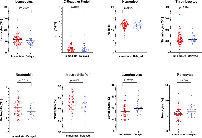
Maternal laboratory parameters. Irrespective of presenting symptoms at diagnosis, upon immediate
admission, women had higher levels of leucocytes (p = 0.006), absolute and relative neutrophils (p = 0.019 and
p = 0.009, respectively) and lymphocytes (p = 0.014). Delayed admission was associated with higher monocyte
counts (p = 0.004). Hemoglobin levels, thrombocytes and CRP were similar in all women, excluding those deliv-
ered by LSCS (Fig. 6).
Discussion
It is widely recognized that the diagnosis of fetal death causes great emotional turmoil in the affected parents.
It usually takes a certain time to come to terms with the sad diagnosis, and every woman adapts differently to
her coping strategies until she feels capable of handling the experience of birth and death of her child collid-
ing, leaving a long-lasting impact on herself and her partner. The admission to hospital for giving birth to the
deceased baby is an important event, and the medical care during this time may have a profound effect on the
future life of the bereaved f amily15.
By this study, we sought to elaborate on the clinical impact of the timing of hospital admission on maternal
and obstetric parameters to gather more evidence regarding care after fetal death during hospitalization. In the
small sample of our retrospective study, the affected women themselves selected the timing of their hospital
admission, unless they presented with immediate signs of labor or as an emergency. The majority of cases of fetal
death in this cohort were diagnosed during routine controls, and more than half of the women were admitted
on the same day of diagnosis. Upon admission, there was no prevalence in presenting symptoms, yet women
Scientific Reports | (2021) 11:18854 | https://doi.org/10.1038/s41598-021-98229-9 5
Vol.:(0123456789)www.nature.com/scientificreports/
Immediate admission Delayed admission
Total (n = 183) (n = 127) (n = 56) p-value
Maternal age (years)a 31 ± 7 31 ± 6 32 ± 7 0.094d
2 b
Body mass index (kg/m ) 24.5 (15.6–43.3) 25.1 (15.6–43.3) 24.2 (18.6–37.6) 0.669e
b
Para (n) 1 (0–9) 0 (0–9) 1 (0–9) 0.529e
None 144 (78.7%) 99 (78.0%) 45 (80.4%)
Previous LSCS (n) 1 30 (16.4%) 21 (16.5%) 9 (16.1%) 0.847f
Maternal 2 9 (4.9%) 7 (5.5%) 2 (3.6%)
None 103 (56.6%) 71 (56.3%) 32 (57.1%)
Fetal 51 (28.0%) 31 (24.6%) 20 (35.7%)
Presenting symptoms Maternal 12 (6.6%) 10 (7.9%) 2 (3.6%) 0.473f
Labor 16 (8.8%) 14 (11.1%) 2 (3.6%)
Not reported 0 (0.0%) 1 (0.1%) 0 (0.0%)
Gestational age (days)b 213 (147–290) 209 (147–290) 221 (152–288) 0.331e
0 17 (11.6%) 16 (15.1%) 1 (2.5%)
Fetal I 20 (13.7%) 17 (16.0%) 3 (7.5%)
Maceration grade < 0.001f
II 43 (29.5%) 34 (32.1%) 9 (22.5%)
III 66 (45.2%) 39 (36.8%) 27 (67.5%)
Vaginal cephalic 110 (60.1%) 78 (61.4%) 32 (57.1%)
Vaginal breech 53 (29.0%) 35 (27.6%) 18 (32.1%)
Delivery modec 0.465f
Ventouse 3 (1.6%) 1 (0.8%) 2 (3.6%)
LSCS 17 (9.3%) 13 (10.3%) 4 (7.1%)
10C 8 (47.1%) 7 (53.8%) 1 (25%)
10B 2 (11.8%) 2 (15.4%) 0 (0.0%)
5C 2 (11.8%) 2 (15.4%) 0 (0.0%)
Robson classification
6C 2 (11.8%) 1 (7.7%) 1 (25.0%) 0.130f
of LSCS
4B 1 (5.9%) 0 (0.0%) 1 (25.0%)
6B 1 (5.9%) 1 (7.7%) 0 (0.0%)
7C 1 (5.9%) 0 (0.0%) 1 (25.0%)
Time length of vaginal delivery (h)b 3 (0–28) 3 (0–28) 4 (0–26) 0.231e
b
2nd stage of labor (min) 10 (0–376) 10 (0–289) 18 (0–376) 0.197e
b
3rd stage of labor (min) 10 (0–90) 10 (0–78) 10 (0–90) 0.323e
b
Obstetrics Blood loss (ml) 200 (30–2500) 200 (30–2500) 150 (50–1000) 0.064e
None 100 (54.6%) 63 (49.6%) 37 (66.1%)
Peridural 36 (19.7%) 26 (20.5%) 10 (17.9%)
Epidural 21 (11.5%) 19 (20.5%) 2 (3.6%)
Anesthesiac 0.089f
Spinal 14 (7.7%) 10 (7.9%) 4 (7.1%)
Intubation 11 (6.0%) 9 (7.1%) 2 (3.6%)
Local 1 (0.5%) 0 (0.0%) 1 (1.8%)
None 181 (98.9%) 126 (99.2%) 55 (98.2%)
Episiotomyc 0.549f
Mediolateral 2 (1.1%) 1 (0.8%) 1 (1.8%)
None 154 (84.2%) 107 (84.3%) 47 (83.9%)
Vaginal tear 9 (4.9%) 8 (6.3%) 1 (1.8%)
1st degree tear 7 (3.8%) 5 (3.9%) 2 (3.6%)
Perineal injuryc 0.358f
Labial tear 6 (3.3%) 2 (1.6%) 4 (7.1%)
2nd degree tear 4 (2.2%) 3 (2.4%) 1 (1.8%)
Cervical tear 3 (1.6%) 2 (1.6%) 1 (1.8%)
Table 2. Comparison of fetomaternal characteristics and obstetric variables after stillbirth delivery between
immediate and delayed admission groups. LSCS lower segment caesarean section. a Mean ± Standard deviation.
b
Median (minimum − maximum). c Frequency (proportion). d Unpaired t-test with level of significance < 0.05.
e
Mann–Whitney U test with level of significance < 0.05. f Fishers’s exact test with level of significance < 0.05.
Robson classification: 10C: All singleton cephalic, ≤ 36 weeks (including previous LSCS): LSCS before labor;
10B: All singleton cephalic, ≤ 36 weeks (including previous LSCS): induced labor; 5C: Previous LSCS, singleton
cephalic, ≥ 37 weeks: LSCS before labor; 6C: All nulliparous breeches: LSCS before labor; 4B: Multipara,
singleton cephalic, ≥ 37 weeks: induced labor; 6B: All nulliparous breeches: induced labor; 7C: All multiparous
breeches (including previous LSCS): LSCS before labor.
Scientific Reports | (2021) 11:18854 | https://doi.org/10.1038/s41598-021-98229-9 6
Vol:.(1234567890)www.nature.com/scientificreports/
Figure 2. Forest plot illustrating the odds ratios (and 95% Confidence Intervals) from multivariate logistic
regression to identify the relationship between timing of hospital admission and possible predictors (i.e.,
weekday, parity, presenting symptom at diagnosis) following intrauterine fetal death (n = 183; level of
significance p < 0.05, two-tailed). IUFD intrauterine fetal death, OR odds ratio, CI confidence interval. Figure
software: Prism 9 for macOS, Version 9.2.0.
Figure 3. Time to stillbirth delivery after immediate (n = 127) and delayed (n = 56) hospital admission (nested
scatter dot plot; width of distribution of points proportional to the number of delivered women; Day 0 = hospital
admission day). Stillbirth delivery occurred in 20.8% (38/183) on Day 0; in 30.1% (55/183) on Day 1, and in
20.2% (37/183) on Day 3. Figure software: Prism 9 for macOS, Version 9.2.0.
with early signs of labor chose to be admitted immediately. Stillbirth delivery most commonly took place on the
first day after admission, yet immediately admitted women equally often delivered on admission day and the
day after, while delayed admitted women more commonly delivered on the day after admission, often following
IOL. Women, who were admitted immediately after diagnosis of fetal death, yet also delivered on the day of
their admission, were either in early gestational weeks and/or bared smaller fetal weight. Also, we observed a
reactive left-shift in the maternal laboratory parameters upon immediate admission: We assume that this might
have either contributed to accelerated laboring and delivery, or may have reflected the inflammatory response of
Scientific Reports | (2021) 11:18854 | https://doi.org/10.1038/s41598-021-98229-9 7
Vol.:(0123456789)www.nature.com/scientificreports/
Figure 4. Presenting symptoms on day of IUFD diagnosis in women who delivered on the same day of
hospitalization (n = 33 immediate admission; n = 5 delayed admission). Figure software: Prism 9 for macOS,
Version 9.2.0.
Figure 5. Methods of induction of labor following fetal death at the Department of Obstetrics and Gynecology,
Medical University of Vienna, Austria, between 2003 and 2019 (“Dinoprostone only” includes Propess, Prostin
E2; “Misoprostol only” includes Cytotec, Misodel, Cyprostol). Figure software: Prism 9 for macOS, Version 9.2.0.
labor itself. Finally, in our cohort, about one in ten women required LSCS following fetal death, the indications
of which were maternal comorbidities in the majority of cases, maternal request, or failed IOL. Whilst the rate
of intrapartum hemorrhage showed no group prevalence, generally, immediately admitted women had a greater
median amount of blood loss after vaginal delivery.
Despite the lack of comparable studies against which we may interpret our findings, our retrospective analysis
shows, that from a clinical standpoint, there seems to be no harm in allowing a delayed hospital admission for
induction following diagnosis of fetal death, neither in labor time, analgesia, birth injuries, and total blood loss,
nor in clinically relevant laboratory parameters, such as maternal hemoglobin, thrombocytes and CRP. These
findings underpin our previous observations, that maternal clotting disorders are uncommon and rarely directly
related to fetal death as such10.
Whilst our study is the first to provide data on the impact of admission timing in a comprehensively investi-
gated cohort of singleton IUFDs, we acknowledge several study limitations inherent to its retrospective design
and single-center nature: First and foremost, the small sample size may limit the power and significance of the
results. The sampling of the subgroups (e.g., mode of delivery; symptoms at diagnosis, methods for IOL) might
have further diminished the strength of the analyses in the compared groups. Also, the length of “delay” of
hospital admission was restricted to one to two days only, therefore, our results may not be generalizable and
translatable into other settings, where a longer watchful waiting or active surveillance is practiced after IUFD.
We, furthermore, acknowledge the failure to control for a possible selection bias in admission timing, either
patient or caregiver induced, and the different lengths of maternal exposure to the deceased fetus in utero, which
may have triggered labor, omitting the need for IOL, yet accelerating admission. Finally, we could not adjust for
errors and inconsistency in the medical documentation resulting in a recall bias by the attending physician at
the time of IUFD diagnosis and hospital admission.
Scientific Reports | (2021) 11:18854 | https://doi.org/10.1038/s41598-021-98229-9 8
Vol:.(1234567890)www.nature.com/scientificreports/
Figure 6. Comparison of maternal laboratory values between immediate versus delayed admission (n = 166,
excluding all women who delivered by caesarean section; Scatter dot plot with line at median; Mann–Whitney U
test; two-tailed Exact p-value with 99% confidence interval). Figure software: Prism 9 for macOS, Version 9.2.0.
After all, these limitations indicate that deeper research is needed. We propose, that in the ViewPoint software,
the electronic establishment of a structured under-category for the rare event of IUFD documentation (with
tick-boxes) would be of great value for future retrospective studies allowing direct and precise retrieval of a set
of important parameters, such as (a) the main circumstance that led to consultation and subsequent diagnosis; (b)
last time when the baby was felt; (c) any precipitating symptoms; (d) any recent illness or deterioration of a specific
co-morbidity which might have contributed to this event; (e) woman’s preference regarding the time of admission/
method of delivery. Also, qualitative evaluation of the motivation for immediate versus delayed admission may
provide important insights to improve care for the affected women.
Conclusion
Our study shows no clinical superiority of immediate over delayed hospital admission for stillbirth delivery.
Under stable medical circumstances, it, therefore, seems feasible to allow the woman to be discharged home
following the diagnosis of fetal death and be readmitted at a later time point for labor and delivery, when
appropriate.
Received: 18 May 2021; Accepted: 6 September 2021
References
1. Statistik Austria. Demographics on Austrian Population—Livebirths and Stillbirths 2020.
2. Royal College of Obstetricians and Gynaecologists. Late Intrauterine Fetal Death and Stillbirth Green-Top Guideline No. 55. (2010).
3. Lockwood, S. & Blackwood, M. Parental reactions to stillbirth. Aust. Fam. Phys. 12, 234–235 (1983).
4. Swinton, A. Grief and stillbirth. BMJ 1, 126 (1977).
5. Royal College of Obstetricians and Gynecologists. When your baby dies before birth. Patients Inf. Leaflets. (2012).
6. Muin, D. A. et al. Institutional guidelines on maternal care and investigations following antepartum stillbirth—A national survey.
BMC Pregnancy Childb. 21, 528 (2021).
7. Gold, K. J., Abdul-Mumin, A. R., Boggs, M. E., Opare-Addo, H. S. & Lieberman, R. W. Assessment of “fresh” versus “macerated”
as accurate markers of time since intrauterine fetal demise in low-income countries. Int. J. Gynaecol. Obstet. 125, 223–227 (2014).
8. Langley, F. A. The perinatal postmortem examination. J. Clin. Pathol. 24, 159–169 (1971).
Scientific Reports | (2021) 11:18854 | https://doi.org/10.1038/s41598-021-98229-9 9
Vol.:(0123456789)www.nature.com/scientificreports/
9. Romero, R., Copel, J. A. & Hobbins, J. C. Intrauterine fetal demise and hemostatic failure: The fetal death syndrome. Clin. Obstet.
Gynecol. 28, 24–31 (1985).
10. Muin, D. A. et al. Impact of fetal maceration grade on risk of maternal disseminated intravascular coagulation after intrauterine
fetal death—A retrospective cohort study. Sci. Rep. 8, 12742 (2018).
11. Robson, M. Classification of caesarean sections. Fetal Maternal Med. Rev. 12, 23–39 (2001).
12. NICE Guideline NG121. Intrapartum Care for Women with Existing Medical Conditions or Obstetric Complications and Their Babies.
(2019).
13. Reddy, U. M. et al. Stillbirth classification-developing an international consensus for research: Executive summary of a National
Institute of Child Health and Human Development workshop. Obstet. Gynecol. 114, 901–914 (2009).
14. Korteweg, F. J. et al. The Tulip classification of perinatal mortality: Introduction and multidisciplinary inter-rater agreement. BJOG
113, 393–401 (2006).
15. Nuzum, D., Meaney, S. & O’Donoghue, K. The impact of stillbirth on bereaved parents: A qualitative study. PLoS ONE 13, e0191635
(2018).
Acknowledgements
DAM is grateful to Prof. DDr. J.C. Huber for his enduring support.
Author contributions
D.M.: Project and Protocol development, Data collection and management, Data analysis, Manuscript writing and
editing.A.S.: Data collection and management, Data analysis.A.F.: Data management, Manuscript editing.H.K.:
Protocol development, Data collection and management.H.H.: Project development, Data collection and man-
agement, Data analysis, Manuscript editing.
Competing interests
The authors declare no competing interests.
Additional information
Supplementary Information The online version contains supplementary material available at https://doi.org/
10.1038/s41598-021-98229-9.
Correspondence and requests for materials should be addressed to D.A.M.
Reprints and permissions information is available at www.nature.com/reprints.
Publisher’s note Springer Nature remains neutral with regard to jurisdictional claims in published maps and
institutional affiliations.
Open Access This article is licensed under a Creative Commons Attribution 4.0 International
License, which permits use, sharing, adaptation, distribution and reproduction in any medium or
format, as long as you give appropriate credit to the original author(s) and the source, provide a link to the
Creative Commons licence, and indicate if changes were made. The images or other third party material in this
article are included in the article’s Creative Commons licence, unless indicated otherwise in a credit line to the
material. If material is not included in the article’s Creative Commons licence and your intended use is not
permitted by statutory regulation or exceeds the permitted use, you will need to obtain permission directly from
the copyright holder. To view a copy of this licence, visit http://creativecommons.org/licenses/by/4.0/.
© The Author(s) 2021
Scientific Reports | (2021) 11:18854 | https://doi.org/10.1038/s41598-021-98229-9 10
Vol:.(1234567890)You can also read