AN OPEN-LABELLED CONTROLLED CLINICAL TRIAL - January 2019 Santa Caterina Hospital
←
→
Page content transcription
If your browser does not render page correctly, please read the page content below
AN OPEN-LABELLED CONTROLLED CLINICAL TRIAL

         END OF TERM PROJECT
               January 2019

                Author: Ana Belén Cendrero Camacho
                       Tutor: Dr. René Robles Cedeño

           Neuroimmunology and Multiple Sclerosis Unit
                                Santa Caterina Hospital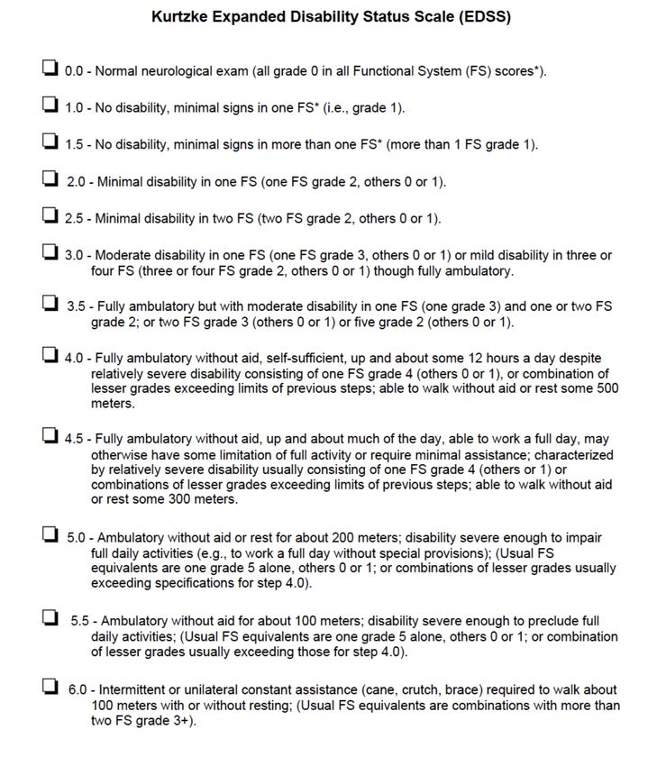
A mi ángel de la guarda
Acknowledgements

Thanks to Dr. René Robles for accompanying me in this last step and for
 showing me that what made me fall in love with Medicine is still alive.

   Y gracias a mi madre, por no rendirse nunca y no dejar que yo lo haga.
            Esto, como todo, como siempre, lo hemos conseguido juntas.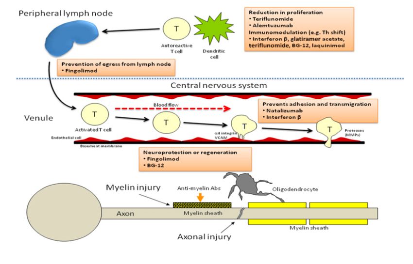
ABSTRACT

BACKGROUND: Multiple sclerosis (MS) is a chronic, immune-inflammatory disease of
the central nervous system. It is the main cause of non-traumatic disability in young
adults, affecting more than 2 million people worldwide. Despite the research carried out
by the scientific community to know the pathophysiology, today it remains uncertain.
Therefore, the only treatments currently available are aimed at preventing relapse and
stopping the progression of the disease, as well as treating the accompanying
symptoms. MS presents a very heterogeneous symptomatology with a great impact on
the quality of life of the patient, and this is the main reason why they are not properly
collected.

OBJECTIVE: The main objective of this trial is to determine if the proper collection and
management of symptoms that afflict MS patients can have a positive effect on their
quality of life (QoL). Furthermore, its influence on the quality of care will be evaluated
secondarily.

DESIGN: multi-centric, open-labelled, randomized controlled clinical trial.

PARTICIPANTS: 450 patients with an age ranged 18 to 65 diagnosed with MS according
to McDonald 2017 criteria that carry out their follow-ups in the Neuroimmunology and
Multiple Sclerosis Unit of Santa Caterina Hospital and other reference hospitals of
Catalonia.

INTERVENTION: participants will be randomly allocated in two groups of equal size. The
members of one group will conduct Patient Reported Outcomes Measures (PROMs)
questionnaires during five consecutive visits and the results of each of them will be
analysed in real time by the neurologist before consultation. Additionally, in the first and
last visit of the study they will have to fill out a QoL form and a Patient Reported
Experience Measures (PREMs) questionnaire to assess the quality of care. The other
group will only have to fill the QoL and PREMs questionnaires at the beginning and at
the end of the study.

KEY WORDS: Multiple sclerosis · PREMs · PROMs · Quality of life · Quality of care
INDEX

1     ABBREVATIONS .................................................................................................... 8

2     INTRODUCTION .................................................................................................. 10

    2.1       BACKGROUND ...................................................................................................... 10
      2.1.1      EPIDEMIOLOGY ............................................................................................................. 10
      2.1.2      AETIOLOGY AND RISK FACTORS ..................................................................................... 11
      2.1.3      PATHOPHYSIOLOGY....................................................................................................... 13
      2.1.4      SYMPTOMS AND CLINICAL PHENOTYPES ....................................................................... 16
      2.1.5      DIAGNOSIS .................................................................................................................... 20
      2.1.6      TREATMENT .................................................................................................................. 22
      2.1.7      PROGNOSIS ................................................................................................................... 27

    2.2       PROMS AND PREMS ............................................................................................. 28

    2.3       JUSTIFICATION ...................................................................................................... 29

3     BIBLIOGRAPHY.................................................................................................... 31

4     HYPOTHESIS ........................................................................................................ 35

    4.1       MAIN HYPOTESIS .................................................................................................. 35

    4.2       SECONDARY HYPOTESIS........................................................................................ 35

5     OBJETIVES ........................................................................................................... 35

    5.1       MAIN OBJETIVES ................................................................................................... 35

    5.2       SECONDARY OBJETIVES ........................................................................................ 35

6     MATERIALS AND METHODS ................................................................................ 36

    6.1       STUDY DESIGN ...................................................................................................... 36

    6.2       STUDY POPULATION ............................................................................................. 36
      6.2.1      INCLUSION CRITERIA ..................................................................................................... 36
      6.2.2      EXCLUSION CRITERIA ..................................................................................................... 36

    6.3       SAMPLE................................................................................................................. 37
      6.3.1      SAMPLE SIZE ................................................................................................................. 37
      6.3.2      SAMPLING METHOD...................................................................................................... 37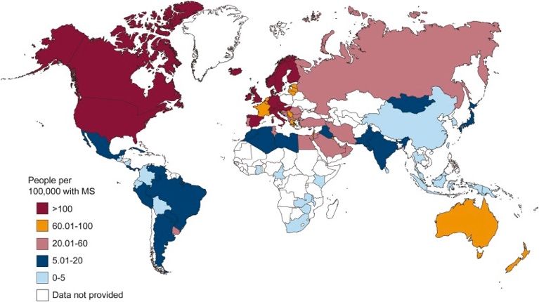
6.4 VARIABLES ............................................................................................................ 38
6.4.1 INDEPENDENT VARIABLES ............................................................................................. 38
6.4.2 DEPENDENT VARIABLE .................................................................................................. 40
6.4.3 COVARIATES.................................................................................................................. 41
6.5 PROCEDURES ........................................................................................................ 42
7 STATISTICAL ANALYSIS ........................................................................................ 43
7.1 DESCRIPTIVE ANALYSIS ......................................................................................... 43
7.2 BIVARIATE INFERENCE .......................................................................................... 43
7.3 MULTIVARIATE ANALYSIS ..................................................................................... 43
8 FEASIBILITY ......................................................................................................... 45
8.1 RESEARCH TEAM ................................................................................................... 45
8.2 WORK PLAN .......................................................................................................... 45
8.2.1 STAGE I: Protocol design................................................................................................ 45
8.2.2 STAGE II: Preparation and initial coordination................................................................ 45
8.2.3 STAGE III: Data collection .............................................................................................. 46
8.2.4 STAGE IV: Data analysis and article elaboration ............................................................. 46
8.2.5 STAGE V: Results publication and dissemination ............................................................ 47
8.3 STUYDY CHRONOGRAM........................................................................................ 47
9 ETHICAL CONSIDERATION ................................................................................... 48
10 STUDY LIMITATIONS ....................................................................................... 49
11 BUDGET .......................................................................................................... 51
12 ANNEXES ......................................................................................................... 53
ANNEX A. Clinical course of MS ......................................................................................... 53
ANNEX B. 2017 McDonald diagnostic criteria .................................................................... 54
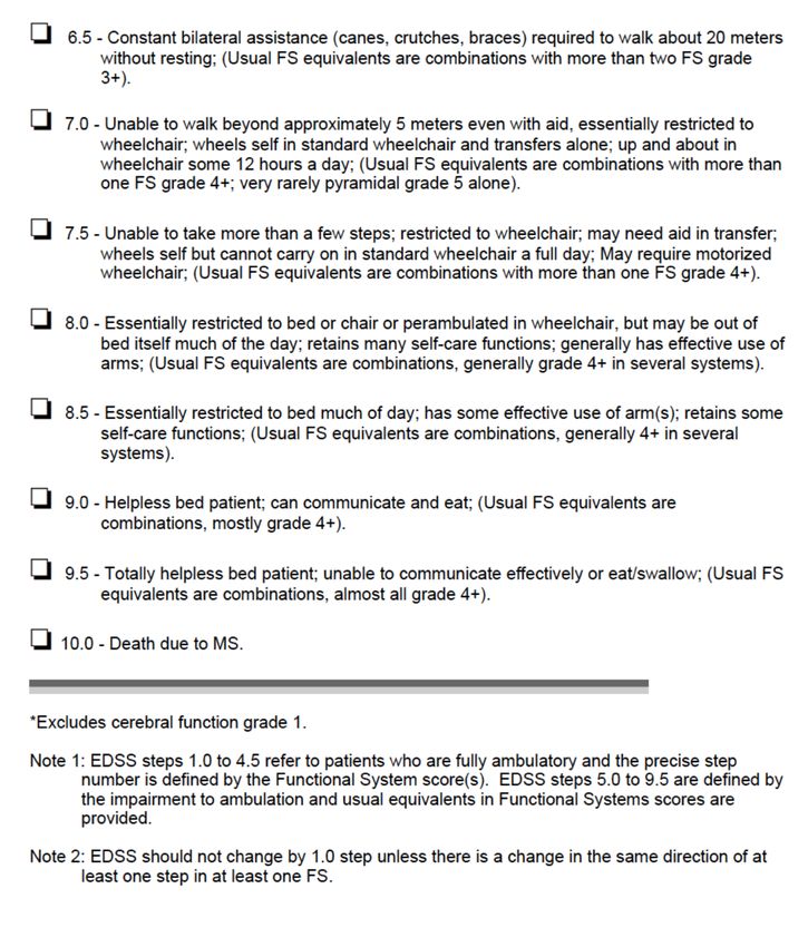
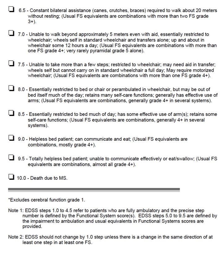
ANNEX C. Kurtzke Expanded Disability Status Scale (EDSS) ............................................... 55
ANNEX D. Patient Reported Outcomes Measures questionnaires..................................... 57
ANNEX E. EQ-5D-5L Health Questionnaire......................................................................... 60
ANNEX F. Picker Patient Experience Questionnaire (PPE-15) ............................................ 62
ANNEX G. Information sheet for participants.................................................................... 64
ANNEX H. Informed consent ............................................................................................. 69

ANNEX I Study chronogram ............................................................................................... 70

ANNEX J Multiple Sclerosis Neuropsychological Questionnaire (MSNQ)........................... 71

1       ABBREVATIONS

AEs         Adverse Effects
APC         Antigen-Presenting Cells
BBB         Blood-Brain Barrier
CEIC        Clinical Research Ethical Committee - “Comitè Ètic d’Investigació Clínica”
CIS         Clinical Isolated Syndrome
CNS         Central Nervous System
CSF         Cerebrospinal Fluid
DIS         Dissemination in Space
DIT         Dissemination in Time
DMT         Disease-Modifying Treatment
EBV         Epstein-Barr Virus
EDSS        Expanded Disability Status Scale
EP          Evoked Potentials
GA          Glatimer Acetate
HLA         Human Leukocyte Antigens
HRQoL       Health-related quality of life
IFN         Interferon beta
IgG         Immunoglobin G
IM          Infectious Mononucleosis
JCV         John Cunningham Virus
MHC         Major Histocompatibility Complex
MOG         Myelin Oligodendrocyte Protein
MRI         Magnetic Resonance Imaging
MS          Multiple Sclerosis
NRL         Neurologist
NRS         Numeric Rating Scale
OCB         Oligoclonal Bands
PI          Principal Investigator
PML         Progressive Multifocal Leukoencephalopathy

                                             8
PPMS   Primary Progressive Multiple Sclerosis
PRMS   Primary relapsing Multiple Sclerosis
QoL    Quality of Life
RRMS   Relapsing-Remitting Multiple Sclerosis
SPMS   Secondary Progressive Multiple Sclerosis
SS     Statistician
Th1    CD4+ T-helper 1
Th2    CD4+ T-helper 2
UVR    Ultra-Violet Radiation

                                   9
2      INTRODUCTION

2.1    BACKGROUND

2.1.1 EPIDEMIOLOGY

Multiple sclerosis (MS) is the most common chronic inflammatory demyelinating
disease of the central nervous system (CNS) and the main cause of neurological disability
in young adults. (1–3)

According to 2013 data 2.3 million people worldwide are diagnosed with MS. It has an
irregular geographical distribution, being more prevalent in Europe (108/100,000
population) and North America (140/100,000). Sub-Saharian Africa and East Asia are the
regions with lowest prevalence rate of MS. Spain is considered a high incidence area
with an estimated prevalence of between 80-125 cases per 100.000 inhabitants and it
presents an important variability between regions. Nowadays, the global incidence and
prevalence of MS tends to increase affecting mainly Europe and the Mediterranean
Basin, although this may be due to a better diagnosis thanks to the use of magnetic
resonance imaging (MRI), new diagnostic criteria and new treatments (1,4,5).

                           Fig. 1 Map of prevalence of MS by country (4)

This pathology is more frequent in women. Furthermore, the incidence has increase
mainly among women and the female-to-male ratio remains around 2:1. This fact can
be explained by the ease of women to consult for milder symptoms than men(1). This

                                               10
difference disappear in older people and the most aggressive forms tend to affect the
male (5).
There seem to be two peaks of incidence according to age, the first one at the third
decade of life and another around 40 years old. The age of onset is rare before 10 and
after 60 (5).
2.1.2 AETIOLOGY AND RISK FACTORS
Even though the aetiology of MS is still unknown, multiple studies point to an
autoimmune cause with a multifactor mechanism that is not completely known.
Apparently, several environmental factors can cause a dysregulation of the immune
system in genetically predisposed individuals (5,6).
2.1.2.1 Genetics factors
MS is not a genetic disorder, but it has a genetic component on its pathogenesis.
Therefore, it is not inherited directly from parents to children (5). This can be de
explanation of why the prevalence of MS varies within the same geographical latitude,
sex or breed.
Several studies support the existence of familial aggregation in MS and higher basal risk
within relatives of patients with MS of suffering the disease than the general population
(7). The incidence of MS among general population is less than 0,5%, whereas the
incidence for first degree relatives is 1,9 to 4,7%. The monozygotic twins display a
concordance of 34%, while the concordance rate between dizygotic twins falls to
approximately 2% (8). All this can vary depending on the sex of the sibling, parental MS
status, and patient onset age (9).
The HLA-DRB1 gene is the strongest genetic factor identified as influencing MS
susceptibility, specifically the DRB1*1501 allele of major histocompatibility complex II
(MHC) represents approximately 50% of the genetic risk of MS (10). Nowadays, genome
wide association studies has identified hundreds of additional variants outside of the
MHC that could be involved in the onset of the disease, all of which have modest
individual effects (7).
112.1.2.2 Environmental factors
- Geographical latitude– VitD. The incidence of MS increases as one moves away
from the equator. This is because the ultra-violet radiation (UVR) exposure and,
consequently, the synthesis of VitD is lower in these areas (8,11). VitD deficiency is
associated with an increased risk of relapse, increased brain atrophy and a more rapid
progression of the disease (12).
VitD modulates the immune response by increasing IL10 production of T cells, causing a
change of antigen presenting cells and CD4+ T cells to a less inflammatory profile. Also,
it decreases the proinflammatory cytokines and the blood-brain permeability (13).
However, different studies have shown contradictory results, so it is believed that VitD
and UVR are independently related to MS (8).
People who migrate during the adolescence acquire the risk of developing MS from the
area they arrive at, while if they do so later the risk is the same as the population of
origin, probably due to childhood infections closely related to MS (9).
- Epstein-Barr virus (EBV). The association between several infectious agents and
MS has been studied, but only EBV has been shown to be strongly associated to MS (14).
The risk of developing the disease is between 15 and 30 times higher in EBV-positive
cases depending of the age of infection, being higher when EVB is acquired during the
adolescence when it is presented symptomatically as a painful pharyngitis and fatigue.
This is the “kissing disease” or infectious mononucleosis (IM). The probability of
triggering MS after IM is 2.17 times higher than when the infection is asymptomatic. The
mechanism by which this occurs is not yet clear (15).
- Smoking. It is considered that smoking is a moderate risk factor for the
development of MS as for many autoimmune diseases. Also, tobacco smoke exposure
during childhood seems to be a risk factor for MS. Smoking can trigger the disease and
its course gets worse (8).
122.1.2.3 Other factors
There are many others risk factors related to MS, but its capacity to trigger the disease
cannot be demonstrated yet. Some of them are: emotional stress, alteration of
microbiota due to the diet or use of high spectrum antibiotics, obesity, age, estrogens
level (1,5,8).
2.1.3 PATHOPHYSIOLOGY
Multiple sclerosis is a complex neurodegenerative autoimmune disorder, characterized
by inflammation, demyelination and axonal degeneration (1)
2.1.3.1 Immunopathological aspects
The loss of self-tolerance toward myelin and other CNS antigens involves CD4+ myelin-
reactive T lymphocytes persistent peripheral activation, spontaneously or by interaction
with some exogenous factor (6,16). These cells disrupt the blood-brain barrier (BBB) on
their way to the CNS and, once there, they are reactivated by the antigen-presenting
cells (APC) , triggering an inflammatory cascade that increases inflation (17). According
to different studies, CD4+ T-helper 1 (Th1) cells releasing proinflammatory cytokines,
are the main inflammatory mediator. In contrast, CD4+ T-helper 2 (Th2) cells regulate
the activity of th1 cells by releasing interleukins. In MS there is a breach of Th1/Th2
balance in favour of Th1 cells (18).
During the process, many other cell lines perform an important role which is not
completely known. The difficulty in determining their function suggests the existence of
several etiopathogenic pathways that could explain the existence of the different
histopathological patterns and evolutionary courses in which the disease occurs (5,18).
One of the most involved cells are the B lymphocytes, which could participate in the
process in different ways: by producing antibodies against myelin and axons that would
explain the appearance of oligoclonal bands in the cerebrospinal fluid (CSF), as APCs or
regulating the inflammatory cascade by recruiting Th2 lymphocytes (19,20).
13Fig. 2 A model of MS pathogenesis from (21)
2.1.3.2 Histological aspects
MS lesions include breakdown of the BBB, multifocal inflammation, demyelination,
oligodendrocyte loss, reactive gliosis and axonal degeneration (22). These lesions can
lead to conduction blocks, neuronal hyperexcitability, and generation of ectopic
potentials responsible for the patient's clinic (5).
The pathological hallmark of MS is the demyelination plaques that appear throughout
the CNS, especially in the optic nerves, brainstem, cerebellum, periventricular white
matter and spinal cord (17).
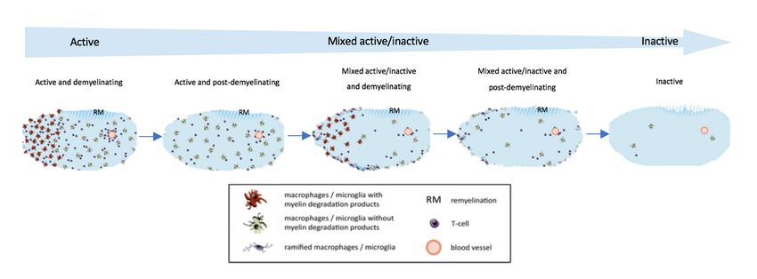
MS plaques can be classified histologically as active, mixed and inactive (6,23). The
active plaques are characterized by an intense infiltration of activated macrophages
loaded with myelin fragments, whereas in the inactive ones the cellular number is low
and there are no active fragmentation signs. However, inactive plaques display an
intense gliosis and a reduction of the axonal density and the number of
oligodendrocytes. Mixed plaques present intermediate characteristics, with a
hypocellular centre and a periphery of activated macrophages (5,23).
14Fig. 3 Temporal devolvement of MS lesions. Adapted from (23)
The plaques vary between patients but remain similar in the same individual. Four
patterns are distinguished (5,22):
1. Demyelination associated with macrophages.
2. Antibody mediated demyelination.
3. Demyelination associated with oligodendropathy.
4. Primary degeneration of oligodendrocytes with secondary destruction of
myelin.
In some cases, the presence of remyelination is possible. In acute plaques a wide
remyelination may occur giving rise to the named “shadow plaques” (6), whereas in
chronic or inactive ones remyelination is usually incomplete inducing an axonal
depletion (18).
Axonal loss is responsible for the loss of functionality and the degree of disability. It may
occur either at the acute point of the outbreak or more slowly on inactive demyelinated
plaques. Axonal loss could occur through a specific immunologic attack on the axon or
by the activation of substances that weaken and damage demyelinated axons. In
addition, these lesions are potential sources of excessive glutamate accumulation that
would activate and metabotropic receptors, resulting in toxic cytoplasmic Ca2+
accumulation and cell death (22).
152.1.4 SYMPTOMS AND CLINICAL PHENOTYPES
MS is a disease with a very heterogeneous clinical presentation due to the different CNS
lesions of sensory, motor, visual, and brainstem pathways (6,16). The type patient is a
young woman who presents a visual or sensory disorder of subacute character (24).
The most frequent are:
• Fatigue: it is the physical tiredness that is not correlated with the degree of
activity performed. Fatigue is the most frequent symptom of multiple sclerosis
and one of the most interfering in the patient daily life, affecting pproximately
90% of patients present it during the course of the disease). It is related to sleep
disorders and many other MS symptoms but not to the severity of the disease
and it is not considered an outbreak. Fatigue increases with body temperature
and during the summer months (24,25).
• Gait difficulties: it is the main cause of disability related to other disease factors
like spasticity but, especially, the weakness that preferentially affects the lower
extremities, and sensory deficits (26).
• Pain: approximately 70% of patients present neuropathic pain during ongoing
disease (24,25). There are studies that suggest that neither the degree of
disability, the age of initiation or the time since diagnosis determine which
patients suffer pain and which do not (26).
• Mental symptoms: cognitive and emotional changes, memory loss, difficulty
concentrating (24).
More than 50% of patients have some affective syndrome, mostly it is a
moderate depression (24). The frequency of depression is independent of the
disability degree (25), suggesting that CNS inflammation is a risk factor of
depression (26).
Frank dementia is not very common, however, between 34 and 65% present a
cognitive deterioration, especially in advanced cases, that mainly affects recent
memory and sustained attention (16).
16• Vision problems: optic neuritis, diplopia, oscillopsia, internuclear
ophthalmoplegia (24).
Optic neuritis (inflammation of the optic nerve) is is the first symptom of MS for
many people (4,26). It is a unilateral decrease in visual acuity, accompanied by
other symptoms such as photophobia, dyschromatopsia and pain that is
exacerbated by movements (24). Almost 100% of patients recover completely
after 2-6 months of onset (24,26).
Internuclear ophthalmoplegia is characterized by the loss of unilateral abduction
and horizontal nystagmus in contralateral abduction with conserved
convergence (24).
• Sensory symptoms: tingling, numbness, burning, tightness (24).
Vibratory sensitivity is the most affected due to lesions of the posterior cords.
The phenomenon of L'hermitte is very characteristic, consisting of an electric
shock-like sensation down the spine and into the limbs evoked by neck flexion
(16).
• Genitourinary, bowel and sexual symptoms: urinary retention or incontinence,
constipation or faecal incontinence, lack of vaginal lubrication, decreased libido,
difficulty in erection and ejaculation. Nearly 80 percent of patients will suffer
from any of these problems. All of them significantly disturb the quality of life
and, in addition, can worsen other symptoms (24,26).
Other less frequent symptoms of MS are: muscular atrophy, speech problems, tremor,
seizures, hearing loss.
According to how these symptoms occur, in 1996, four MS disease phenotypes were
defined: relapsing-remitting MS (RRMS), primary progressive (PPMS), secondary
progressive MS (SPMS) and progressive-relapsing MS (PRMS). Due to advances in
diagnostic imaging, this classification was revised and published updated in 2013. New
classification is based on central nervous system (CNS) lesion activity, according to
clinical relapses and MRI findings, and progression of disability and allows to know the
17evolution of the neurodegenerative process, determining in some way the prognosis
and the possible therapeutic interventions (27–29) (see ANNEX A).
- Relapsing-Remitting MS spectrum: RRMS is the most common clinical form.
Approximately 85% of people suffering MS have outbreaks of the disease (4) with full
recovery, or with sequelae upon recovery and periods between relapses characterized
by a lack of disease progression (6,27).
Clinically isolated syndrome (CIS) is now included in the RRMS disease spectrum. The
first clinical manifestation of MS and consistent with MS but isolated in time; may or
may not be isolated in space. It affects mainly the optic nerves, brainstem, or spinal cord
(6,27,29).
Fig. 4 The 1996 vs 2013 multiple sclerosis phenotype descriptions from (28)
- Progressive MS spectrum: includes any form of the disease characterized by
continuous worsening of neurological impairment over at least 6–12 months, this is
PPMS and SPMS (PRMS has being eliminated of the new classification) (20,27).
- Secondary progressive MS (SPMS): is the second most frequent phenotype of the
disease since 80% of patients with RRMS will go on to develop a progressive form (4)
with or without relapses. There is no test to determine the transition from RRMS to
SPMS (28).
18- Primary Progressive MS (PPMS): is the less frequent clinical form of MS, accounting for
about 5% of cases (4). Is characterized by a continuous progression of disability from the
onset. However, PPMS can present plateaus and temporary minor improvements during
it course (2). It usually affects in patients over 40% and there is no distinction by sex (24).
Fig. 5 The 1996 vs 2013 multiple sclerosis phenotype descriptions for progressive disease from (28)
- Assessment activity
• Clinically: showing evidence of new relapses or outbreaks, those symptoms or
signs of neurological dysfunction that last more than 24 hours or the marked
deterioration of a previously stabilized or absent symptom for at least 30 days
after excluding any other possible cause (24,25).
A “pseudo-outbreak” is the one that occurs in the context of fever (Uhthoff
phenomenon) or systemic disease with a variable duration from hours to days
(24,25).
• Imaging: new gadolinium enhancing lesions and/or new or enlarging T2 lesions
on MRI over a specified time period (30).
- Assessment progression
At this point it is necessary to differentiate between two concepts (25):
19• Worsening: increased disability confirmed over a specified time period as result
of a relapse or progressive disease.
• Disease progression: objective worsening of the disease confirmed over certain
period of time, with or without relapses. It is only used in cases of progressive-
phase disease.
2.1.5 DIAGNOSIS
There is no pathognomonic test for the diagnosis of MS. It continues to be based on the
clinical presentation, supported by the results of neuroimaging and, in some cases, by
the results of CSF analysis .and evoked potentials studies (6).
Different criteria have been proposed for the diagnosis of the disease. The most used in
current clinical practice are the McDonald criteria (see ANNEX B). These criteria are
based on the demonstration of dissemination in space (DIS) and time (DIT) of neurologic
signs and symptoms using clinical, laboratory and/or MRI data (31). It required
elimination of other possible diagnoses (32).
Fig. 6 Differential diagnosis of MS from (6)
20- Blood test: there is no definitive serum biomarker for MS, but it may reflect the
immune response situation and, what is more important, rule out other possible
diagnoses like infections, some hereditary diseases, or collagen-vascular diseases
among others that could mimic MS (33) (see Fig. 6). The most studied serum immune
biomarkers are immunoglobulin M against extracellular domain of myelin
oligodendrocyte protein (MOG) and antibodies specific for myelin basic protein (MBP)
(30).
- Magnetic resonance imaging (MRI): it is the most sensitive method that currently
exists for the assessment of DIT/DIS and the monitoring of the course of the disease
(7,33). Approximately 5 percent of people with MS do not initially show lesions on MRI
at the time of diagnosis (33).
MRI is particularly helpful in patients with CIS (33). About 70% of brain lesions and 30%
of spinal lesions develop without clinical evidence of relapse. MRI allows us to identify
new asymptomatic lesions (radiological relapses) that would confirm diagnosis of early
MS (30). In addition, the number of lesions that are seen on the MRI may establish the
risk of developing a second attack which allows to diagnosticate a “clinically-definite
MS”(30,33).
A brain MRI protocol includes several sequences necessary for the evaluation of the
patient with possible (34,35). Spinal cord MRI is recommended in patients with
symptoms at the spinal cord level or in patients with focal neurological signs and
negative brain MRI if the diagnosis of MS is still being weighed (30,34).
• T1- weighted images the acute lesions appear as hypointense areas. At times
they show dark areas called “black holes”, that are thought to indicate areas of
chronic nerve damage and disability (25). In order to differentiate them, black
holes should be persistent for at least 6 months (34). Gadolinium enhanced T1
supplies information about disease activity. When there is active inflammation,
the BBB is disrupted and gadolinium can enter and highlight the inflamed areas
(7,25,30,33).
• T2-weighted images provide information about the total amount of lesion area,
both old and new (33,34).
21• FLAIR (fluid attenuated inversion recovery) images are used to better identify
brain lesions associated with MS (33).
MS lesions seen on MRI are typically ovoid in shape and small size located mainly in the
periventricular white matter, although they can be found in other locations, and usually
arranged perpendicularly to the ventricles (7).
Once the disease is diagnosed, annual follow-up MRIs are recommended (33,34).
- Cerebrospinal fluid (CSF): It is obtained through a lumbar puncture and is not specific
for MS (31). Further, 5-10 percent of patients with MS never show CSF abnormalities,
hence it cannot confirm or rule out a diagnosis of MS by itself (33).
The findings of the CSF analysis that could lead to MS diagnosis are the result of an
abnormal immune response. These are (30,33):
• Elevated levels of immunoglobin G (IgG) antibodies and/or
• IgG oligoclonal bands. (OCB)
• Proteins that are the breakdown products of myelin.
- Evoked potential (EP): EP testing has been eliminated from the 2017 revised
McDonald criteria for the diagnosis MS (33) but it continues to be done in clinical
practice. Up to 50 percent of patients with MS present al slowdown of electrical
conduction caused by damage (demyelination) along different sensory pathways (30).
2.1.6 TREATMENT
Due to the wide range of symptoms, the approach has to be multidisciplinary and
includes three different aspects (36):
Fig. 7 Scheme of the MS approach adapted from (36)
222.1.6.1 Outbreak treatment
Not all outbreaks require treatment (36). The therapeutic options are:
• Methylprednisolone: high-dose corticosteroids shorten the duration of
symptoms and accelerate the recovery of function after relapses, without
modifying the progression of the disease (37). Its adverse effects (AEs) are
usually mild and transient (euphoria, depression, acne, insomnia, facial flushing,
transient HTA, fatigue) (36,38).
• Plasmapheresis: it is used in severe relapses or when there is no response to
methylprednisolone (37,39).
2.1.6.2 Disease-modifying treatment (DMT)
MS has no cure (40). The currently authorized treatments only act on the inflammatory
phase of MS and their aim is to reduce the activity of the disease, by decreasing the
number of relapses and preventing the appearance of new lesions in MRI, and delay the
progression of the same (41). To achieve this, it is important that the patient becomes
aware of the transcendence of adherence to treatment (25).
- First-line drugs: the effectiveness of these treatments is moderate, but they have
a good safety profile (36).
- Interferon beta (IFN): is obtained through the biotechnological processing of
one of the natural interferons, and it acts modulating the activity of T and B cells
and reducing the disruption of the BBB by a mechanism that is not known exactly
(42). All IFNs are a once or several times a week injectable treatments,
subcutaneously (Betaseron® and Rebif®) or intramuscularly (Avonex®) (41),
except pegylated form of subcutaneous IFN (Plegridy®), whose long half-life
allows administration every 2-4 weeks (43). It is approved for CIS and active
progressive forms of MS, decreasing relapse rate by one-third (44). Its main AEs
are flu-like symptoms, hepatotoxicity, inflammatory reactions and pain at the
injection site, anemia and thrombocytopenia (36).
- Glatiramer acetate (GA): it is an acetate of synthetic polypeptides that mimic
and compete with the myelin basic protein, blocking myelin-damaging T-cells
23(41). GA (Copaxone®) is approved for CIS and RRMS with an efficacy similar to
IFN (44). It is an injectable solution three times a week with few adverse effects
(45). The most common are inflammatory reactions and at the injection site,
lipoatrophy and skin necrosis (important to rotate the injection area with each
injection) and post-injection reaction (46). Due to its safety profile, it is the most
indicated during pregnancy, although none of them is authorized for it (44).
- Teriflunomide (Aubagio®): it is once-daily oral DMT. It has an anti-inflammatory
effect by inhibiting a pyrimidine synthesis, which in turn reduces the
proliferation of T and B immune cells (47). In general, it is a well-tolerated drug.
Its main AEs are: gastrointestinal symptoms, weak hair and analytical alterations
(hepatotoxicity and lymphopenia) (48). Aubagio has an efficacy of 34% and it is
indicated for patients with relapsing forms of MS as well as for patients with a
MS clinical first episode of, but no during pregnancy and lactation due to its
prolonged half-life and teratogenicity (41).
- Dimethyl fumarate/BG-12 (Tecfidera®): it is twice-daily oral treatment (41). BG-
12 activates the nuclear-related factor 2 transcriptional pathway, which is
related to anti-inflammatory and anti-oxidant properties (49). Several studies
show an efficiency greater 50% in the treatment of relapsing MS (41). It is a safe
DMT with self-limited AEs: gastrointestinal intolerance (nausea, pain, dyspepsia)
and flushing (50).
- Second-line drugs: are high-efficacy (60-70%) drugs indicated in case of
therapeutic failure of first-line drugs or as first-line therapy for early aggressive
MS. These treatments present a slightly more complex security profile
(36,41,44).
- Fingolimod: it is an oral therapy taken once per day (41). Finglolimod (Gilenya®)
is a sphingosine 1-phosphate receptor modulator and acts trapping lymphocytes
in the lymph nodes. This way, those cells cannot cross the BBB into the central
nervous system, thereby reducing inflammatory damage (51). This DMT
produces lymphopenia (41), which makes the organism more susceptible to viral
24infections, especially varicella zoster. It has a first dose effect consisting of
bradycardia that requires clinical observation for 6 hours after (36,41).
- Natalizumab (Tysabri®): it is a humanized monoclonal antibody that blocks
α4-integrine and prevents lymphocytes from attaching to the cerebral vascular
endothelium and reaching the CNS (52). This treatment is administered
intravenously once a month and have a good safety profile, except for a risk of
developing progressive multifocal leukoencephalopathy (PML) in selected cases,
an infection of the CNS with the John Cunningham virus (JCV)(53). To know the
individual risk of developing PML, a serological anti-JCV test is routinely
performed and it is recommended to repeat the test every 6 months(54).
- Alemtuzumab (Lemtrada®): is a humanized monoclonal antibody directed
against CD52, a protein on the surface of lymphocytes and monocytes (41). The
aim of this intravenous therapy is to “reset” the immune system. The treatment
consists of an initial cycle of 5 doses of alemtuzumab and a new cycle of 3 doses
one year later. After that, it may be extended annually (55). Up to 20% of patients
develop autoimmune thyroid disease and almost 1% have idiopathic
thrombocytopenic purpura (41).
- Ocrelizumab (Ocrevus®): it has recently been approved and it is the only DMT
that has shown efficacy in the treatment of both the remitting and PPMS forms
(56). Ocrelizumab is a humanized monoclonal antibody that targets CD20
positive B lymphocytes similar to rituximab (another anti-CD20 agent)(41,56),
but less immunogenic with repeated infusions and with better benefit–risk
profile than rituximab (56). Ocrelizumab completely decreases the CD19+
(marker of B-cell counts in anti-CD20-treated patients) B-cell count in blood after
2 weeks and maintains it for 6 to 9 months. It is administered intravenously
every 6 months after a second dose in the second week of the start of treatment.
More common AEs of this drug are respiratory tract infections and infusion
reactions (it is recommended to pre-medicate with methylprednisolone and
antihistamine approximately 30 minutes prior to each Ocrevus infusion to
reduce the frequency and severity of these reactions) (56,57).
25Fig. 8 Immunopathogenic mechanisms in MS and proposed targets of
different disease modifying therapies from (6)
- Third-line therapies: in patients not responding to any of the previous treatment
lines or in patients with a PPMS form, other strategies can be used, such as
rituximab, cyclophosphamide or an autologous hematopoietic stem cell
transplantation (36).
The selection of the treatment is based on the consensus developed by the Spanish
Society of Neurology in 2016 (44). The main aspects to consider are patient preference,
MS activity and the degree of neurological impairment, always bearing in mind the
benefit-risk profile of the drug (36,41,44).
2.1.6.3 Symptomatic treatment
The objective is to treat the accompanying symptoms of MS that make the daily life of
patients difficult and thus promote well-being and improve their quality of life (QoL)
(58).
Fatigue and gait difficulties are the most disabling symptoms of the disease (24). Physical
exercise and physiotherapy are baseline to improve the patient autonomy, but it is also
important to avoid habits that excessively increase body temperature. If these strategies
26fail it will be necessary to resort to pharmacological treatments such as amantadine,
modafinil, fluoxetine, fampridine and others (25).
Besides, it is important to avoid the risk factors that intervene in the pathogenesis of the
disease. To avoid tobacco and any other toxic substance, control classic cardiovascular
risk factors and maintain a diet that follows the recommendations of the Mediterranean
diet can positively influence the situation of patients. However, VitD supplementation is
not recommended, except in case of deficiency (38).
2.1.7 PROGNOSIS
The great clinical variability of MS prevents knowing the possible evolution of the
disease on an individual level (41). Development of a progressive course is the most
important factor associated with long-term outcome. The clinical and demographic
feature that the patient presents at the onset of the disease can be used as progression
predictors (see Table 1), but none of the are able to predict the rate of this progression.
Paraclinical tests are the best tool for foretell the risk of CIS conversion into clinically
definite multiple sclerosis, relapses, recovery level and later disability. MRI is the most
sensitive, but also immunological markers such as OCB and IgM anti-MOG, and EP
studies (59).
Table 1 Prognosis factors summary adapted from (1,59)
GOOD PROGNOSIS FACTORS BAD PROGNOSIS FACTORS
Age < 25 Age > 25
Female Male
Relapsing-remitting phenotype Progressive phenotype
Onset: optic neuritis, sensory problems Onset: motor, cerebellar or spinal problems
Unifocal onset Polysymptomatic onset
EDDS < 3 EDDS >3
Full recovery from the initial attack Incomplete recovery from the initial attack
Low relapse frequency in the first 2-5 years High relapse frequency in the first 2-5 years
Low disability after 5 years High disability after 5 years
Longer interval between first two attacks Shorter interval between first two attacks
27Two-stage disability progression in MS is defined by using two scores on the Kurtzke
Extended Disability Status Scale (EDSS) (see ANNEX C) as benchmarks of neurological
impairment accumulation (1,41): an early “phase 1” from onset to irreversible EDSS 3
during which focal inflammatory lessons influence disability progression; and later
“phase 2” from EDSS 3 to EDSS 6 during which focal inflammatory lessons influence
disability progression and, therefore, independent of “phase 1”. Predictive factors of
progression only influence first phase (60).
Fig. 9 Two-stage disability progression in MS from(1)
Although patients with MS have a life expectancy similar to that of the general
population, especially in the first 20 years of the disease, survival seems to be reduced
from 6 to 14 years. The average time from onset of symptoms to death varies from 24
to 45 years (1). Progressive disability leads to severe handicaps, which increase the risk
of infections and respiratory-related diseases, first cause of death followed by
cardiovascular problems and cancer (38,61). It should also be noted that suicide is from
1.6 to 7.5 times more common than in the general population (62).
2.2 PROMS AND PREMS
Patient reported outcome measures (PROMs) are a tool that provides a quantification
of symptoms which cannot be measured objectively using validated generic and MS
specific scales (63). They are a method to assess patients QoL identified by themselves
(64). Furthermore, PROMs make possible to detect worsening of symptoms, provide
information that may have otherwise been missed and enhance shared decision making
and patient engagement (65,66).
28Some studies also suggest that PROMs improve the quality of health care, care
coordination and even reduce costs and increase efficacy (67). In this way, patient
reported experience measures (PREMs) reflect patient perception of their experience
with health care through questionnaires that evaluate items such as waiting time,
quality of communication or knowledge about their own process. At the same time,
better experiences seem to associated with better outcomes (68).
2.3 JUSTIFICATION
Multiple sclerosis is an autoimmune demyelinating chronic, and potentially progressive,
disorder of the CNS. It has a complex pathophysiological mechanism that is not
completely known, which makes it difficult to predict the possible evolution and
prognosis of the patient, as well as the formulation of a curative treatment. Its incidence
is increasing, especially in developed countries, that affects young people, mainly
women, and presents a wide range of symptoms that the patient identifies as disabling
and, therefore, with a great impact on his quality of life.
Considering that the MS tends to debut at an early age and that, despite not having
curative treatment, life expectancy is close to that of the general population,
therapeutic efforts should be directed to try to stop the progression of the disease and
normalize the patient daily life as much as possible.
In routine clinical practice, due to the heterogeneity of MS symptoms, many of them are
obviated or not picked up correctly. PROMs questionnaires make it possible to collect
more quickly and effectively of this information and minimize the burden of data
collection during the visit.
This study aims at capturing the symptoms that the patient identifies as more disabling,
by using PROMs questionnaires, in order to evaluate how they relate to the patient QoL,
and if the correct management of them has a positive impact on the health-related QoL.
According to the literature, electronic systems linked to a registry enable an easier
collection of this data and afford timely feedback to clinicians so that could take
measures that improve the functionality and quality of life of the patient and,
29consequently, improve the quality of care by focusing the visit on the problems that
really afflict the patient and promoting a better patient-clinician communication.
303 BIBLIOGRAPHY
1. Leray E, Moreau T, Fromont A, Edan G. Epidemiology of Multiple Sclerosis. Neurol
Clin. 2016;34(4):919–39.
2. Pugliatti M, Rosati G, Carton H, Riise T, Drulovic J, Vécsei L, et al. The epidemiology
of multiple sclerosis in Europe. Eur J Neurol. 2006;13(7):700–22.
3. Kingwell E, Marriott JJ, Jetté N, Pringsheim T, Makhani N, Morrow SA, et al.
Incidence and prevalence of multiple sclerosis in Europe: A systematic review.
BMC Neurol. 2013;13.
4. Atlas of MS 2013: Mapping Multiple Sclerosis around the world [Internet].
Sclerosis International Federation. 2013. Available from: www.msif.org
5. Costa-Frossard L. Esclerosis múltiple: introducción, epidemiología, fisiopatogenia.
In: Manejo del paciente con esclerosis múltiple en atención primaria: diagnóstico,
pronóstico y tratamiento. 2017. p. 5–16.
6. Garg N, Smith TW. An update on immunopathogenesis, diagnosis, and treatment
of multiple sclerosis. Brain Behav. 2015;5(9).
7. Milo R, Miller A. Revised diagnostic criteria of multiple sclerosis. Autoimmun Rev.
2014;13(4–5):518–24.
8. Pantazou V, Schluep M, Du Pasquier R. Environmental factors in multiple
sclerosis. Vol. 44, Presse Medicale. 2015.
9. B. K, F.D. L. The genetics of multiple sclerosis. A review. Biomed Pharmacother.
1999;53(8):358–70.
10. Westerlind H, Ramanujam R, Uvehag D, Kuja-Halkola R, Boman M, Bottai M, et al.
Modest familial risks for multiple sclerosis: A registry-based study of the
population of Sweden. Brain. 2014;137(3):770–8.
11. Gale CR, Martyn CN. Migrant studies in multiple sclerosis. Prog Neurobiol. 47(4–
5):425–48.
12. Ascherio A, Munger KL, White R, Köchert K, Simon KC, Polman CH, et al. Vitamin
D as an Early Predictor of Multiple Sclerosis Activity and Progression. JAMA
Neurol. 2014 Mar 1;71(3):306.
13. Hernández-Pedro NY, Espinosa-Ramirez G, de la Cruz VP, Pineda B, Sotelo J. Initial
Immunopathogenesis of Multiple Sclerosis: Innate Immune Response. Clin Dev
Immunol. 2013;2013:1–15.
14. Ascherio A, Munger KL. Environmental risk factors for multiple sclerosis. Part I:
The role of infection. Ann Neurol. 2007;61(4):288–99.
15. Handel AE, Williamson AJ, Disanto G, Handunnetthi L, Giovannoni G,
Ramagopalan S V. An Updated Meta-Analysis of Risk of Multiple Sclerosis
following Infectious Mononucleosis. Jacobson S, editor. PLoS One. 2010 Sep
1;5(9):e12496.
16. Hauser SL, Oksenberg JR. The Neurobiology of Multiple Sclerosis: Genes,
Inflammation, and Neurodegeneration. Neuron. 2006 Oct 5;52(1):61–76.
17. Mahad DH, Trapp BD, Lassmann H. Pathological mechanisms in progressive
multiple sclerosis. Lancet Neurol. 2015;14(2).
18. Yadav SK, Mindur JE, Ito K, Dhib-Jalbut S. Advances in the immunopathogenesis
of multiple sclerosis. Curr Opin Neurol. 2015;28(3):206–19.
3119. Lehmann Horn K, Kronsbein HC, Weber MS. Targeting B cells in the treatment of
multiple sclerosis: Recent advances and remaining challenges. Ther Adv Neurol
Disord. 2013;6(3):161–73.
20. Correale J, Gaitán MI, Ysrraelit MC, Fiol MP. Progressive multiple sclerosis: from
pathogenic mechanisms to treatment. Brain. 2017;140(3):527–46.
21. Large-scale gene-expression studies and the challenge of multiple sclerosis
[Internet]. [cited 2019 Jan 11]. Available from: http://www.mult-
sclerosis.org/news/Oct2002/FullTextLargeScaleGeneExpressionStudiesOfMS.ht
ml
22. Dutta R, Trapp BD. Relapsing and progressive forms of multiple sclerosis: Insights
from pathology. Curr Opin Neurol. 2014;27(3):271–8.
23. Kuhlmann T, Ludwin S, Prat A, Antel J, Brück W, Lassmann H. An updated
histological classification system for multiple sclerosis lesions. Acta Neuropathol.
2017 Jan 17;133(1):13–24.
24. Martínez M. Manifestaciones clínicas de la esclerosis múltiple. In: Manejo del
paciente con esclerosis múltiple en atención primaria: diagnóstico, pronóstico y
seguimiento. Luzán 5; 2017. p. 17–21.
25. Yusta A. Evaluación y control del paciente con esclerosis múltiple. In: Manejo del
paciente con esclerosis múltiple en atención primaria: diagnóstico, pronóstico y
seguimiento. Luzán 5; 2017. p. 33–41.
26. MS Symptoms : National Multiple Sclerosis Society [Internet]. [cited 2019 Jan 13].
Available from: https://www.nationalmssociety.org/Symptoms-Diagnosis/MS-
Symptoms
27. Klineova S, Lublin FD. Clinical Course of Multiple Sclerosis. Cold Spring Harb
Perspect Med. 2018 Sep;8(9):a028928.
28. Lublin FD, Reingold SC, Cohen JA, Cutter GR, Sørensen PS, Thompson AJ, et al.
Defining the clinical course of multiple sclerosis: the 2013 revisions. Neurology.
2014 Jul 15;83(3):278–86.
29. Lublin FD. New Multiple Sclerosis Phenotypic Classification. Eur Neurol.
2014;72(s1):1–5.
30. Karussis D. The diagnosis of multiple sclerosis and the various related
demyelinating syndromes: A critical review. J Autoimmun. 2014;48–49:134–42.
31. Kister I, Bacon TE, Chamot E, Salter AR, Cutter GR, Kalina JT, et al. Natural history
of multiple sclerosis symptoms. Int J MS Care. 2013;15(3):146–58.
32. National Multiple Sclerosis Society. Updated McDonald Criteria Expected to
Speed the Diagnosis of MS and Reduce Misdiagnosis.
33. Diagnosing MS : National Multiple Sclerosis Society [Internet]. [cited 2019 Jan 13].
Available from: https://www.nationalmssociety.org/Symptoms-
Diagnosis/Diagnosing-MS
34. Li D, Traboulsee A, Coyle PK, Arnold DL, Barkhof F, Grossman R, et al. Standardized
MR Imaging Protocol for Multiple Sclerosis : Consortium of MS Centers
Consensus. Am J Neuroradiol. 2006;27(2):455–61.
35. Chen JJ, Carletti F, Young V, Mckean D, Quaghebeur G. MRI differential diagnosis
of suspected multiple sclerosis. Vol. 71, Clinical Radiology. 2016.
36. García Domínguez J. Tratamiento de la esclerosis múltiple. In: Manejo del
paciente con esclerosis múltiple en atención primaria: diagnóstico, pronóstico y
seguimiento. Luzán 5; 2017. p. 23–32.
3237. Managing Relapses : National Multiple Sclerosis Society [Internet]. Available
from: https://www.nationalmssociety.org/Treating-MS/Managing-Relapses
38. Arrieta E. Comorbilidad en esclerosis múltiple. In: Manejo del paciente con
esclerosis múltiple en atención primaria: diagnóstico, pronóstico y tratamiento.
Luzán 5; 2017. p. 53–9.
39. Meca-Lallana JE, Rodríguez-Hilario H, Martínez-Vidal S, Saura-Luján I, Carretón-
Ballester A, Escribano-Soriano JB, et al. [Plasmapheresis: its use in multiple
sclerosis and other demyelinating processes of the central nervous system. An
observation study]. Rev Neurol. 37(10):917–26.
40. Huang W-J, Chen W-W, Zhang X. Multiple sclerosis: Pathology, diagnosis and
treatments. Exp Ther Med. 2017 Jun;13(6):3163–6.
41. Wingerchuk DM, Carter JL. Multiple sclerosis: Current and emerging disease-
modifying therapies and treatment strategies. Mayo Clin Proc. 2014;89(2):225–
40.
42. Dhib-Jalbut S. Mechanisms of action of interferons and glatiramer acetate in
multiple sclerosis. Neurology. 2002 Apr 23;58(8 Suppl 4):S3-9.
43. Newsome SD, Scott TF, Arnold DL, Nelles G, Hung S, Cui Y, et al. Long-term
outcomes of peginterferon beta-1a in multiple sclerosis: results from the
ADVANCE extension study, ATTAIN. Ther Adv Neurol Disord.
2018;11:1756286418791143.
44. Garcia-Merino JA. Consenso para el tratamiento de la esclerosis múltiple nola de
Neurología 2016. 2017;32(2).
45. Khan O, Rieckmann P, Boyko A, Selmaj K, Zivadinov R, GALA Study Group. Three
times weekly glatiramer acetate in relapsing-remitting multiple sclerosis. Ann
Neurol. 2013 Jun;73(6):705–13.
46. FDA. COPAXONE HIGHLIGHTS OF PRESCRIBING INFORMATION [Internet]. 2018.
Available from:
https://www.copaxone.com/Resources/pdfs/PrescribingInformation.pdf
47. Claussen MC, Korn T. Immune mechanisms of new therapeutic strategies in MS:
teriflunomide. Clin Immunol. 2012 Jan;142(1):49–56.
48. AUBAGIO® (teriflunomide) | Official Healthcare Professional Site [Internet].
Available from: https://www.aubagiohcp.com/
49. Albrecht P, Bouchachia I, Goebels N, Henke N, Hofstetter HH, Issberner A, et al.
Effects of dimethyl fumarate on neuroprotection and immunomodulation. J
Neuroinflammation. 2012 Jul 7;9(1):163.
50. FDA. TECFIDERA HIGHLIGHTS OF PRESCRIBING INFORMATION [Internet]. 2017
[cited 2019 Jan 15]. Available from:
https://www.tecfidera.com/content/dam/commercial/multiple-
sclerosis/tecfidera/pat/en_us/pdf/full-prescribing-info.pdf
51. Cohen JA, Chun J. Mechanisms of fingolimod’s efficacy and adverse effects in
multiple sclerosis. Ann Neurol. 2011 May;69(5):759–77.
52. Ransohoff RM. Natalizumab for Multiple Sclerosis. N Engl J Med. 2007 Jun
21;356(25):2622–9.
53. Ho P-R, Koendgen H, Campbell N, Haddock B, Richman S, Chang I. Risk of
natalizumab-associated progressive multifocal leukoencephalopathy in patients
with multiple sclerosis: a retrospective analysis of data from four clinical studies.
Lancet Neurol. 2017 Nov;16(11):925–33.
3354. Gorelik L, Lerner M, Bixler S, Crossman M, Schlain B, Simon K, et al. Anti-JC virus
antibodies: implications for PML risk stratification. Ann Neurol. 2010
Sep;68(3):295–303.
55. FDA. LEMTRADA HIGHLIGHTS OF PRESCRIBING INFORMATION [Internet]. 2001
[cited 2019 Jan 15]. Available from:
https://www.accessdata.fda.gov/drugsatfda_docs/label/2018/103948s5160_51
65lbl.pdf
56. Mulero P, Midaglia L, Montalban X. Ocrelizumab: a new milestone in multiple
sclerosis therapy. Vol. 11, Therapeutic Advances in Neurological Disorders. 2018.
57. FDA. OCREVUS HIGHLIGHTS OF PRESCRIBING INFORMATION [Internet]. 2017
[cited 2019 Jan 15]. Available from:
https://www.gene.com/download/pdf/ocrevus_prescribing.pdf
58. Dalgas U. Rehabilitation and multiple sclerosis: hot topics in the preservation of
physical functioning. J Neurol Sci. 2011 Dec 1;311 Suppl 1:S43-7.
59. Swanton J, Fernando K, Miller D. Early prognosis of multiple sclerosis. In:
Handbook of clinical neurology. 2014. p. 371–91.
60. Leray E, Yaouanq J, Le Page E, Coustans M, Laplaud D, Oger J, et al. Evidence for
a two-stage disability progression in multiple sclerosis. Brain. 2010 Jul
1;133(7):1900–13.
61. Lalmohamed A, Bazelier MT, Van Staa TP, Uitdehaag BMJ, Leufkens HGM, De Boer
A, et al. Causes of death in patients with multiple sclerosis and matched referent
subjects: a population-based cohort study. Eur J Neurol. 2012 Jul 1;19(7):1007–
14.
62. Scalfari A, Knappertz V, Cutter G, Goodin DS, Ashton R, Ebers GC. Mortality in
patients with multiple sclerosis. Neurology. 2013 Jul 9;81(2):184–92.
63. Clark R, Welk B. Patient reported outcome measures in neurogenic bladder.
Transl Androl Urol. 2016;5(1):22–30.
64. Giovannetti AM, Pietrolongo E, Giordano A, Cimino V, Campanella A, Morone G,
et al. Individualized quality of life of severely affected multiple sclerosis patients:
practicability and value in comparison with standard inventories. Qual Life Res.
2016;25(11):2755–63.
65. Worthen VE, Lambert MJ. Outcome oriented supervision: Advantages of adding
systematic client tracking to supportive consultations. Couns Psychother Res.
2007;7(1):48–53.
66. Marshall S, Haywood K, Fitzpatrick R. Impact of patient-reported outcome
measures on routine practice: A structured review. J Eval Clin Pract.
2006;12(5):559–68.
67. Conway PH, Mostshari F, Clancy C. The Future of Quality Measurement for
Improvement and Accountability. J Am Med Assoc. 2013;309(21):2215–6.
68. Black N, Varaganum M, Hutchings A. Relationship between patient reported
experience (PREMs) and patient reported outcomes (PROMs) in elective surgery.
BMJ Qual Saf. 2014;23(7):534–43.
34You can also read