A comprehensive review on the carcinogenic potential of bisphenol A: clues and evidence
←
→
Page content transcription
If your browser does not render page correctly, please read the page content below
Environmental Science and Pollution Research
https://doi.org/10.1007/s11356-021-13071-w

    REVIEW ARTICLE

A comprehensive review on the carcinogenic potential of bisphenol
A: clues and evidence
Nadeem Ghani Khan 1 & Jacinta Correia 1 & Divya Adiga 1 & Padmalatha Satwadi Rai 2 & Herman Sunil Dsouza 3 &
Sanjiban Chakrabarty 1,4 & Shama Prasada Kabekkodu 1,4

Received: 1 December 2020 / Accepted: 17 February 2021
# The Author(s) 2021

Abstract
Bisphenol A [BPA; (CH3)2C(C6H4OH)2] is a synthetic chemical used as a precursor material for the manufacturing of plastics
and resins. It gained attention due to its high chances of human exposure and predisposing individuals at extremely low doses to
diseases, including cancer. It enters the human body via oral, inhaled, and dermal routes as leach-out products. BPA may be
anticipated as a probable human carcinogen. Studies using in vitro cell lines, rodent models, and epidemiological analysis have
convincingly shown the increasing susceptibility to cancer at doses below the oral reference dose set by the Environmental
Protection Agency for BPA. Furthermore, BPA exerts its toxicological effects at the genetic and epigenetic levels, influencing
various cell signaling pathways. The present review summarizes the available data on BPA and its potential impact on cancer and
its clinical outcome.

Keywords Bisphenol A, . Endocrine disruptor, . Carcinogen, . Environmental toxicant, . Human cancer

Introduction                                                            (more than 6 × 10⁹ lb/year) (Gao et al. 2015). BPA was
                                                                        synthetized by Alexander Dianin and has been commercially
Bisphenol A (BPA) is an endocrine-disrupting synthetic                  available since 1957 (Hoque 2019). BPA is a colorless, solid
chemical used to manufacture consumer products such as wa-              organic compound with the chemical formula
ter bottles, water pipes, and food cans. It is one of the most          (CH3)2C(C6H4OH)2. It is a 4,4’-methanediyldiphenol with
abundant industrial synthetic chemicals produced globally               moderate solubility in water, whereas it is completely soluble
                                                                        in an organic solvent. BPA is widely present in hard plastics,
                                                                        epoxy resins, medical devices, dental sealants, baby toys,
Nadeem Ghani Khan and Jacinta Correia contributed equally to this
work.
                                                                        kitchenware, thermal receipt paper, and internal coatings in
                                                                        food and beverage packing cans/containers (Kubwabo et al.
Responsible Editor: Lotfi Aleya                                         2009). Thus, BPA has ubiquitously been found in the domes-
                                                                        tic environment around the world.
* Shama Prasada Kabekkodu
                                                                            Exposure to BPA is a major health concern due to its ability
  shama.prasada@manipal.edu
                                                                        to disrupt endocrine signaling pathways and cause varieties of
1
     Department of Cell and Molecular Biology, Manipal School of Life   human diseases even at very low doses (den Braver-Sewradj
     Sciences, Manipal Academy of Higher Education,                     et al. 2020). BPA is a derivative of diphenylmethane contain-
     Manipal, Karnataka 576104, India                                   ing two hydroxyphenyl groups owing to its structural similar-
2
     Department of Biotechnology, Manipal School of Life Sciences,      ity to synthetic estrogen. Many studies have reported that BPA
     Manipal Academy of Higher Education,                               mimics and competes with estrogen to bind to the estrogen
     Manipal, Karnataka 576104, India
                                                                        receptors α and β (ERα and ERβ) and modulate estrogen-
3
     Department of Radiation Biology and Toxicology, Manipal School     responsive gene expression (Paris et al. 2002; Lee et al. 2012).
     of Life Sciences, Manipal Academy of Higher Education,
                                                                        Pupo et al. demonstrated that BPA activates G protein-
     Manipal, Karnataka 576104, India
4
                                                                        coupled estrogen receptor 1 (GPER, ERK 1/2, EGFR) signal-
     Center for DNA repair and Genome Stability (CDRGS), Manipal
                                                                        ing pathway in cancer cells via inducing the expression of
     Academy of Higher Education, Manipal, Karnataka 576104, India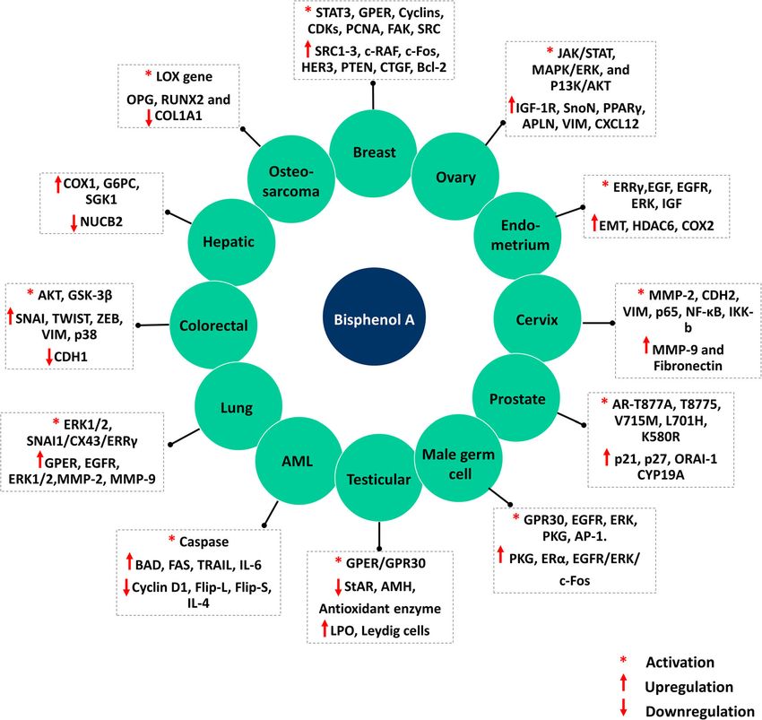
Environ Sci Pollut Res
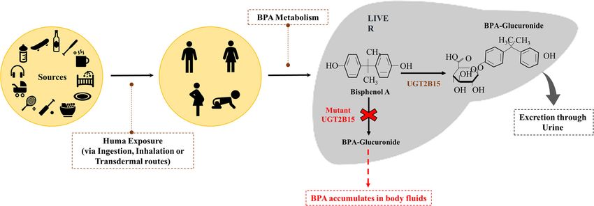
target genes coupled with G protein receptor (Pupo et al. in UGTs enzyme lead to the elevation of unconjugated BPA
2012). In vitro and in vivo studies have shown that BPA ex- concentration in the body. Many studies have reported the
posure has a pro-carcinogenic influence in hormone- presence of the unconjugated forms of BPA in human body
dependent and hormone-independent cancers (Gao et al. fluids, such as milk, maternal urine, amniotic, and placental
2015; Seachrist et al. 2016). BPA exposure is reported to alter fluids, and in neonates that corresponds with the occurrence of
the cancer cells’ biological behaviors, notably, proliferation, many hormonal abnormalities (Vandenberg et al. 2010;
invasion, growth, survival, migration, and apoptosis Inadera 2015) (Fig. 1).
(Chevalier et al. 2012; Prins et al. 2014; Ge et al. 2014; BPA exposure affects several signaling pathways, includ-
Wang et al. 2015, 2017, 2019; Ma et al. 2015; Song et al. ing interference with cellular receptors (nuclear, steroid hor-
2015; Pfeifer et al. 2015; Shi et al. 2017; Jeong et al. 2017; mone, and orphan) functioning. Several studies have demon-
Sauer et al. 2017; Li et al. 2017; Huang et al. 2018; Hui et al. strated estrogen receptors, androgen receptor, estrogen-related
2018; Qu et al. 2018; Hanafi et al. 2019). Besides, in vitro receptors, thyroid hormone receptor, peroxisome proliferator-
studies have shown that low doses of BPA exposure have activated receptors, pregnane X receptor, and aryl hydrocar-
reported inducing resistance to anticancer drugs such as ta- bon receptor and downstream signaling as targets of BPA.
moxifen (TAM), carboplatin, poly ADP ribose polymerase Besides, many enzymatic pathways related to steroid biosyn-
(PARP) inhibitors, doxorubicin, bevacizumab, vinblastine, thesis and metabolism associated with endocrine and/or repro-
cisplatin, and others by modulating the expression of many ductive systems are targeted by BPA. Modulation of these
oncogenic signaling pathways in both hormone-dependent pathways has been linked to cancer development. For exam-
and hormone-independent human cancers (Hafezi and ple, abnormal expression of estrogen receptors plays an essen-
Abdel-Rahman 2019). MAPK, PI3K/AKT, NFκB and JNK, tial role in the development of carcinoma of the breast, ovary,
and Ca2+ homeostasis are the most widely studies pathways in liver, and low-grade endometrium (Gao et al. 2015). BPA has
relation to BPA and cancer (Zhang et al. 2016; Qu et al. 2018). a higher affinity to bind to various cell surface receptors such
The current experimental evidence demonstrates that BPA can as ER (ERα and ERβ), androgen receptor (AR), membrane
significantly increase the risk of both hormone-dependent and receptor GPER (GPR30), epidermal growth factor receptor
hormone-independent cancers via inducing epigenetic chang- (EGFR), and estrogen-related receptors (ERRs) (Acconcia
es such as altered promoter methylation of tumor suppressors et al. 2015; Gao et al. 2015). The primary mechanism of
or oncogenes, global hypomethylation, and histone modifica- BPA-stimulated carcinogenesis may be due to its estrogenic
tions and by modulating the expression of noncoding RNAs activity. BPA binds to membrane estrogen receptors (mERs),
such as miRNA, lncRNA, and snoRNA (Ho et al. 2015; nuclear ERs, and receptor GPR30 and alters the genomic and
Şenyildiz and Özden 2015; Şenyıldız et al. 2016; Senyildiz non-genomic signaling pathways differently in different cell
et al. 2017; Prins et al. 2017; Cheong et al. 2018; Fatma types and alters the normal biological functions leading to
Karaman et al. 2019). Herein, we review the current literature carcinogenesis (Wang et al. 2010). Taken together, BPA acts
on the role of BPA in hormone-dependent and hormone- through both estrogen-dependent and estrogen-independent
independent human cancers, their mechanism of action, and pathways in cancer (Fig. 2). The sections below describe the
their potential impact on cancer development. association between BPA and cancer.
Human exposure, metabolism, Association of BPA with cancer
and mechanism of action
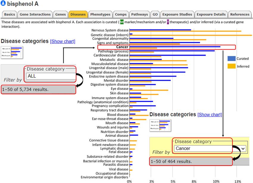
We have used comparative toxicogenomics database (CTD;
Human exposure to BPA occurs when it leaches from plastic- http://ctdbase.org/) to predict the association of BPA with
lined food and drink containers, water bottles, and dental seal- human health (Davis et al. 2021). Analysis using CTD iden-
ants when they are repeatedly heated or washed with harsh tified association of BPA with 5734 diseases across 36 disease
detergents, or when they contain acidic liquids. It enters the categories. Interestingly, among cancer classes, BPA was as-
human body via inhalation, ingestion (consumption of con- sociated with 464 entries (Fig. 3). Exposure to BPA has been
taminated food and water), and through dermal exposure linked with increased cancer risk (Gao et al. 2015). The car-
(TSAI 2006). BPA is absorbed by the gastrointestinal tract cinogenic potential of BPA was demonstrated in several pre-
and gets metabolized in the liver through glucuronidation or viously published studies using in vitro and in vivo models. A
sulfonation. The liver enzyme UDP-glucuronosyltransferases study by Seachrist and co-workers in 2016 showed that early-
2B15 (UGTs) is responsible for the glucuronidation of BPA, life exposure to BPA is a risk factor for breast and prostate
followed by its excretion from the body either through bile or cancer and proposed to classify BPA as a chemical carcinogen
urine in the form of BPA glucuronide (Kurebayashi 2003; belonging to “group 2A” (Seachrist et al. 2016). There are
Genuis et al. 2012; Thongkorn et al. 2019). The abnormalities numerous evidences to demonstrate that exposure to BPA,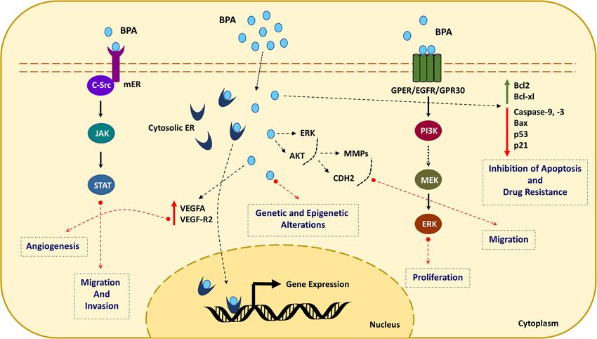
Environ Sci Pollut Res

Fig. 1 Bisphenol A exposure to humans and its metabolism. BPA is       glucuronidation of BPA followed by its excretion through sweat or urine
metabolized in the liver through glucuronidation. The liver enzyme     in the form of BPA glucuronide. Deregulated activity of this enzyme
UDP-glucuronosyltransferases 2B15 (UGTs) is responsible for the        results in BPA accumulation leading to aberrant oncogenic signaling

even at a very low level, can either increase cancer risk or           pathways. As per the literature, BPA can induce proliferation,
aggravate cancer. Some of the hormone-dependent cells are              growth, migration, and invasion in various cell types such as
highly sensitive to BPA as it induces pro-proliferative path-          cervical (SiHa, HeLa, C33A), breast (MCF7, MDA-MB-231,
ways. Several earlier studies demonstrated the pro-                    BT-549), prostate (LNCaP), human trophoblasts (HTR-8/
carcinogenic activity of BPA. Both in vitro, in vivo, and ani-         SVneo), mesenchymal stem cells (hUM-MSCs), ovarian
mal studies have shown the ability of BPA to facilitate the            (OVCAR-3, SkBr3), lung (A549), and colorectal (SW480)
acquisition of cancer hallmarks via altering the biological be-        via activation of signaling pathways, notably JAK-STAT,
havior of the cells and induction of pro-carcinogenic signaling        PI3-AKT, MAPK, and others (Wang et al. 2013; Nomiri

Fig. 2 Mechanism of action of bisphenol A and associated cancer        (mERs), nuclear ERs, and receptor GPR30 and alters the genomic and
hallmarks. Mechanism of BPA-stimulated carcinogenesis may be due       non-genomic signaling pathways differently in different cell types and
to its estrogenic activity. BPA binds to membrane estrogen receptors   alters the normal biological functions
Environ Sci Pollut Res

Fig. 3 Bisphenol A and associated diseases (CTD disease landscape). Across 36 different groups, BPA is associated with 5735 diseases. Among the
cancer classes, bisphenol A is linked with 466 of the cancers

et al. 2019). In addition to its carcinogenic role, several studies      pathways play a crucial role in the growth, development, and
have demonstrated the genotoxic effect of BPA using in vitro             functioning of the mammary gland. EDCs have been reported
systems. By modulating the epigenetic enzymes, BPA can                   to alter endocrine-mediated signaling pathways, thus affecting
bring about genome-wide epigenetic changes leading to al-                the functions of the mammary gland. Epidemiological reports
tered expression. The epigenetic changes induced upon expo-              indicate that elevated levels of estrogen, prolactin, and proges-
sure to BPA are suspected of playing a key role in disease               terone are associated with the development of BC (Bernstein
pathophysiology, including cancer. The following section de-             2002; Gao et al. 2015). BPA can be absorbed and stored in
scribes the association of BPA with different cancer types               human fat tissue and breast stroma, due to its lipophilic nature,
(Table 1 and Fig. 4).                                                    and may subsequently activate cancer-promoting signaling
                                                                         pathways to induce BC (Cimmino et al. 2020).
                                                                            Inappropriate activation of estrogen signaling plays a
Breast cancer                                                            key role in BC development. Estrogen exerts its effects
                                                                         via nuclear ERα and ERβ or via mERs such as GPER/
As per the National Cancer Institute and The Institute of                GPR50 (Prossnitz and Barton 2011). BPA-induced chang-
Medicine (IOM), BPA is declared as a significant risk fac-               es in the mammary gland tissues include enhanced estra-
tor for breast cancer (BC). Surveillance, epidemiology, and              diol sensitivity and increased progesterone receptors. BPA
experimental studies have evaluated the association between              exposure during mammary gland development is associ-
BPA and BC and demonstrated the potential of low doses of                ated with an increased risk of tumorigenesis (Fenton
BPA to induce neoplastic lesions. Estrogen, progesterone, and            2006). Low doses of BPA have been shown to stimulate
prolactin play an essential role in the development of mammary           proliferation in both ER-positive and ER-negative cancer
glands (Rachoń et al. 2008). Endocrine-mediated signaling                cells (LaPensee et al. 2009).
Environ Sci Pollut Res
Table 1 Mechanism of action of bisphenol A on various cancers
Cancer Targets Hallmarks References
Breast cancer • Activation of STAT3, GPER, Cyclins (A, D3), • Increases proliferation, migration, and (Zhang et al. 2014, LaPensee et al.
CDKs (2 and 6), PCNA, FAK, SRC, ERK1/2 invasion in vitro and induces 2009, Pupo et al. 2012, Dairkee
• Upregulation of GPER, EGFR, PR-A, SRC1-3, epithelial-mesenchymal transition et al. 2013, Wang et al. 2015)
AKT, c-RAF, ERK1/2, AKT, c-Fos, HER3, (EMT)
PTEN, ERRγ, P38, MMP-2, MMP-9, CTGF, • Increase in the levels of progesterone
Bcl-2 receptors
• Downregulation of FOXA1, Fork head Family • Reduction in the efficacy of multiple
Transcription Factor, P53, BAX and BIM, chemotherapeutic agents
PDCD5 and BCL2L11 • Emphasizing the pathway of ER
• Hypermethylation of BCL2L11, PARD6G, receptor–DNMTs-TET2-DNA
FOXP1, and SFRS11, NUP98, and CtIP hydroxymethylation
(RBBP8)
• Decreases the expression of TET2 among the three
TET dioxygenases. Decrease the level of
genomic 5hmC
Ovarian cancer • Activates JAK/STAT, MAPK/ERK, and • Increases cellular growth, migration, (Kim et al. 2015, Hoffmann et al.
PI3K/AKT invasion, and proliferation 2017, Ptak et al. 2014, Ptak et al.
• Phosphorylates IRS, CCND1, • Increases intracellular ATP, lactate, 2011, Hall and Korach 2013)
• Upregulates mRNA levels of ERα, IGF-1R, and pyruvic acid levels
SnoN, PPARγ, APLN, VIM, CXCL12
• Downregulates SMAD3, CDH1, ZO-1
• Inhibits TGF-β, CASP3, CASP7, and CASP9
Endometrial • Increases expression of EMT markers (VIM, • Enhances cell proliferation, growth, (Wang et al. 2015, Klotz et al. 2000,
cancer CD90, CD44, CD105), HDAC6 and COX2 migration, and invasion Gertz et al. 2012, Chou et al. 2017,
through MAPK pathway by estrogenic effect • Affects hedgehog signaling via Xiong et al. 2020, Yaguchi 2019)
• Downregulates the expression of CDH1, increasing miR-107 expression
HOXA10, and decidual markers PRL and
IGFBP-1
• Activates ERRγ/EGF/EGFR/ERK signaling
pathway in Ishikawa cells
• Activates the IGF signaling pathway via ERα
• Decreases miR-149 expression and downregulates
DNA repair gene (ARF6) and p53 and
upregulates CCNE2
Cervical cancer • Activates MMP-2, CDH2, VIM, p65, NF-κB, and • Induces cell migration and invasion (Ma et al. 2015)
IKK-b
• Upregulates MMP-9 and Fibronectin
Prostate cancer • Stimulates the transcriptional activity of • Increases cell proliferation, migration, (Wetherill et al. 2006, Bilancio et al.
AR-T877A most likely through AR-T877A 2017, Derouiche et al. 2013, Fatma
• It activates AR mutant alleles such as T877A, • Changes cell morphology Karaman et al. 2019)
T8775, V715M, L701H, and K580R • Cell cycle arrest
• Activation of ERK • Induction and amplification of calcium
• Downregulation of ERK, cyclin D1, and entry in LNCaP cells
chromatin-modifying enzymes • Alters methylation of tumor
• Upregulation of p21 and p27 and ion channel suppressor genes
protein ORAI1 • Induces prostate cancer progression
• Increases aromatase (CYP19A) activity, androgen
receptor (AR) expression in the ventral prostate,
and also increases centrosome number
• Increases DNA methylation and downregulates
p16
Male germ cell • Activates GPR30, EGFR, ERK, PKG, and AP-1 • • Enhances proliferation of (Sheng et al. 2013)
cancer genes present in the 5′-flanking regions of the spermatogonial GC-1 cells
GPR30
• Upregulates PKG, ERα, and EGFR/ERK/c-Fos
pathways through increased expression of GPER
Testicular • Decreases the testis weight and downregulate the • Reduces testicular size in male pups (Xi et al. 2012, Kawai et al. 2003,
cancer expression of StAR, AMH • Reduces the daily sperm production, Chevalier et al. 2012)
• Inhibits antioxidant enzyme and elevates lipid sperm count, fertility and motility
peroxidation which in turn enhance oxidative • Induces proliferation in testicular
stress in the testis seminoma cells through
GPER/GPR30Environ Sci Pollut Res
Table 1 (continued)
Cancer Targets Hallmarks References
• Increases the number of Leydig cells in adult
Long-Evans rats
Acute myeloid • Activates caspase-3, caspase-8, and caspase-9 • Induces proliferation and (Terasaka et al. 2005, Bontempo et al.
leukemia • Increases phosphorylation of BAD and acetylation chemoresistance of AML cells 2009, S. Zhang et al. 2020)
(AML) of Histone H3 • DNA fragmentation
• Upregulates FAS and TRAIL, IL-6 • Cell cycle arrest and apoptosis
• Downregulates Cyclin D1, Flip-L, Flip-S, IL-4
• Decrease phosphorylation of ERK, Rb, and AKT
Lung Cancer • Activates ERK1/2 through GPER/EGFR and • Cell migration and invasion (Zhang et al. 2014, Ryszawy et al.
SNAI1-1/CX43/ERRγ-dependent EMT signal- • Increases motility of lung 2019)
ing pathway in A549 lung cancer cells adenocarcinoma cells and induces
• Upregulates GPER, EGFR, ERK1/2, MMP-2, cytoskeleton remodeling
MMP-9, • Stimulates invasion in A549 tumor
cells through the
SNAI1-1/CX43/ERRγ-dependent
EMT signaling pathway
Colorectal • Phosphorylates AKT, GSK-3β • Increases migration and invasion (Chen et al. 2015, Qu et al. 2018)
cancer • Increases expression of SNAIL, TWIST, ZEB and • Induces toxicity in human colon
VIM and p38 phosphorylation cancer cells at higher concentration
• Decreases CDH1 expression • Causes oxidative damage and
• Impairs E2-induced extranuclear activities of ERb increases mitochondrial and
• Depolarizes MMP and results in loss of intracellular ROS
mitochondrial integrity • Increased intracellular release of Ca2+
Hepatic cancer • Increases COX1 and G6PC expression while, • Induces mitochondrial dysfunction in (Ilagan et al. 2017, Weinhouse et al.
NUCB2 expression was decreased in female mice liver 2014, Moon et al. 2012)
• Induces the ACSS2 expression • Alteration in the liver miRNome and
• Elevates SGK1 expression in primary liver cancer transcriptome that causes adverse
health effect including cancer
• Acts as a partial/competitive agonist
for estrogen
Oral and • Downregulates OPC signaling pathways • Involved in the progression of (Li et al. 2017)
Oropharyng- • Promotes OC and OPC through estrogenic and endoderm-derived carcinogenesis
eal cancer non-estrogen-dependent pathway
Thyroid cancer • Higher concentrations of urinary BPA were • High BPA concentration in the body (Zhou et al. 2017, Li et al. 2019,
observed in study participants underwent thyroid has been associated with an increased Zhang et al. 2017)
ultrasonography risk of thyroid cancer
• Increased concentration of BPA in the blood in • Enhances thyroid cancer cell
patients with thyroid cancer proliferation
• Upregulation of ER and GPR30 expression in
BHP10-3 cells
Osteosarcoma • Interacts with LOX gene and enhances the risk of • Increases the risk of Osteosarcoma (Jia et al. 2013, Fic et al. 2015, Kidani
the osteosarcoma • Changes cell morphology, motility and et al. 2017)
• Downregulates OPG, RUNX2, and COL1A1 filopodia formation
• Inhibits CDC42 expression
Adrenocortical • Stimulates adrenal cell proliferation via • Stimulates adrenal cell proliferation (Medwid et al. 2018, Lan et al. 2015)
carcinoma ERβ-mediated activation of the Shh pathway • Increases adrenal development
• Activates CYP11A1 gene expression and
increases corticosterone production through the
JNK/c-Jun signaling pathway
• Enhances PCNA, cyclin D1 and D2, sonic
hedgehog (shh) protein expression
Studies have reported exposing breast cancer cell lines and is proposed as a mechanism for increased risk of BC
to BPA causes cell proliferation, migration, and invasion (Fernandez 2012). The study demonstrated that exposure
(Kim et al. 2017). BPA alters the morphogenesis of the to low doses of BPA induces aberrant methylation of
fetal mammary gland through epigenetic modification. genes such as lysosomal-associated membrane protein 3
Besides, BPA were detected in maternal milk (Mandrup (LAMP3). The role of LAMP3 is well established in BC
et al. 2016). Exposure to BPA alters DNA methylation (Weng et al. 2010). BPA promotes migration andEnviron Sci Pollut Res Fig. 4 Role of bisphenol A in the development of hormone-related cancers. BPA to facilitate the acquisition of cancer hallmarks via modulating the expression of many oncogenic signaling pathways invasion of BT-549 and MD-MB-231 cells. Exposure of including BC. BPA upregulates the EERγ in estrogen MCF-7 cell lines to low doses of BPA triggers cell pro- receptor-positive (ER+) breast cancer cells by phosphorylation liferation (Kim et al. 2017). In TNBC cells, the pro- of the ERK1/2. These data suggested EERγ/ERK1/2 axis to proliferative and pro-survival effects of BPA depend on promote cell proliferation in BC. ERK1/2 and AKT activation (Zhang et al. 2016). Homeobox B9 (HOXB9) is activated in response to Besides, BPA also stimulates the expression of MMPs. BPA exposure in BC (Hafezi and Abdel-Rahman 2019). The role of ERK1/2 and AKT activation and MMPs is HOXB9, located at 17q21.32, participates in cell cycle pro- very well established in BC. BPA enhanced migration- gression, embryonic patterning, mammary gland develop- related protein and mRNA expression including matrix ment, and cell proliferation. HOXB9 is upregulated in BC metalloproteinase-2 (MMP2), and MMP9 is independent and has been demonstrated to stimulate neovascularization, of vimentin (VIM) and fibronectin expression in TNBC tumor invasion, and disease progression. The HOXB9 gene cells (Zhang et al. 2016). promoter contains a potential estrogen response element ERRs, the nuclear receptor superfamily, are also known as (ERE4) which mediates its response to both E2 and BPA orphan receptors because they do not have endogenous li- (Deb et al. 2016). A study showed that BPA is susceptible gands. ERRs have estrogen receptor sequence homology but to mammotrophic hormones and increases breast cancer do not bind to estrogen. By binding to ERRγ, BPA stimulates risk later in life. This hypothesis was supported by a study the growth and proliferation of various types of cancer cells that showed the role of BPA in the differentiation of stem
Environ Sci Pollut Res

cells by altering their sensitivity to BMP signaling, by         and PIK3CA pathways (Gertz et al. 2012). Besides, BPA also
downregulating the mammary fibroblast development of             activates ERα and ERβ depending in a cell type and
BMPs, and by altering the localization and expression of         concentration-dependent manner. Further, BPA also activates
type 1 BMP receptors. BPA altered the BMP signaling by           the IGF signaling pathway via ERα in the ovary of adult mice
SMAD1/5/8 phosphorylation (Clément et al. 2017; Bach             and increases mitotic cells (Klotz et al. 2000; Gertz et al.
et al. 2018). Taken together, various published studies on       2012).
BC suggest that BPA may impart its pro-carcinogenic ef-              BPA exposure alters microRNA (miRNA) expression to
fects via inducing epigenetic modifications, DNA damage,         contribute to EC. Chou et al. carried out a transcriptomic anal-
stem cell differentiation, and breast microenvironment al-       ysis to discover altered mRNA and miRNAs in response to
teration through activation of pro-carcinogenic signaling        BPA exposure in human endometrial cells. The results
pathways.                                                        showed that exposure to BPA decreases the expression of
                                                                 miR-149, downregulates DNA repair gene (ARF6) and
                                                                 TP53, upregulates CCNE2, impairs the cell cycle, and initiates
Endometrial cancer                                               cell migration and invasion. Further, BPA affects hedgehog
                                                                 signaling via an increase in miR-107 expression (Chou et al.
Endometrial cancer (EC) is the fourth most common cancer in      2017).
women reported to originate from a hyperestrogenic pattern           A recent study that determined the effect of BPA on endo-
(Brooks et al. 2019). Many scientific investigations have pro-   metrial stromal cell decidualization suggested the potential
posed endocrine disruptors, including BPA, to be included as     crosstalk between BPA and epigenetic modifications. The
a hormonal risk factor category. Due to its estrogen-            study elaborates on the crosstalk between BPA and histone
mimicking properties, BPA is considered a key risk factor        modifications during endometrial stromal cell decidualization
for EC. In vitro, in vivo, and human studies indicated BPA       resulting in the downregulation of HOXA10, PRL, and
as a risk factor of EC. For example, a study by Aquino et al.    IGFBP-1 (Xiong et al. 2020). BPA exposure induces the pro-
demonstrated a high concentration of BPA in the urine and        liferation of HEC265 cells and Ishikawa cells by nuclear trans-
blood of EC patients compared to healthy controls. The same      location of ERRγ and increased BPA/ERRγ-target gene ex-
study proposes that BPA can directly upregulate the ER genes     pression. In addition, BPA facilitated the Ca2+ influx in
leading to hyperestrogenism and EC (Aquino et al. 2019).         Ishikawa cells and EGF secretion to the extracellular space,
Thus, BPA can favor the hormonal dysregulation at the base       activating the EGFR/ERK pathway. BPA enhanced the ex-
of the endometrial neoplasm and contribute to proliferation      pression of BPA/ERγ-target genes in HEC265 cells, without
effects on neogenesis acting on EGF or microRNAs regula-         affecting Ca2+ mobilization and EGF secretion (Yaguchi
tion. Overall, estrogen may regulate miRNA transcription         2019). These data suggest that BPA might activate multiple
through ERα and ERβ in a specific and cell-dependent man-        signaling pathways to stimulate the proliferation of EC cells.
ner and the fact that EDCs such as BPA would be involved in      However, more detailed mechanistic studies are needed to
endometrial carcinogenesis. BPA exposure during critical pe-     understand the impact of BPA in EC.
riods of growth and development can cause an adverse effect
at later stages of life. CD-1 female mice exposed to BPA by
subcutaneous injection have shown cystic ovaries and endo-       Ovarian cancer
metrial hyperplasia when compared to control mice when ex-
amined at 18-month postinjection (Newbold et al. 2007).          Ovarian cancer (OVC) ranks the seventh most common cause
    The upregulation of cyclooxygenase 2 (COX-2) and EMT         of cancer mortality among women with the worst prognosis
pathway genes is related to tumor development and                and high mortality rate (https://gco.iarc.fr/). In addition to
progression. Wang et al. (2015) have reported that 10-8 M        family history, the risk factors for OVC include obesity,
concentration of BPA enhanced the expression of mesenchy-        smoking, alcoholism, late menopause, hormone replacement
mal cell surface markers (CD44, CD90, CD105, and VIM) and        therapy, early menarche, nulliparity, and mutations in BRCA1/
a cell-cell interaction regulator (HDAC6) and downregulates      2 (Brett et al. 2017; Jammal et al. 2017). Besides this, expo-
the expression of epithelial cell-cell adhesion molecule         sure to EDCs is reported as one of the significant risk factors
(CDH1) in RL95-2 cells. The same study demonstrated that         for OVC (Rachoń 2015). Sex steroids play an important role
BPA could stimulate the growth, invasion, and migration of       in OVC development and progression (Gómora et al. 2018).
RL95-2 cells via the mitogen-activated protein kinase            There is an elevated level of ER expression in epithelial ovar-
(MAPK) pathway, which possibly leads to the upregulation         ian cancer cells compared to benign and normal ovarian epi-
of COX-2 expression (Wang et al. 2015). BPA elevates ER1         thelial cells (Ajani et al. 2017). Estrogen consumption, either
binding sites and alters the expression of a subset of genes     as an oral contraceptive or hormone replacement therapy, in-
affected by E2, leading to the activation of MAPK1, AKT1,        creases the risk of OVC. Estrogen is reported to facilitate
Environ Sci Pollut Res tumor progression via activation of pro-proliferative and pro- and pyruvic acid leading to augmented proliferation in survival cellular mechanisms. BPA mimics estrogen. Hence, OVCAR-3 cells via ERα pathway (Shi et al. 2017). BPA exposure may mimic the effects of estrogen to promote Exposure of SKOV3 cells to low doses of BPA induced OVC. Based on the epidemiological data and molecular in- EMT via canonical Wnt pathway activation. The same study vestigation, BPA is proposed as a potential risk factor in OVC. has shown that low dose of BPA exposure significantly in- BPA is reported to interrupt steroidogenesis in the ovary, duced migration without altering cell proliferation in SKOV3 facilitating the development of polycystic ovary syndrome and A2780 cells (Lin et al. 2018). BPA is an activator of (Konieczna et al. 2018). Various studies in the rodent model phosphatidylinositol-3-kinase (PI3K) signaling in OVC. suggested that BPA exposure induces morphological alter- SnoN is a negative regulator of TGF-β signaling and an acti- ations in the ovary, increase in the number of atretic follicles vator of the tumor suppressor p53 in response to cellular and farm cystic ovaries, endometrial hyperplasia, reduced pri- stress. BPA promotes the growth and proliferation of OVC mordial follicles, and other problems associated with ovary cells by upregulating SnoN and APLN expression (Park and development (Pivonello et al. 2020). Exposure to BPA was Choi 2014). Caspases are required for completing apoptosis, reported to induce gene expression, some of which are activa- and caspase-9 and caspase-3 are involved in apoptosis induc- tors of oncogenic signaling. Both estrogenic-dependent and tion and execution, respectively. By downregulation of estrogenic-independent signaling mechanisms are proposed caspase-3 and caspase-9, BPA inhibits apoptosis in OVC for BPA in ovarian tissues. BPA promotes proliferation, inva- cells. Transplacental exposure to BPA induced changes in sion, angiogenesis, and chemoresistance in ovarian cancer via the expression of genes linked to estrogenic activity in phosphorylation of signal transducer and activator of tran- Sprague-Dawley rat ovaries (Naciff 2003). Furthermore, scription 3 (STAT3), extracellular signal-regulated kinases BPA regulated the expression of BCL2-associated X, apopto- (ERK1/2), and activation of CDH2 (N-cadherin), MMP2, sis regulator (BAX), BCL2, CDK4, and CCNE1 through the and MMP9 leading to the induction of epithelial- genomic estrogenic pathway. BCL-2/BAX plays an important mesenchymal transition (EMT) (Ptak et al. 2014; Hafezi and role in regulating caspase-dependent and caspase-independent Abdel-Rahman 2019). Derailed expression of CDH1 (E- apoptosis. CDK4 plays an essential role in the G1 to S phase cadherin) is implicated in the invasion and metastasis of of the cell cycle (Ptak et al. 2011; Peretz et al. 2012; Gao et al. OVC. BPA downregulates CDH1 expression in ovarian tu- 2015). In OVC, BPA stimulates ERα expression and en- mors (Ptak et al. 2014). Likewise, the presence of BPA in hances the growth of OVC cells by activating ER and aug- OVC cells has been shown to increase SNAI1 response via menting the CXC motif, chemokine 12 (CXCL12)- CXC che- ER-α and ER-β (Kim et al. 2015). The same study showed mokine receptor 4 (CXCR4) signaling axis (Hall and Korach the impact of BPA on OVCAR-3 cell migration compared to 2013). Chemokine receptor CXCR4 and its ligand CXCL12 17β estradiol (E2). Ptak et al. (2014) have reported that ele- play an essential role in the metastatic homing of tumor cells. vated BPA level induces matrix metalloproteinase-2 (MMP-2) BPA exposure resulted in an elevated level of SnoN ex- and MMP-9 and CDH2 in turn facilitates cell migration (Ptak pression and decreased phosphorylation of Smad3 in BG-1 et al. 2014). Studies have reported that treatment with BPA cells via inhibition of the TGF-β signaling pathway. can upregulate cyclin-dependent kinase 4 (CDK4), cyclin E1 Likewise, transplanted ovarian BG-1 cancer cells in the xeno- (CCNE1), cyclin D1 (CCND1), insulin-like growth factor 1 graft mouse model showed increased expression of SnoN and receptor (IGF-1R), B cell lymphoma 2 (BCL2), and ER-α and reduced phosphorylation of SMAD3 after treatment with BPA downregulate aryl hydrocarbon receptor nuclear translocator 2 (Park and Choi 2014). Despite the evidence which shows that (ARNT2) and CDKN1A, resulting in cell proliferation and ap- BPA exposure can affect ovarian function and can stimulate optosis inhibition (Ptak et al. 2011). BPA cooperates with the oncogenic signaling pathway in OVC cell lines, there is leptin to inhibit caspase-3 expression and OVC cell function. still a lack of epidemiological data to support BPA’s role in BPA was shown to increase the expression of leptin receptors OVC incidence. Hence, more detailed epidemiological func- and to induce proliferation by STAT3, ERK1/2, and AKT tional studies are required. phosphorylation (Ptak and Gregoraszczuk 2012). Interestingly, Hoffmann et al. showed that low concentrations of BPA induced OVC progression by upregulation of Apelin Cervical cancer (APLN) expression through peroxisome proliferator-activated receptor gamma (PPARγ) (Hoffmann et al. 2017). APLN is an Cervical cancer (CC) is not an estrogen-dependent cancer. endogenous ligand for G protein-coupled APJ receptors. The However, various experimental and epidemiology data APLN gene is enhanced by tumor necrosis factor-α (TNF-α). showed that exposure to EDCs is a potential risk factor for Interestingly, reports also show that low exposure of BPA CC. The association between diethylstilbestrol and vaginal leads to increased cell viability, cell proliferation, and glycol- clear cell adenocarcinoma was the first report linking EDCs ysis, resulting in elevated levels of intracellular ATP, lactate, and cancer (Reed and Fenton 2013; Tournaire et al. 2015).
Environ Sci Pollut Res

Subsequently, studies have shown diethylstilbestrol as a risk        human stem cells expressing ER and GPR30 in dose-
factor for CC. A study performed by Ma et al. demonstrated           dependent manner (Prins et al. 2014).
BPA as a promoter of migration and invasion of SiHa, HeLa,               Defective calcium signaling is associated with the
and C-33A cells via modulation of estrogen signaling (Ma             cell migration in different cancer types, including PC. BPA
et al. 2015). Nevertheless, Den et al. demonstrated the role         is reported to enhance the migration of PC cells via modulat-
of stromal estrogen signaling in CC development (den Boon            ing calcium signaling. For example, Derouiche et al. (2013)
et al. 2015). Interestingly, cervical tissue is hormone-sensitive,   have shown that BPA modulates calcium signaling to promote
and it shows a high response in the presence of estrogen. Thus,      the migration of LNCaP cells. Treatment of LNCaP cells with
the pro-migratory and pro-invasive response to BPA exposure          1 and 10 nM of BPA showed a significant increase in cell
may be due to the hormone-sensitive nature of cervical tissue.       migration. The same study also showed that BPA induces
Studies showed that the BPA level in the human body corre-           PC cell migration via modulating the expression of an ion
sponds with the occurrence of cervical CC. High BPA levels           channel protein associated with calcium entry, namely,
are detected in urine samples of CC patients (Ma et al. 2015).       ORAI1 (Derouiche et al. 2013). Very interestingly, in mice,
BPA activates nuclear factor kappa B (NF-κB) and                     the treatment resulted in high levels of AR and ER when
upregulates MMP-9 and fibronectin (FN) in SiHa and Hela              compared with the respective controls. This suggests that
cells. Upon BPA exposure, IKKβ activates NF-κB signaling             BPA exposure may lead to inappropriate stimulation of AR
leading to its nuclear translocation and results in cell migration   and ER activation pathways (Di Donato et al. 2017). The
and upregulation of FN and MMP-9. Thus, in CC, NF-κB,                centrosome plays an important role in cell cycle regulation
FN, and MMPs are the target genes of BPA (Ma et al. 2015).           and is currently being recognized as a key target for cancer
BPA also enhances the incidence of proliferative lesions of the      therapy. Centrosome aberration is highly prevalent in cancer
uterus as well as sarcoma of the uterine cervix (Pivonello et al.    as it affects cell cycle progression. Exposure to BPA has been
2020). A study by Newbold et al. reported that BPA exposure          reported to enhance the centrosome number in both normal
enhances the incidence of stromal sarcoma of the cervix              and PC cells (Tarapore et al. 2014). Besides, low doses of
(Newbold et al. 2009). Although BPA is linked to CC, its             BPA disturb the centrosome duplication cycle. Bilancio and
precise role, exact molecular mechanism, biological function,        co-workers demonstrated BPA-induced cell cycle arrest in
and signaling pathways regulated by BPA in CC are yet to be          both prostate cancer (LNCaP) and normal prostate epithelial
identified and established. Hence, further epidemiological and       (EPN) cells. The treatment of LNCaP and EPN with 10 to
functional studies are required.                                     100 μM of BPA induced cell cycle arrest by lowering
                                                                     CCND1 and concomitant upregulation of p21 and p27.
                                                                     Besides, the same study also showed the activation of
                                                                     EGFR- and ERK-dependent pathways (Bilancio et al. 2017).
Prostate cancer                                                          BPA induces a variety of epigenetic modifications in a
                                                                     wide array of cells. Studies on PC cells have suggested
Prostate cancer (PC) is the cancer of the male reproductive          that BPA induces posttranscription and posttranslational
tissue. Epidemiology and experimental studies have demon-            modifications, both globally and regionally. Thus, BPA-
strated the role of BPA as a risk factor for PC. Besides, in vitro   induced epigenetic modification may contribute to the
and in vivo models have shown that BPA can influence the             abnormal biological behavior of the cell. To unravel this,
progression of PC. Early-life exposure to BPA increases the          a research examined the impact of BPA on the expres-
susceptibility to hormone-related carcinogenesis in the pros-        sion of chromatin-modifying enzymes, promoter methyl-
tate gland (Prins et al. 2015). Various preclinical model-based      ation of tumor suppressor genes, and histone modifica-
studies demonstrated the role of estradiol levels in PC patho-       tions in PC-3 cells. Treatment with BPA induced hyper-
genesis. Reports have suggested an increase in the level of          methylation of the p16 promoter, leading to its downreg-
estradiol during aging and related studies conducted in an           ulation. The same study reported significant changes in
animal models have shown BPA exposure can lead to an                 global histone modifications (H3K9ac, H3K9me3,
increase in estradiol levels in aging male rats, contributing to     H3K27me3, and H4K20me3) in PC-3 cells possibly via
PC’s susceptibility (Prins et al. 2017). Many studies have also      downregulation of chromatin-modifying enzymes includ-
reported BPA exposure to induce an abnormal epigenetic               ing nuclear receptor binding SET domain protein 1
modification in genes belonging to multiple signaling path-          (NSD1) and lysine demethylase 5B (KDM5B) and altered
ways (Ho et al. 2006). Defective calcium signaling is associ-        promoter methylation of tumor suppressor genes (BCR,
ated with cell migration in different cancer types including         GSTP1, LOX, MGMT, NEUROG1, PDLIM4, PTGS2,
PC. Besides, BPA also activates key genes linked to prolifer-        PYCARD, TIMP3, TSC2, and ZMYDN10). The ChIP re-
ation and cell survival. For instance, Prins et al. showed that      sults showed a significant increase (1 and 10 μM of
BPA enhanced the phosphorylation of AKT and ERK in                   BPA) in histone modifications (Fatma Karaman et al.
Environ Sci Pollut Res
2019). Neonatal or early-life exposure to BPA is a risk a PKA-PKG-GPER–dependent manner (Cariati et al.
factor for PC later in life. This could be because of the 2019). Compromised immunity plays an important role
developmental reprogramming of the prostate gland in- in cancer development and progression. A study by
duced by epigenetic reprogramming. A study by Prins Nava-Castro et al. investigated the role of BPA on im-
and co-workers using rat models proposed that BPA ex- mune response and TC. The same study showed that
posure can increase PC susceptibility to epigenetic mod- exposure to BPA in pregnant female mice increases tes-
ification through the induction of global hypomethylation ticular tumor size via effecting the immune component
of key cancer susceptibility genes (Prins et al. 2017). and immune response in male offspring’s (Nava-Castro
Overall, BPA is a potent regulator of epigenetic enzymes et al. 2019). Overall, studies show that BPA can affect
and can bring about abnormal epigenetic changes in PC the male reproductive system, induce reproductive dys-
and may facilitate its progression. function, and may contribute to testicular germ cell can-
cer. However, further detailed investigations are required
before conclusions are drawn.
Testicular germ cell cancer
Male germ cell cancer represents nearly 2% of all can- Acute myeloid leukemia
cers at risk. Occupational exposure to BPA is proposed
as a potential risk factor for male germ cell cancer. Acute myeloid leukemia (AML) is a hematopoietic stem cell
In vitro studies have demonstrated that BPA can alter malignancy. The estrogenic signals can play a key role in the
proteins and affect the functional characteristics of the progression of AML. Studies have reported the presence of
germ cells of the testis and may affect male fertility. BPA in the serum sample of AML patients (Zhang et al.
Reports indicated an exposure of male germ cells (sper- 2020). Both cell proliferation and apoptosis induction proper-
matogonial GC-1 cells) to 1000-μM BPA for 48 h acti- ties are reported for BPA (Bontempo et al. 2009). One of the
vates various genes such as GPR30, EGFR, ERK, and earliest studies has shown the cell cycle arrest and apoptosis-
PKG. Low doses of BPA boost the proliferation of sper- inducing properties of BPA in AML cells treated with a mi-
matogonial GC-1 cells via EFGR/ERK/GPR30/c-Fos/ cromolar concentration of BPA (Terasaka et al. 2005). BPA
ER-α/PKG axis (Nomiri et al. 2019). Male germ cell treatment induced apoptosis in NB4 cells via activation of
tumors show overexpression of GPR30. Similar to ERs, caspases involving FAS/TRAIL/BAD axis (Bontempo et al.
GPR30 controls the cellular response towards 17β-estra- 2009). It induces inter-nucleosomal DNA fragmentation and
diol. Besides, GPR30 is proposed as a molecular target activates caspase-9 and caspase-3, implicating the induction
in male germ cell cancer. Sheng et al. (2013) showed of apoptosis (Terasaka et al. 2005). In contrast to this study,
that a low dose of BPA induces the progression of male another study demonstrated BPA to induce cell proliferation
germ cell cancer and activates the expression of GPR-30 and resistance to daunorubicin and cytarabine in AML (Zhang
via EFGR/ERK/GPR30/c-Fos/ER-α/PKG pathway et al. 2020). Daunorubicin and cytarabine are chemotherapeu-
(Sheng et al. 2013). Fetal exposure to BPA has an ad- tic drugs. Specifically, it is used for the treatment of AML.
verse effect on the male reproductive system. BPA is Abnormal expression of cytokines and chemokines plays an
known to affect the quality and quantity of sperms. important role in AML progression. BPA-induced cell prolif-
Gestational exposure of female mice to BPA results in eration involves the upregulation of interleukin 6 (IL6) and
a decrease in testis weight and size, downregulation of downregulation of IL4 (Zhang et al. 2020). Activation of
anti-Mullerian hormone (AMH), and steroidogenic acute NF-κB upregulates IL6 while NFAT contributes to IL4 down-
regulatory protein (StAR) in male pups (Kawai et al. regulation. More recent studies report BPA to trigger AML
2003; Xi et al. 2012). Many studies have established a (Terasaka et al. 2005).
relationship between environmental pollutants, impair-
ment in male germ cell development, and testicular tu-
mor development. As per the study by Delbès et al., Lung cancer
BPA exposure resulted in the reduction of sperm produc-
tion and enhanced the incidence of testicular cancer (TC) The endocrine-disrupting property of BPA is reported to en-
(Delbès et al. 2006). BPA increases seminoma cells pro- hance the susceptibility to lung cancer (LC). Accumulating
liferation by activating GPR30 (Chevalier et al. 2012). evidences have suggested that estrogen and its receptors can
Besides, fetal exposure to BPA has been reported to in- contribute to LC. The lung alveolar cells get exposed to BPA
duce reproductive dysfunction and contribute to male in- via inhalation and may alter the functional properties of the
fertility and TC (Adiga et al. 2020). The low-dose BPA cells. A Chinese population-based case-control study demon-
exposure induced seminiferous tubule cell proliferation in strated that BPA levels were significantly higher in non-smallEnviron Sci Pollut Res
cell lung cancer cases compared to control samples (Li et al. et al. 2018). It has been reported that BPA causes oxidative
2020). Zhang and colleagues demonstrated the role of BPA in damage to the colonic epithelium as shown by increased
altering the biological behavior and function of lung cancer mitochondrial and intracellular ROS and increased levels
cells. BPA concentration less than 10-4 M enhances the mi- of hydrogen peroxide and malondialdehyde (Wang et al.
gration and invasion of A549 cells (Zhang et al. 2014). 2019). BPA increased the intracellular release of Ca2+ and
Besides this, BPA also upregulated the expression of MMP2 is responsible for the depolarization of MMPs and the loss
and MMP9 by ERK1 activation through GPER/EGFR (Zhang of mitochondrial integrity in HCT116 cell lines (Qu et al.
et al. 2014). ERK1 and MMPs activation are potential pro- 2018).
carcinogenic signals in numerous cancers, including LC.
Similarly, another study involving the cross talk between
BPA and induction of pro-metastatic signaling suggested the Hepatic cancer
activation of SNAI1/Cx43/ERRγ-dependent EMT signaling
pathway upon BPA exposure in A549 cells (Ryszawy et al. Hepatic tissue is the nonreproductive target of estrogen. Many
2019). BPA induces morphological changes and enhances environmental agents are reported to cause hepatocellular car-
motility by cytoskeletal rearrangements (Zhang et al. 2014). cinoma (HCC). The primary risk factor for HCC is chronic
Some studies reported BPA to induce EMT in lung cancer via hepatitis B and C virus infection, environmental exposure, and
a switch from CDH1 to CDH2 and vimentin/SNAI1/connexin excessive alcohol consumption (Balogh et al. 2016). The liver
(Cx) 43 upregulation (Ryszawy et al. 2019). Besides inducing expresses ERs and responds to signaling related to steroid
migration and invasion, BPA exposure is responsible for the hormones. BPA is demonstrated to cause hepatic toxicity
inflammation and oxidative stress in rat lung tissues. The same and liver injury (Hassan et al. 2012; Thoene et al. 2017).
study showed that BPA upregulated malondialdehyde and BPA can induce liver damage by oxidative stress (Esplugas
IL18 with a reduction in superoxide dismutase (SOD) levels. et al. 2018). Studies have demonstrated that ~70% of the fetal
Oxidative stress and inflammatory pathways in lung cells are liver in humans shows the presence of detectable BPA levels
promoters of LCs (Abedelhaffez et al. 2017). and is linked to altering the methylation levels of xenobiotic-
metabolizing enzymes. The first systematic study, conducted
by Weinhouse et al., demonstrated the potential role of an
Colorectal cancer environmentally relevant dose of BPA in inducing hepatic
tumors in AvyC3HeJ/C57BL/6 mice (Weinhouse et al.
Studies have shown that estrogens and their structural analo- 2014) BPA exposure also induced both tumor and precancer-
gous along with their receptors are involved in intestinal dis- ous conditions. Mitochondrial dysfunction is potentially relat-
eases and development and progression of colorectal cancer. ed to carcinogenesis. Many previous studies have clearly dem-
E2-induced caspase activation is essential for apoptosis in onstrated that epigenome-wide changes, oxidative stress, in-
many cell types. Apoptosis induced by activation of E2 is flammation, and mitochondrial damage may lead to liver dam-
impaired in colon cancer cells. Chen and co-workers in 2014 age and subsequently to HCC later in life. BPA induces mi-
investigated the effect of BPA on colorectal cancer using a tochondrial dysfunction and hepatic injury by enhancing oxi-
proteomic approach. In SW80 cells, BPA treatment altered the dative stress in the liver (Moon et al. 2012). BPA is reported to
expression of 56 proteins related to structure, movement, pro- alter mitochondrial structure, increase malondialdehyde
liferation, and others. Besides, they showed EMT, spindle- levels, and decrease glutathione peroxidase 3 expression
shaped mesenchymal morphology with upregulation of (Moon et al. 2012). The increase in IL6 and TNF-α levels
CDH2 and SNAI1. CDH2 is a transmembrane protein, and it was reported, suggesting that intraperitoneal administration
functions to mediate cell-cell adhesion, and it is a hallmark of of BPA can induce inflammation of liver cells in mice
EMT. SNAI1 is a typical transcription factor that could induce (Moon et al. 2012). In HepG2 cells, nanomolar concentration
EMT and cancer progression with concomitant downregula- of BPA has shown to reduce oxygen consumption rate, ATP
tion of CDH1 (Chen et al. 2015). This suggests that, in colon level, and mitochondrial membrane potential (Huc et al.
cancer, BPA may participate in the induction of aggressive 2012). Studies conducted in a zebrafish models have revealed
phenotypes. The BPA effect on colon cancer is diverse. that BPA can induce hepatic epigenetic alterations. The same
Glycogen synthase kinase 3β (GSK3β) is a multifunctional study has also reported altered expression of genes related to
protein involved in various cellular activities such as develop- mitochondrial functions including oxidative phosphorylation,
ment, differentiation, and disease. By altering GSK3β expres- in response to BPA exposure (Renaud et al. 2017). BPA en-
sion, BPA promotes migration and invasion of colon cancer hances the expression of COX1 and G6PC genes in female
cells via activating of BCL-xl and inhibits apoptosis (Chen mice compared to male mice, while nucleobindin-2 (NUCB2)
et al. 2015). A recent study showed that 250 μM of BPA expression was decreased in female mice. BPA induces
has induced toxicity in human colorectal cancer cells (Qu ACSS2 expression, which is a cancer susceptibility gene.Environ Sci Pollut Res
Serum/glucocorticoid-regulated kinase 1 expression is elevat- of urinary BPA with an increased risk of TC (Li et al. 2019).
ed in primary liver cancer (Ilagan et al. 2017). BPA affects BPA treatment enhances the H2O2 generation in PCCL3 cells
liver functions that are evident by the reduction in the activi- and suggests that BPA may promote oxidative stress and dam-
ties of several enzymes, including catalase, glutathione, and age thyrocytes leading to thyroid disorders (da Silva et al.
others (Aboul Ezz et al. 2015). BPA is reported to cause liver 2018). BPA enhances thyroid cancer cell proliferation
injury, which included necrosis, vacuolization of the cyto- through the regulation of ER and GPR30 expression
plasm, and decreased hepatocellular compactness (Sangai (Zhang et al. 2017). Taken together, there may be a con-
et al. 2014). The epigenome-wide changes, oxidative stress, nection between BPA and thyroid nodules or cancer, but
inflammation, and mitochondrial damage may lead to liver further investigations are required to correlate BPA and
damage and subsequently to HCC later in life. thyroid carcinogenesis.
Head and neck cancer Osteosarcoma
Oral cancer (OC) and oropharyngeal cancer (OPC) are one of Environmental BPA accounts for the pathogenesis of osteo-
the groups of head and neck cancer. The risk factor for OC sarcoma. In vitro and in vivo studies have shown that expo-
includes the use of tobacco products, excess alcohol intake, sure to BPA is associated with the occurrence of osteoporosis.
and diet. Oral cavity and oropharyngeal space are the first sites A Chinese hospital-based case-control study has reported that
of exposure for ingested environmental toxicants. Thus, the BPA exposure induces a genetic variation −22G/C polymor-
oral cavity and oropharyngeal space are at high risk for BPA- phism of the lysyl oxidase gene (LOX) and enhances the risk
induced carcinogenesis. BPA can promote OC and OPC of osteosarcoma (Jia et al. 2013). BPA downregulate the os-
through the estrogenic- and non-estrogen-dependent pathway. teoprotegerin (OPG), runt-related transcription factor 2
Li and colleagues reported that BPA facilitates proliferation, (Runx2), and protein-coding collagen type I alpha 1
migration, and invasion of laryngeal squamous cell carcinoma (COL1A1) genes in HOS cells (Fic et al. 2015). OPG and
(LSCC). There was an upregulation of MMP2 and IL6, sug- Runx2 are the major transcription factors playing an important
gesting the activation of inflammatory pathways by BPA. role in adult bone remodeling. COL1A1 is a bone matrix pro-
Further, the proliferation and migration induced by BPA tein gene; it is regulated by Runx2. BPA exposure increased
is a GPER- and IL-6-dependent process (Li et al. 2017). the plasma levels of procollagen type I N-terminal propeptide
The current literature survey suggests a lack of data on (P1NP) associated with the risk of bone metastasis (Lind et al.
BPA and head and neck cancer. Hence, more detailed func- 2019). Further studies are needed to understand further the
tional studies are needed to assess the toxic effects of BPA molecular mechanisms of BPA in osteosarcoma.
on OC and OPC.
BPA and therapeutic resistance
Thyroid cancer (TCs)
Therapeutic resistance is a major problem in cancer therapy
EDCs have emerged as a major public health problem glob- and contributes to a high mortality rate. Metastatic cancer
ally. EDCs have been reported to modify the natural endocrine patients develop chemoresistance to several drugs. Several
function as they can directly interact with steroid hormone in vitro studies showed that BPA exposure could promote
receptors. One of the earliest studies demonstrated that BPA, therapy-resistant phenotypes in BC. Several reports have dem-
by acting as an antagonist, disrupts the action of thyroid hor- onstrated that anticancer drugs have been shown to be antag-
mone. Several investigators have evaluated the relationship onistic to estrogen. BPA has a high binding affinity towards
between EDCs and TCs. Being a member of EDC, BPA that of estrogen-related receptor-γ (ERRγ). BC patients have
may affect the thyroid hormone and its action. Li et al. report- shown resistance to tamoxifen, lapatinib, doxorubicin, and
ed the higher incidence of thyroid nodules in Chinese women cisplatin; BPA have been shown to inhibit the efficacy of
who are exposed to BPA (Li et al. 2019). A study by Lee and doxorubicin by enhancing the levels of BCL-2 and BCL-xL
co-workers demonstrated the positive correlation between the (Barret 2009). The study confers that, in both ERα-positive
BPA levels in blood with that of ANXA6 and valosin- and ERα-negative BC cells, BPA antagonizes the cytotoxic
containing protein expression in TC patients (Lee et al. effect of chemotherapy agents such as doxorubicin, cisplatin,
2018). The relationship between higher urinary BPA levels or vinblastine. The same study also reported that BPA exhibits
along with higher iodine intake was linked to papillary thyroid its anticytotoxic function by inhibiting ERα or ERβ, suggest-
carcinoma (Zhou et al. 2017). Another case-control study ing the potential role of BPA to activate nonclassical ER(s)
demonstrated the association between higher concentrations (LaPensee et al. 2009). Both MDA-MB-468 and T47D cellsYou can also read