A Review of Systemic Lupus Erythematosus (SLE): Symptoms, Risk Factors, Treatment, and Health Related Quality of Life Issues - Scientific ...
←
→
Page content transcription
If your browser does not render page correctly, please read the page content below
Open Journal of Rheumatology and Autoimmune Diseases, 2021, 11, 115-143
                                                                                                https://www.scirp.org/journal/ojra
                                                                                                          ISSN Online: 2164-005X
                                                                                                            ISSN Print: 2163-9914

A Review of Systemic Lupus Erythematosus
(SLE): Symptoms, Risk Factors, Treatment, and
Health Related Quality of Life Issues

Sarah N. Al-Gahtani

Department of Internal Medicine, King Fahd Medical City, Riyadh, Saudi Arabia

How to cite this paper: Al-Gahtani, S.N.        Abstract
(2021) A Review of Systemic Lupus Ery-
thematosus (SLE): Symptoms, Risk Factors,       In this paper, we present a thorough review of one of the most life-threatening
Treatment, and Health Related Quality of
                                                autoimmune diseases, Systemic lupus erythematosus (lupus). Symptoms, risk
Life Issues. Open Journal of Rheumatology
and Autoimmune Diseases, 11, 115-143.           factors, including genetic and epidemiological factors are discussed. Treat-
https://doi.org/10.4236/ojra.2021.114014        ment, life expectancies, and Health Related Quality of Life of patients with
                                                SLE will be discussed as well. Special attention will be given to Lupus Nephri-
Received: August 5, 2021
Accepted: September 13, 2021
                                                tis.
Published: September 16, 2021
                                                Keywords
Copyright © 2021 by author(s) and
Scientific Research Publishing Inc.             Autoimmune Disorders, Systemic Lupus Erythematosus, Environmental Risk
This work is licensed under the Creative        Factors, Familial Aggregation, Lupus Nephritis, Treatment of SLE, Health
Commons Attribution International
                                                Related Quality of Life
License (CC BY 4.0).
http://creativecommons.org/licenses/by/4.0/
               Open Access

                                              1. Introduction
                                              Autoimmune system disorders cause abnormally low activity or overactivity of
                                              the immune system. In cases of immune system overactivity, the body attacks
                                              and damages its own tissues (autoimmune diseases). Immune deficiency diseases
                                              decrease the body’s ability to fight invaders, causing vulnerability to infections.
                                              In response to an unknown trigger, the immune system may begin producing
                                              antibodies that instead of fighting infections, and attack the body’s own tissues.
                                              When the immune system determines that healthy cells are foreign, it begins to
                                              produce antibodies to fight off the healthy cells. It is believed to be the source of
                                              an illness or infection. When an autoimmune disease is suspected, a rheumatol-
                                              ogist will administer tests to determine what antibodies are being produced.
                                              Treatment for autoimmune diseases generally focuses on reducing immune sys-

DOI: 10.4236/ojra.2021.114014 Sep. 16, 2021                        115      Open Journal of Rheumatology and Autoimmune Diseases
S. N. Al-Gahtani
tem activity. Examples of autoimmune diseases include Systemic lupus erythe-
matosus (lupus). Lupus is defined as an inflammatory disease in which the
body’s immune system attacks its own tissues and organs. The result of this
hyperactive immune system is inflammation, swelling, and damage to the joints,
skin, kidneys, blood, heart, and lungs. For some people, the symptoms of lupus
are relatively minor. For others, the disease leads to lifelong disability. People
with lupus develop autoimmune antibodies that can attach to tissues throughout
the body. The joints, lungs, blood cells, nerves, and kidneys are commonly af-
fected by lupus. Treatment often requires daily oral prednisone, a steroid that
reduces immune system function. Lupus nephritis occurs when lupus autoanti-
bodies affect structures in your kidneys that filter out waste. This causes kidney
inflammation and may lead to blood in the urine, protein in the urine, high
blood pressure, impaired kidney function or even kidney failure. In this review,
we shall discuss history of the disease, symptoms, risk factors including genetics,
treatments, and quality of life and treatment of SLE with special emphasis on
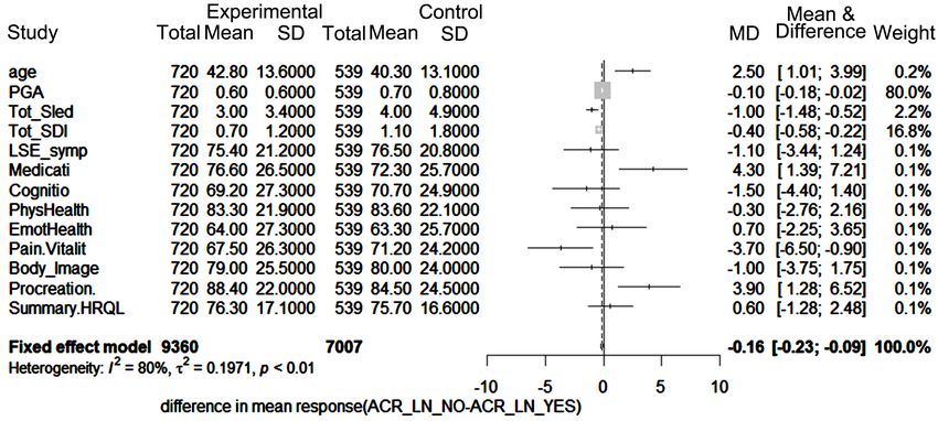
lupus nephritis. We provide a flowchart for the plan of work in the Appendix.
2. History of Lupus
In a seminal paper by Smith and Cyr [1] (1988), the history of lupus was divided
into three periods, classical, neoclassical, and modern. In this section we sum-
marize the medical research history to uncover the mystery of this autoimmune
disease.
2.1. Lupus in the Classical Period (1230-1856)
The authors of this important article by Smith and Cyr identified the term “lu-
pus” (Latin for “wolf”) which was used to describe erosive facial lesions that
were reminiscent of a wolf’s bite. The reader is referred to the list of references in
the paper. The lesions now referred to as discoid lupus were described in (1833)
by Cazenave [2] under the term “erythema centrifugum,” while the butterfly
distribution of the facial rash was noted by Von Hebra in (1846) [3].
2.2. Lupus in the Neoclassical Period (1872-1948)
The Neoclassical era of the history of lupus began in 1872 when Kaposi [4] first
described the systemic nature of the disorder. He proposed that there were two
types of lupus erythematosus; the discoid form and a disseminated (systemic)
form. Furthermore, he enumerated various signs and symptoms which characte-
rized the systemic form of the disease.
The existence of a systemic form of lupus was firmly established in (1904) by
the work of Osler [5] in Baltimore and Jadassohn in Vienna [6]. Over the next
thirty years, pathologic studies documented the existence of nonbacterial ver-
rucous endocarditis (Liebman-Sacks disease) and wire-loop lesions in individu-
als with glomerulonephritis; such observations at the autopsy table led to the
construct of collagen disease proposed by Klemperer and colleagues [7] in
DOI: 10.4236/ojra.2021.114014 116 Open Journal of Rheumatology and Autoimmune Diseases
S. N. Al-Gahtani
(1941). This terminology, “collagen vascular disease,” persists in usage more
than seventy years after its introduction.
2.3. Lupus in the Modern Era (1948-Present)
The most important discovery which characterized the modern era was the dis-
covery of the LE cell by Hargraves and colleagues [8] in (1948). The investigators
observed these cells in the bone marrow of individuals with acute disseminated
lupus erythematosus and postulated that the cell “…is the result of… phagocyto-
sis of free nuclear material with a resulting round vacuole containing this par-
tially digested and lysed nuclear material…”
This discovery ushered in the present era of the application of immunology to
the study of lupus erythematosus; it also allowed the diagnosis of individuals
with much milder forms of the disease. This possibility, coupled with the dis-
covery of cortisone as a treatment, changed the natural history of lupus as it was
known prior to that time.
Two other immunologic markers were recognized in the 1950s as being asso-
ciated with lupus: the biologic false-positive test for syphilis and the immunof-
luorescent test for antinuclear antibodies. Moore [9], working in Baltimore,
demonstrated that systemic lupus developed in 7 percent of 148 individuals with
chronic false-positive tests for syphilis and that a further 30 percent had symp-
toms consistent with collagen disease.
2.4. The Development of Animal Model
Two other major advances in the modern era have been the development of
animal models of lupus and the recognition of the role of genetic predisposition
to the development of lupus [10]. The mouse model has provided many insights
into the immunopathogenesis of autoantibody formation, mechanisms of im-
munologic tolerance, the development of glomerulonephritis, the role of sex
hormones in modulating the course of disease, and evaluation of treatments in-
cluding recently developed biologic agents such as anti-CD4, among others.
3. Signs and Symptoms of Lupus
Lupus can affect almost any organ in your body. The symptoms of lupus also
differ from person to person [11] [12] [13]. For example, one woman with lupus
may have swollen knees and fever. Another woman may be tired all the time or
have kidney trouble. Someone else may have rashes. Over time, new symptoms
may develop, or some symptoms may happen less often.
Lupus symptoms also usually come and go, meaning that you don’t have them
all of the time. Lupus is a disease of flares, and remissions (the symptoms im-
prove, and you feel better).
Lupus symptoms include:
Muscle and joint pain. You may experience pain and stiffness, with or with-
out swelling. This affects most people with lupus. Common areas for muscle
DOI: 10.4236/ojra.2021.114014 117 Open Journal of Rheumatology and Autoimmune DiseasesS. N. Al-Gahtani
pain and swelling include the neck, thighs, shoulders, and upper arms.
Fever. A fever higher than 100 degrees Fahrenheit affects many people with
lupus. The fever is often caused by inflammation or infection. Lupus medi-
cine can help manage and prevent fever.
Rashes. You may get rashes on any part of your body that is exposed to the
sun, such as your face, arms, and hands. One common sign of lupus is a red,
butterfly-shaped rash across the nose and cheeks.
Chest pain. Lupus can trigger inflammation in the lining of the lungs. This
causes chest pain when breathing deeply.
Hair loss. Patchy or bald spots are common. Hair loss could also be caused
by some medicines or infection.
Sun or light sensitivity. Most people with lupus are sensitive to light, a con-
dition called photosensitivity. Exposure to light can cause rashes, fever, fati-
gue, or joint pain in some people with lupus.
Kidney problems. Half of people with lupus also have kidney problems, called
lupus nephritis. Symptoms include weight gain, swollen ankles, high blood
pressure, and decreased kidney function.
Mouth sores. Also called ulcers, these sores usually appear on the roof of the
mouth, but can also appear in the gums, inside the cheeks, and on the lips.
They may be painless, or you may have soreness or dry mouth.
Prolonged or extreme fatigue. You may feel tired or exhausted even when
you get enough sleep. Fatigue can also be a warning sign of a lupus flare.
Anemia. Fatigue could be a sign of anemia, a condition that happens when
your body does not have red blood cells to carry oxygen throughout your
body.
Memory problems. Some people with lupus report problems with forgetful-
ness or confusion.
Blood clotting. You may have a higher risk of blood clotting. This can cause
blood clots in the legs or lungs, stroke, heart attack, or repeated miscarriages.
Eye disease. You may get dry eyes, eye inflammation, and eyelid rashes.
Lupus flare. Flare is defined by International Consensus as a measurable in-
crease in disease activity in one or more organ systems involving new or worse
clinical signs and symptoms and/or laboratory measurements. Flares have been
associated with more hospitalizations [14] and more organ and system damage,
which in turn can lead to poorer prognosis and increased mortality [15] [16]
[17]. In addition, the prolonged use of CS in the presence of persistence of dis-
ease or during flares can contribute to damage [18]. Flares, damage, and pro-
longed use of CS can contribute to poor health-related quality of life. Therefore,
flare prevention is an important treatment goal in patient management. Under-
standing the pattern of flares in SLE patients would be informative not only in
the day-to-day management of these patients, but also in the interpretation of
clinical trials of new medications where frequency and type of flares are included
as outcome measures.
DOI: 10.4236/ojra.2021.114014 118 Open Journal of Rheumatology and Autoimmune DiseasesS. N. Al-Gahtani
4. Risk Factors for SLE
Scientists divide risk factors associated with SLE into three groups; environmen-
tal, we divide the risk factors for Lupus into two groups; the first is related to en-
vironmental and the other is genetic factors.
4.1. Environmental Risk Factors
Aggregate data from population base studies reported that approximately 90% of
patients with SLE are female, and the incidence of SLE among African Ameri-
cans is increased 3 - 4-fold compared with that among Caucasians [19] [20]. It is
therefore postulated that gender is a non-modifiable risk factor of prime impor-
tance.
There is strong epidemiologic evidence linking environmental factors, including
current cigarette smoking, vitamin D level infections such as Epstein-Barr virus,
dietary factors. In the following section we list some of these environmental risk
factors.
Cigarette Smoking
Mechanistic evidence exists implicating smoking in SLE pathogenesis. Expo-
sure to toxic components from cigarette smoke (e.g. tars, nicotine, carbon mo-
noxide, polycyclic aromatic hydrocarbons and free radicals) can induce oxida-
tive stress and directly damage endogenous proteins and DNA, leading to genet-
ic mutations and gene activation, which could be involved in development of
SLE [19]. Cigarette smoking stimulates the expression of CD95 on B and CD4 T
cell surfaces, potentially inducing autoimmunity [20], and augments production
of pro-inflammatory cytokines [21]. In a retrospective case-control study of SLE
patients, current smokers were significantly more likely to have anti-double
stranded DNA antibodies compared to never smokers [21]. Additionally, smok-
ing leads to the formation of immunogenic DNA adducts with a half-life of 9 to
13 weeks, which may explain why current smoking has been more strongly asso-
ciated with increased SLE risk [21]. However, two additional case-control studies
performed since then have demonstrated an elevated risk for both current and
former smokers compared to never smokers [22] [23].
Vitamin D
Further complicating our understanding of the relationship between UV radi-
ation and SLE pathogenesis is the controversial role of Vitamin D. While expo-
sure to solar UV radiation may trigger SLE disease flares, UV light exposure is
also the main source of vitamin D production [24]. Vitamin D may be immu-
nosuppressive once metabolized to 1α, 25(OH)2D, and it has been suggested that
UV-B radiation could reduce SLE risk via stimulation of cutaneous vitamin D
synthesis [25]. Many cross-sectional and case-control studies have reported low
25(OH) vitamin D concentrations in SLE patients compared to controls, howev-
er, it is not clear whether low vitamin D is a cause or consequence of chronic dis-
ease.
Infections
(EBV) seropositivity rates are much higher in adults and children with SLE
DOI: 10.4236/ojra.2021.114014 119 Open Journal of Rheumatology and Autoimmune DiseasesS. N. Al-Gahtani
than age-matched controls [26]. Potential mechanisms involve Epstein-Barr vi-
rus protein complexes inducing type 1 interferon via Toll-like receptor 3 and
molecular mimicry between EBV and SLE antigens [27]. Additionally, SLE pa-
tients have impaired CD8+ cytotoxic T cells, and irregular cytokine production
in plasmacytoid dendritic cells and CD69+ CD4+ T cells in response to EBV.
However, no conclusive data have established that EBV infection is linked to fu-
ture risk of SLE. Notably, in a large population-based Danish cohort, EBV-serologic
negative individuals had a sustained increased risk for SLE highest in the 1 to 4
years after testing (standardized incidence rate, 6.6; 95% CI, 3.3 - 13.2), but this
finding may have been due to a surveillance bias as EBV testing is likely to be
performed during the work-up for early SLE symptoms [28]. In that study, no
associations were found with EBV serologic positivity, infectious mononucleosis,
or severe infectious mononucleosis requiring hospitalization [28] [29]. There-
fore, the association between EBV and incident SLE, in particular the question of
causality, remains to be fully elucidated.
Air Pollution
Particulate air pollution has effects like those of inhaled cigarette smoke and
silica on the immune system and has been linked to asthma, chronic bronchitis,
cardiovascular disease, and lung and laryngeal cancers [30] [31].
Heavy Metals
Data from experimental studies suggest that heavy metals may increase sys-
temic autoimmunity, and that co-exposure to certain heavy metals may increase
the risk associated with other exposures [32]. Mercury-exposed gold miners
were demonstrated to have a higher prevalence of detectable ANA as compared
to diamond and emerald miners with no occupational mercury exposure [33].
Dietary Factors and Medications
While there are very few prospective studies of dietary intake and SLE, several
lines of evidence implicate dietary factors in SLE pathogenesis. First, murine
models suggest that dietary exposures can induce epigenetic changes and SLE
autoimmunity [34] [35]. When genetically-SLE predisposed C57BL/6 × SJL mice
were fed methyl donor poor diets, they developed lupus nephritis, whereas those
fed diets rich in methyl group micronutrients did not [36] [37] [38]. As oxidative
stress, inflammation and cytokine dysregulation are central to SLE pathogenesis,
diet may play an important accelerating role. Prior studies suggest that high in-
take of certain antioxidants; fish, olive oil and cooked vegetables may confer a
protective effect against chronic disease development [38]. Women consuming >
200 mL of coffee per day had increased inflammation markers, such as interleu-
kin-6 and tumor necrosis factor-α, compared with coffee non-drinkers [38]. A
prior case-control study suggests a significant increased SLE risk with black tea
consumption and borderline increased risk with coffee consumption, but not
green tea [38].
4.2. Genetic Risk Factors
During the past few years extensive research on the genetic bases of SLE has
DOI: 10.4236/ojra.2021.114014 120 Open Journal of Rheumatology and Autoimmune DiseasesS. N. Al-Gahtani
emerged. Genetic variation was first shown to be important in SLE in the 1970s
with associations in the human leukocyte antigen region. Almost four decades
later, and with the help of increasingly powerful genetic approaches, more than
25 genes are now known to contribute to the mechanisms that predispose indi-
viduals to lupus [39]. Over half of these loci have been discovered in the past 2
years, underscoring the extraordinary success of genome-wide association ap-
proaches in SLE. Well-established risk factors include alleles in the major histo-
compatibility complex region (multiple genes), IRF5, ITGAM, STAT4, BLK,
BANK1, PDCD1, PTPN22, TNFSF4, TNFAIP3, SPP1, some of the Fcg receptors,
and deficiencies in several complement components, including C1q, C4 and C2.
As reviewed here, many susceptibility genes fall into key pathways that are con-
sistent with previous studies implicating immune complexes, host immune sig-
nal transduction and interferon pathways in the pathogenesis of SLE. Other loci
have no known function or apparent immunological role and have the potential
to reveal novel disease mechanisms. Certainly, as our understanding of the ge-
netic etiology of SLE continues to mature, important new opportunities will emerge
for developing more effective diagnostic and clinical management tools for this
complex autoimmune disease.
In the past 2 years, a series of landmark studies have been reported that have
revolutionized our understanding of the genetics of lupus. These are composed
of insightful candidate gene studies and are anchored by genome-wide associa-
tion (GWA) studies that include more than 10,000 individuals of European des-
cent genotyped at over 300,000 single nucleotide polymorphisms (SNPs). Suc-
cessful mapping of risk loci for lupus is comparable to inflammatory bowel dis-
ease where over 30 loci have now been identified [40]. SLE has long been consi-
dered a prototypic autoimmune disease, and from the genetics perspective, has
served that title well. The coming years will most certainly continue to reveal
additional associations, refinements to our current understanding of those estab-
lished thus far and provide new clues to explain the clinical heterogeneity that is
a hallmark of SLE. Additional work will include detailed genetic studies in mul-
tiple ethnic groups, the development of models of disease that incorporate envi-
ronmental influences, and studies to determine the overlapping and unique rela-
tionships among genetic variants associated with other related autoimmune
phenotypes. Every robust, authentic association has a compulsory role in patho-
genesis, which promises to change profoundly our capacity to diagnose, predict
and treat human disease [40] [41].
A recent paper [42] focusing on the Genome-wide association studies of Sys-
temic Lupus Erythematosus (SLE) nominated 3073 genetic variants at 91 risk lo-
ci. To systematically screen these variants for allelic transcriptional enhancer ac-
tivity, the authors construct a massively parallel reporter assay (MPRA) library
comprising 12,396 DNA oligonucleotides containing the genomic context around
every allele of each SLE variant. Collectively, their approach provided a blueprint
for the discovery of allelic gene regulation at risk loci for any disease and offers
DOI: 10.4236/ojra.2021.114014 121 Open Journal of Rheumatology and Autoimmune DiseasesS. N. Al-Gahtani
insight into the transcriptional regulatory mechanisms underlying SLE.
4.3. Genetic Epidemiology of SLE: Studies on the Familial
Aggregation of SLE
Family studies are the only design that helps quantifying the inheritance of SLE
and its clustering among family members. Only a few studies have looked at fa-
milial aggregation of SLE on a nationwide level. In a recent cohort study from
Taiwan, 18 283 [43] persons were identified as having SLE. Among these, 607
first-degree relatives had SLE, corresponding to an overall 17 times increased
risk of SLE in first-degree relatives and a 315 times increased risk in twins [43]
[44]. The cohort was based on national health insurance data, where the insured
person had to claim relatives as dependents. Hence, 20% of the cohort members
were registered alone without any identifiable relative. However, the diagnosis of
SLE could only be agreed upon by an expert panel from the Taiwan National.
The study [43] which is a nation-wide registry-based assessed the impact of
having a family history of SLE on the risk of developing autoimmune-diseases
(AD) in a population-based Danish cohort. The important findings were that the
risk of developing any AD was significantly 51% and 28% elevated in individuals
with a first-degree or second- or third-degree relative with SLE, respectively. The
risk was substantially elevated for SLE, but also for RA, IBD, type 1 diabetes mel-
litus and the combined group of other ADs in individuals with an SLE-affected
first degree relative. Individuals who had a co-twin or more than one first-degree
relative with SLE were at a 76- and 61-times increased risk of developing SLE,
respectively. There was no clear influence of the sex of the SLE-affected family
member on the risk of SLE, although male cohort members with an SLE-affected
male relative were at a 10-fold elevated relative risk of developing SLE as com-
pared with the 8-fold increased risk in female cohort members. In one previous
cohort study, there was a trend that men with SLE-affected male relatives were at
a higher risk of SLE [44]. In summary, the study showed that a family history of
SLE constitutes a major risk factor for subsequent development of SLE and other
ADs in a manner that depends on the degree of relatedness. Our findings may be
useful when counselling families affected by SLE. Many close family members of
SLE patients may find it reassuring to learn that although their relative risk of
developing SLE themselves is markedly elevated; their absolute risk remains low;
according to the present national cohort study only around 2% of individuals
with an SLE-affected non-twin first-degree relative developed SLE during an av-
erage of 22 years of follow-up.
Another population-based study on the familial aggregation of SLE has re-
ported the Hazard of SLE and other AD. The study assessed the impact of having
a family history of SLE on the risk of developing an AD in a population-based
Danish cohort. We summarized and modified their findings in Table 1. We also
calculated the correlation between the risk of SLE and that of AI for all family
relations. The Pearson’s correlation between the HR of SLE and the HR of AI
DOI: 10.4236/ojra.2021.114014 122 Open Journal of Rheumatology and Autoimmune DiseasesS. N. Al-Gahtani
Table 1. Familial aggregation of risk of SLE and other Auto-Immune (AI) system diseas-
es.
Family relation HR_SLE HR_AI P-SLE p-AI
MZT 86.70 (149.5) 3.84 (3.6) 0.655 0.431
DZT 49.70 (86.5) 0.95 (1.7) 0.0001 0.972
CHILD 17.00 (5.6) 1.45 (0.2) 0.004 0.022
SIB 11.10 (2.38) 1.68 (0.11) 0.0001 0.0001
PARENT 8.72 (1.3) 1.44 (0.06) 0.0001 0.0001
GRAND 2.10 (1.2) 1.44 (0.08) 0.362 0.0001
AUNT_UNC 3.05 (5.3) 1.17 (0.23) 0.70 0.460
HALF_SIB 5.66 (3.9) 1.08 (0.17) 0.237 0.638
N_NEPH 4.57 (2.3) 1.06 (0.1) 0.116 0.516
COUSIN 5.16 (4.8) 1.22 (0.2) 0.391 0.265
HALF_A_U 4.75 (8.3) 1.35 (0.17) 0.650 0.207
was 0.78, which is highly significant. This high correlation underscores the rela-
tionship between Lupus and other autoimmune disorders, and their aggregation
among family relatives.
5. Lupus Nephritis
Lupus nephritis (LN) occurs in ~50% of patients with SLE and is the most
common, but not the only, cause of kidney injury in SLE. Men with SLE tend to
have more aggressive disease with higher rates of renal and cardiovascular in-
volvement and are more likely to develop kidney failure than women [45]. Pa-
tients with SLE who develop LN present at a younger age than patients with SLE
without nephritis [46]. Additionally, LN typically develops early in the disease
course, generally within the first 6 to 36 months, and may be present at initial
diagnosis. Risk factors for the development of LN include younger age, male sex,
and non-European ancestry. In the United States, the incidence of LN is higher
in black (34% - 51%), Hispanic (31% - 43%), and Asian (33% - 55%) compared
with white (14% - 23%) patients. Black and Hispanic patients have worse out-
comes and are more likely to progress to kidney failure than white patients.
Black and Hispanic patients tend to have more severe underlying histopatholo-
gy, higher serum creatinine levels, and more proteinuria than white patients at
LN diagnosis. Additionally, autoantibodies strongly associated with LN, includ-
ing anti-Sm, anti-Ro, and anti-ribonucleoprotein antibodies, are more frequently
positive in black compared with white patients. The reasons for these racial and
ethnic differences are not completely understood, but genetic and socioeconom-
ic factors likely contribute Mortality associated with lupus is significantly higher
in those with LN compared with those without LN, and death directly attributa-
ble to kidney disease occurs in 5% to 25% of patients with proliferative LN
within 5 years of onset. Furthermore, 10% to 30% of patients with LN progress
DOI: 10.4236/ojra.2021.114014 123 Open Journal of Rheumatology and Autoimmune DiseasesS. N. Al-Gahtani
to kidney failure requiring kidney replacement therapy (KRT). Patients with
proliferative forms of LN (class III, IV, or III/IV + V) are at highest risk for re-
quiring KRT. Achieving a complete clinical response to treatment is critical to
preserving long-term kidney health. In one study, patients who achieved a com-
plete clinical response had 92% kidney survival at 10 years compared to 43% in
partial responders and 13% in non-responders. Overall, the kidney failure risk
associated with LN improved from the 1970s to 2000. However, since 2000, the
rate of LN requiring KRT has remained consistent and there is evidence to sug-
gest that these rates are increasing now, particularly in black populations.
Genetic and environmental factors play important roles in the pathogenesis of
SLE [45] [46]. The incidence and prevalence of SLE is higher in non-European
ancestry, especially in African ancestry. The severity of SLE also varies among
the ethnic groups, being more severe in non-European populations [47] [48]
[49]. Supporting a genetic contribution to disease, monozygotic twins are much
more likely to be concordant for SLE than dizygotic twins (concordance rate
24% and 2%, respectively) [50]. Familial aggregation of SLE has also been clearly
documented, and most pedigrees support a non-Mendelian complex inheritance
[51]. These facts strongly support notion of a polygenic genetic contribution to
SLE pathogenesis.
Among the various organ manifestations of SLE, lupus nephritis (LN) is one
of the most feared, potentially resulting in organ damage and renal insufficiency
those results in poor clinical outcomes despite recent improvements in SLE treat-
ment.
Genome-Wide-Association Studies (GWAS) and candidate gene association
studies have revealed numerous risk genes for SLE, including loci which contain
genes that function in the innate and adaptive immune system [52] [53]. Some
of these genes are also closely associated with LN. However, most of these pre-
vious studies were not primarily focused on the nephritis phenotype, and less is
known about which genes predispose to LN. Some recent studies which have
focused on identifying the genes specifically responsible for LN have identified
intrarenal genes that are associated with LN, but not associated with the suscep-
tibility of SLE in general [54] [55]. While the exact functional mechanisms of
these renal-related candidate genes remain unclear, it seems that the genetic ba-
sis of LN involves a combination of general SLE susceptibility genes which func-
tion in the immune system and genes which are more renal-specific that predis-
pose specifically to LN.
5.1. Difference in Incidence Rate and Severity of LN between
Ethnicities
Among various organ manifestations of SLE, LN is one of the most severe, and
can progress to end-stage renal disease (ESRD) leading to increased morbidity
and mortality. LN affects about 40% of SLE patients throughout their lifetime
[56]. Despite recent advances in treatment, patients with LN still have higher
morbidity and mortality compared with those without LN [56] [57] [58]. It is
DOI: 10.4236/ojra.2021.114014 124 Open Journal of Rheumatology and Autoimmune DiseasesS. N. Al-Gahtani
well known that African, Asian, and Hispanic populations are more likely to de-
velop LN as compared to European ancestry [48] [56] [59] [60] [61]. There are
also differences in severity of LN among the ethnicities, with LN being more se-
vere in non-European populations [48] [56]. This disparity between ancestral
backgrounds could be related to genetic or environmental factors [62]. To inves-
tigate the importance of genetic factors, Sanchez et al. conducted a study eva-
luating the genetic impact of the proportion of Amerindian vs. European genetic
ancestry in admixed populations living in South America. This is an informative
way to study the contribution of genetics to LN with some control over envi-
ronment, as different individuals living in the same population and same loca-
tion will have different proportional genetic ancestry. This study revealed that an
increased proportion of Amerindian genetic ancestry correlated with increased
risk of developing LN [47]. Another study demonstrated familial clustering of
ESRD African ancestry SLE patients with LN, suggesting shared genetic factors
contributing to LN in these families [63]. These studies support the idea that ge-
netic factors contribute to the pathogenesis of LN.
Not only genetic changes, but epigenetic changes (i.e., post-translational mod-
ifications) also play an important role in the pathogenesis of SLE. DNA methyla-
tion is one of the important post-translational regulatory modifications, typically
occurring at CG dinucleotides. DNA methylation results in gene silencing by
tightening the chromatin structure and limiting the access of transcriptional
factors, while DNA hypomethylation induce transcription of genes.
Impaired DNA methylation status in CD4+ T cells of SLE patients was re-
ported more than 20 years ago [64] [65] [66]. As next-generation sequencing
technology has advanced, genome-wide methylation studies have demonstrated
the differences in methylation profiles of CD4 T cells in SLE patients compared
to those of healthy controls. Some studies have shown a difference in methyla-
tion profiles between different groups of SLE patients [67] [68]. Of note, hypo-
methylation of type I IFN-regulated genes known to play important roles in the
pathogenesis of SLE are reported in SLE patients [69] [70]. More recently, Coit
et al. [70] identified that there are more robust differences in methylation status
of type I IFN-regulated genes when compared between SLE patients with LN
and SLE patients without LN [71].
These studies shed light to another aspect of genetic involvement in the pa-
thogenesis of SLE and LN, although there is still much work to be done to clarify
their specific role to LN and take advantage of this knowledge to design treat-
ments.
5.2. Trend in Mortality of Patients with Lupus Nephritis
A recent study on the survival rate of patients with renal diseases reported very
important findings from a community-based study [72]. The rates of renal sur-
vival (i.e., survival without dialysis) in patients with lupus nephritis in the 1990s
ranged from 83% to 92% over 5 years of follow up and from 74% to 84% over 10
years of follow up [73] [74] [75]. The risk of end-stage renal disease (ESRD) has
DOI: 10.4236/ojra.2021.114014 125 Open Journal of Rheumatology and Autoimmune DiseasesS. N. Al-Gahtani
been particularly high in patients with diffuse proliferative glomerulonephritis,
with risk estimates ranging from 11% to 33% over 5 years of follow up [76] [77]
[78]. The prognosis of lupus nephritis depends on many demographics, racial,
genetic, histopathologic, immunologic, and time-dependent factors [79]. Renal
disease that fails to remit with immunosuppressive therapies is a major risk fac-
tor for subsequent deterioration of renal function and poor outcome [80] [81].
Recent studies have reiterated that lupus nephritis patients of African, Hispanic,
or Asian ethnicity have generally experienced poorer outcomes [82] [83] [84]
[85].
More recent studies have focused on the relative mortality of patients with
lupus renal disease as compared to different reference groups [86]-[91]. Howev-
er, the effect of different histologic classes of lupus glomerulonephritis on the
relative mortality of SLE, as compared to mortality rates in the general popula-
tion, has been largely unreported. Moreover, data on the life expectancy of pa-
tients with lupus nephritis are not available in the literature. Therefore, the present
study was carried out to evaluate the effect of renal disease, histologic class of
lupus nephritis, renal damage, and renal failure on the standardized mortality
ratio (SMR) and life expectancy in a longitudinal cohort of SLE patients from
China. The findings of the above research concluded that patients with SLE have
increased mortality. This is due to multiple factors that include an increased
susceptibility to infection, accelerated atherosclerosis, and malignancies, as well
as organ damage due to treatment failure or complications [92]. The survival of
patients with SLE has improved tremendously in the past 3 - 4 decades, which is
attributed to earlier diagnosis and treatment, more judicious use of corticoste-
roids, the emergence of novel treatments, and better supportive care for organ
failure and infection-related complications [92]. However, the improvement in
the SLE survival rate appears to have reached a plateau since the 1990s [93]. Pa-
tients with SLE still have a mortality rate higher than that of the general popula-
tion [87] [91], although a dropping trend has been observed [94]. Renal disease
is a major organ manifestation of SLE, and its presence further increases the risk
of death, because the disease still progresses to ESRD in a constant proportion of
patients over time [86]-[91].
5.3. Treatment of the Diseases
The significant diversity of lupus nephritis (LN) has been the subject of intense
investigation for a long time [95]. Several attempts were made to classify the pa-
thologic features of LN. This classification represented distinctive characteristics
and hence differences in response to treatment.
Intravenous cyclophosphamide (IVCY) has been widely used as a form of
therapy to induce remission of diffuse proliferative LN (also known as class IV
LN) for more than 20 years [96]. Since 1997, mycophenolate mofetil (MMF) has
also been used successfully for the treatment of class IV LN [97] [98] [99].
A combined therapy consisting of steroids, MMF, and tacrolimus has been
applied in the field of organ transplantation for years. It was shown to be an ef-
DOI: 10.4236/ojra.2021.114014 126 Open Journal of Rheumatology and Autoimmune DiseasesS. N. Al-Gahtani
fective treatment for early mixed cellular and humoral renal allograft rejections
[100] [101] [102] [103] [104]. Does it work in LN? Considering both the phar-
macologic differences between tacrolimus and MMF and their efficacy for the
treatment of renal allograft rejections, we investigated the therapeutic efficacy
and adverse effects of this combined therapy (steroid + MMF + tacrolimus) in
the induction treatment of class V + IV LN, in comparison with IVCY therapy.
An extensive clinical trial [95] documented important findings regarding the
treatment of LN and its classes. It was noted that the overlap of class V with III
(Vc) and IV (Vd) was described as a subcategory of membranous LN in the 1982
World Health Organization classification but was eliminated in the 2003 ISN/RPS
classification [105].
Although the cause of systemic lupus erythematosus remains elusive, the un-
deniable fact is that different types of LN may involve different immune patho-
geneses. LN is characterized by the deposition of IgG4 and the absence of de-
layed type hypersensitivity effectors [106].
In summary, the therapeutic goal for patients with LN is to achieve prompt
remission and avoid disease flare and chronic renal impairment. As studies have
shown [107] [108] relapses of LN may be common after the induction treatment.
A prolonged follow-up period is needed for the exploration of this treatment’s
impact on long-term prognosis and the recurrence rate during the maintenance
therapy period [96].
Even with treatment, loss of kidney function sometimes progresses. If both
kidneys fail, people with lupus nephritis may need dialysis. Dialysis involves fil-
tering the blood through a machine to remove waste products from the body.
Ultimately, it may be necessary to have a kidney transplant. In those cases,
people will need additional drugs to keep their immune system from rejecting
the transplanted kidney.
Why is Health-Related Quality of Life & Well-Being Important?
Healthy People 2020 emphasizes the importance of health-related quality of
life and well-being by including it as one of the initiative’s 4 overarching goals,
“promoting quality of life, healthy development, and health behaviors across all
life stages [109] [110]. It also was established as one of the HP2020 4 foundation
health measures [111].
The significance of quality of life and well-being as a public health concern is
not new. Since 1949, the World Health Organization (WHO) has noted that health
is “a state of complete physical, mental, and social well-being and not merely an
absence of disease and infirmity.” [112]. In 2005, WHO recognized the impor-
tance of evaluating and improving people’s quality of life in a position paper
[113]. Because people are living longer than ever before, researchers have changed
the way they examine health, looking beyond causes of death and morbidity to
examine the relationship of health to the quality of an individual life.
When quality of life is considered in the context of health and disease, it’s
commonly referred to as health-related quality of life (HRQOL). Researchers to-
day agree that HRQOL is multidimensional and includes domains that are re-
DOI: 10.4236/ojra.2021.114014 127 Open Journal of Rheumatology and Autoimmune DiseasesS. N. Al-Gahtani
lated to physical, mental, emotional, and social functioning and the social con-
text in which people live [114].
HRQOL is a subjective and multidimensional concept that includes aspects of
physical, mental, and social health [115] [116]. For Healthy People 2020, the Pa-
tient-Reported Outcomes Measurement Information System (PROMIS) Global
Health Items were identified as reliable and valid measures of self-reported physi-
cal and mental health and are currently being considered to monitor these 2
domains across the decade. PROMIS is an NIH Roadmap initiative designed to
develop an electronic system to collect self-reported HRQOL data from diverse
populations of individuals with a variety of chronic diseases and demographic
characteristics [117] [118].
An important study [119] on HRQOL used cross sectional data obtained from
patients with systemic lupus erythematosus (SLE) during psychometric evalua-
tion studies of Lupus from various countries were compared between those: 1)
with and without LN and 2) with active and inactive-LN. Data compared included
demographics, disease characteristics, and Lupus PRO constructs. Presence of
LN was present if listed among the ACR classification criteria (ACR-LN), while
presence of active LN was based on presence of urinary casts, hematuria, prote-
inuria or pyuria in the disease activity assessment (SELENA-SLEDAI) performed
at the time of the study visit. Lupus PRO has Health related QOL (HRQOL) and
non-HRQOL constructs. HRQOL domains include lupus symptoms, cognition,
medication, procreation, physical health, emotional health, pain-vitality, and body
image. Non-HRQOL domains include desires-goals, social support, coping and sa-
tisfaction with care. Non-parametric tests were used to make comparisons, and p
values ≤ 0.05 were considered significant.
The results of the study are summarized here. There were 1259 SLE patients.
Five-hundred and thirty-nine had ACR-LN. These patients were younger, had
greater disease activity (PGA, Total SELENA-SLEDAI) and damage (SLICC/ACR)
than those without LN. Summary HRQOL and non-HRQOL scores were similar
in both groups; however, those with ACR-LN had significantly worse scores on
medications and procreation domains, while those without ACR-LN had worse
scores on Pain-Vitality domains.
129/540 ACR-LN patients had active LN. Patients with active LN were young-
er, had significantly greater disease activity (PGA, Total SELENA-SLEDAI),
worse HRQOL and non-HRQOL than patients with inactive LN. Specific do-
mains scores adversely affected among active LN patients were lupus symptoms,
medications, procreation, emotional health, body image and desires-goals. Satis-
faction with care was significantly higher among patients with active LN as com-
pared to inactive LN patients. We conducted meta-analytic approach using com-
ponents of the study (study factors) [119]. In Figure 1: study = components of
the HRQOL, Experimental = LN_NO, and Control = LN_YES.
In Figure 2, study = components of the HRQOL, Experimental = ACR_LN
(NO), control = ACR_LN (YES). These notations are the default naming of
groups provided by the software program R.
DOI: 10.4236/ojra.2021.114014 128 Open Journal of Rheumatology and Autoimmune DiseasesS. N. Al-Gahtani
Figure 1. Confidence levels for LN_ACTIVE (NO) as compared to the LN_ACTIVE
(YES).
Figure 2. Confidence levels for ACR_LN (NO) as compared to the ACR_LN (YES).
The Pearson’s correlation among the HRQOL scores between ACR_LN (NO)
and ACR_LN (YES) is 0.998.
The Pearson’s correlation among the HRQOL scores between LN_ACTIVE
(NO) and LN_ACTIVE (YES) is 0.997.
Several important components of the HRQOL instruments need explanation
in the above figures; they are “The Physician Global Assessment” (PGA). The
PGA [120] is a visual analogue score that reflects the clinician’s judgment of
overall SLE disease activity.
Construct validity was demonstrated by a good correlation (r ≥ 0.50) between
the PGA with the SLEDAI [121]. The SLEDAI is a global index that was devel-
oped and introduced in 1985 as a clinical index for the assessment of lupus dis-
ease activity in the preceding 10 days. It consists of 24 weighted clinical and la-
boratory variables of nine organ systems. This instrument was derived by con-
sensus among experts in rheumatology followed by application of regression
models to assign relative weights to each parameter. SLEDAI was modeled based
on clinician global judgment.
A modified version of the SLEDAI (SELENA-SLEDAI) was devised for use in
the Safety of Estrogens in Lupus National Assessment (SELENA) study. A glos-
sary was added, and the scoring was modified to account for persistent active
disease in some descriptors (rash, mucosal ulcers, and alopecia), which were pre-
DOI: 10.4236/ojra.2021.114014 129 Open Journal of Rheumatology and Autoimmune DiseasesS. N. Al-Gahtani
viously not scored unless they were new or recurrent.
In the SELENA-SLEDAI, researchers accepted the presence of either objective
or subjective findings to score the descriptor as present [19]. The SELENASLEDAI
version awaits rigorous validation with other measures related to disease activity
in SLE.SLEDAI-2000 SLEDAI-2000 (SLEDAI-2 K) was introduced in 2002 as a
measure of global disease activity. SLEDAI-2 K is a modification of the original
SLEDAI to allow the documentation of persistent.
6. Discussion
The fundamental goals of treatment of patients with SLE are to improve long-term
patient outcomes. Management should aim at remission of disease symptoms and
signs, prevention of damage accrual and minimization of drug side-effects, as well
as improvement of quality of life [122] [123]. Complete remission (absence of clin-
ical activity with no use of GC and IS drugs) is infrequent [124] [125] [126] [127]
[128]. To this end, newly defined low disease activity states (based on a SLEDAI
score ≤ 3 on antimalarials, or alternatively SLEDAI ≤ 4, PGA ≤ 1 with GC ≤ 7.5
mg of prednisone and well-tolerated IS agents) have shown comparable rates with
remission, regarding halting of damage accrual [129] [130] [131] [132]. According-
ly, treatment in SLE should aim at remission or, if this state cannot be achieved, at
low disease activity in all organ systems.
Prevention of disease flares is an additional milestone of SLE treatment. Al-
though a universally accepted definition is lacking, most experts agree that a flare
is a measurable increase in disease activity usually leading to change of treatment
[133] [134] [135].
Another issue related to the human burden of the disease is “Risk of infection”
associated with both disease-related and treatment-related factors; high-dose GC
therapy, CYC, MMF and RTX are all associated with an increased risk for infec-
tion, while high disease activity, severe leucopenia and presence of renal involve-
ment in nephrotic syndrome also contribute independently. Protection against in-
fections should be proactive, focusing both on primary prevention, as well as
timely recognition and treatment. Patients with lupus should receive vaccina-
tions according to the EULAR [136] recommendations for vaccination of patients
with autoimmune rheumatic diseases [137] [138]. Immunization against season-
al influenza and pneumococcal infection (both PCV13 and PPSV23) should be
strongly considered, preferably during stable disease.
SLE is an independent risk factor for cardiovascular disease (CVD), due to
both traditional and disease-related risk factors, such as persistent disease activ-
ity, LN, presence of aPL and use of GC [139] [140] [141]. Low-dose aspirin may
be considered for primary prevention of CVD, as it may reduce the risk for in-
cident CVD in SLE [142].
However, this must be viewed considering recent large studies in diabetics and
elderly showing that the benefits of aspirin for primary CVD prevention are
counterbalanced by the larger bleeding hazard [143] [144].
DOI: 10.4236/ojra.2021.114014 130 Open Journal of Rheumatology and Autoimmune DiseasesS. N. Al-Gahtani
As a final statement, there is no cure for LN. The reference [145] provides a
recent update to the recommended treatment of LN. The main objectives of the
treatment are to reduce symptoms or make symptoms disappear (remission), keep
the disease from getting worse, maintain remission, and avoid the need for kid-
ney transplant. For severe LN, [145] recommends, to stop the immune system
from attacking healthy cells, several treatment options such as: Cyclosporine,
Tacrolimus, Cyclophosphamide Azathioprine (Imuran), Mycophenolate, and
Rituximab (Rituxan).
In addition to the human suffering attributed to the disease, there is the eco-
nomic impact and the cost of disease management. These issues warrant research
within the framework of the field of “Health Economics”.
Acknowledgements
The author would like to acknowledge the constructive comments for an ano-
nymous reviewer.
Conflicts of Interest
The author declares no conflicts of interest regarding the publication of this pa-
per.
References
[1] Smith, C.D. and Cyr, M. (1988) The History of Lupus Erythematosus, from Hippo-
crates to Osler. Rheumatic Disease Clinics of North America, 14, 1-14.
https://doi.org/10.1016/S0889-857X(21)00942-X
[2] Cazenave, P.L.A. and Schedel, H.E. (1852) Manual of the Diseases of the Skin. Bur-
gess, Trans., Henry Renshaw, London.
[3] Hebra, F. (1866) On Diseases of the Skin Including the Exanthemata. Vol. 1.
Adams, F., Trans., The New Sydenham Society, London.
[4] Kaposi, M. (1875) Lupus vulgaris. In: Hebra, F. and Kaposi, M., Eds., On Diseases of
the Skin Including the Exanthemata, Vol. IV. Tay, W., Trans., The New Sydenham
Society, London.
[5] Osler, W. (1895) On the Visceral Complications of Erythema Exudativum Multi-
forme. The American Journal of the Medical Sciences, 110, 629-646.
[6] Jadassohn, J. (1904) Lupus Erythematodes. In: Mracek, F., Eds., Handbuch der
Hautkrankheiten, Alfred Holder, Wein, 298-404.
[7] Klemperer, P., Pollack, A. and Baehr, G. (1984) Diffuse Collagen Disease: Acute Dis-
seminated Lupus Erythematosus and Diffuse Scleroderma. JAMA, 251, 1593-1594.
https://doi.org/10.1001/jama.1984.03340360059032
[8] Hargraves, M.M. (1969) Discovery of the LE Cell and Its Morphology. Mayo Clinic
Proceedings, 44, 579-599.
[9] Moore, J.E. and Lutz, W.B. (1955) The Natural History of Systemic Lupus Erythe-
matosus: An Approach to Its Study through Chronic Biological False Positive Reac-
tions. Journal of Chronic Diseases, 2, 297-316.
https://doi.org/10.1016/0021-9681(55)90039-4
[10] Bielschowsky, M., Helyer, B.J. and Howie, J.B. (1959) Spontaneous Hemolytic
DOI: 10.4236/ojra.2021.114014 131 Open Journal of Rheumatology and Autoimmune DiseasesS. N. Al-Gahtani
Anemia in Mice of the NZB/BL Strain. Proceedings of the University of Otago
Medical School, 37, Article No. 9.
[11] Longo, D.L., Kasper, D.L., Larry, J.J., Fauci, A.S. and Hauser, S.L. (2011) Harrison’s
Principles of Internal Medicine. 18th Edition, McGraw-Hill Professional Pub, New
York.
[12] Cojocaru, M., Cojocaru, I.M., Silosi, I. And Vrabie, C.D. (2011) Manifestations of
Systemic Lupus Erythematosus. Maedica: A Journal of Clinical Medicine, 6, 330-336.
[13] Lee, Y.H., Woo, J.H., Choi, S.J., Ji, J.D. and Song, G.G. (2010) Induction and Main-
tenance Therapy for Lupus Nephritis: A Systematic Review and Meta-Analysis. Lu-
pus, 19, 703-710. https://doi.org/10.1177%2F0961203309357763
[14] Edwards, C.J., Lian, T.Y., Badsha, H., The, C.L., Arden, N. and Chng, H.H. (2003)
Hospitalization of Individuals with Systemic Lupus Erythematosus: Characteristics
and Predictors of Outcome. Lupus, 12, 672-676.
[15] Ugarte-Gil, M.F., Acevedo-Vásquez, E., Alarcón, G.S., Pastor-Asurza, C.A., Alfa-
ro-Lozano, J.L., Cucho-Venegas, J.M., et al., on behalf of GLADEL (2015) The
Number of Flares Patients Experience Impacts on Damage Accrual in Systemic Lu-
pus Erythematosus: Data from a Multiethnic Latin American Cohort. Annals of the
Rheumatic Diseases, 74, 1019-1023.
https://doi.org/10.1136/annrheumdis-2013-204620
[16] Bruce, I.N., O’Keeffe, A.G., Farewell, V., Hanly, J.G., Manzi, S., Su, L., et al. (2015)
Factors Associated with Damage Accrual in Patients with Systemic Lupus Erythe-
matosus: Results from the Systemic Lupus International Collaborating Clinics (SLICC)
Inception Cohort. Annals of the Rheumatic Diseases, 74, 1706-1713.
https://doi.org/10.1136/annrheumdis-2013-205171
[17] Yee, C.S., Su, L., Toescu, V., Hickman, R., Situnayake, D., Bowman, S., et al. (2015)
Birmingham SLE Cohort: Outcomes of a Large Inception Cohort Followed for Up
to 21 Years. Rheumatology, 54, 836-843.
https://doi.org/10.1093/rheumatology/keu412
[18] Al Sawah, S., Zhang, X., Zhu, B., Magder, L.S., Foster, S.A., Iikuni, N., et al. (2015)
Effect of Corticosteroid Use by Dose on the Risk of Developing Organ Damage over
Time in Systemic Lupus Erythematosus—The Hopkins Lupus Cohort. Lupus Science
& Medicine, 2, Article ID: e000066. https://doi.org/10.1136/lupus-2014-000066
[19] Pryor, W.A. and Stone, K. (1993) Oxidants in Cigarette Smoke. Radicals, Hydrogen
Peroxide, Peroxynitrate, and Peroxynitrite. Annals of the New York Academy of
Sciences, 686, 12-27. https://doi.org/10.1111/j.1749-6632.1993.tb39148.x
[20] Bijl, M., Horst, G., Limburg, P. and Kallenberg, C. (2001) Effects of Smoking on Ac-
tivation Markers, Fas Expression and Apoptosis of Peripheral Blood Lymphocytes.
European Journal of Clinical Investigation, 31, 550-553.
https://doi.org/10.1046/j.1365-2362.2001.00842.x
[21] Freemer, M.M., King Jr., T.E. and Criswell, L.A. (2006) Association of Smoking
with dsDNA Autoantibody Production in Systemic Lupus Erythematosus. Annals of
the Rheumatic Diseases, 65, 581-584. https://doi.org/10.1136/ard.2005.039438
[22] Kiyohara, C., Washio, M., Horiuchi, T., Asami, T., Ide, S., Atsumi, T., et al. (2012)
Cigarette Smoking, Alcohol Consumption, and Risk of Systemic Lupus Erythema-
tosus: A Case-Control Study in a Japanese Population. The Journal of Rheumatolo-
gy, 39, 1363-1370. https://doi.org/10.3899/jrheum.111609
[23] Washio, M., Horiuchi, T., Kiyohara, C., Kodama, H., Tada, Y., Asami, T., et al.
(2006) Smoking, Drinking, Sleeping Habits, and Other Lifestyle Factors and the
Risk of Systemic Lupus Erythematosus in Japanese Females: Findings from the
DOI: 10.4236/ojra.2021.114014 132 Open Journal of Rheumatology and Autoimmune DiseasesS. N. Al-Gahtani
KYSS Study. Modern Rheumatology, 16, 143-150.
https://doi.org/10.3109/s10165-006-0474-6
[24] Kuhn, A. and Beissert, S. (2005) Photosensitivity in Lupus Erythematosus. Au-
toimmunity, 38, 519-529. https://doi.org/10.1080/08916930500285626
[25] Cantorna, M.T. and Mahon, B.D. (2004) Mounting Evidence for Vitamin D as an
Environmental Factor Affecting Autoimmune Disease Prevalence. Experimental Bi-
ology and Medicine, 229, 1136-1142.
https://doi.org/10.1177%2F153537020422901108
[26] McClain, M.T., Poole, B.D., Bruner, B.F., Kaufman, K.M., Harley, J.B. and James,
J.A. (2006) An Altered Immune Response to Epstein-Barr Nuclear Antigen 1 in Pe-
diatric Systemic Lupus Erythematosus. Arthritis & Rheumatism, 54, 360-368.
https://doi.org/10.1002/art.21682
[27] Ulff-Moller, C.J., Nielsen, N.M. and Rostgaard, K. (2010) Epstein-Barr Virus-Associated
Infectious Mononucleosis and Risk of Systemic Lupus Erythematosus. Rheumatol-
ogy, 49, 1706-1712. https://doi.org/10.1093/rheumatology/keq148
[28] Cooper, G.S., Dooley, M.A., Treadwell, E.L. and William St Clair, E. (2002) Risk
Factors for Development of Systemic Lupus Erythematosus: Allergies, Infections,
and Family History. Journal of Clinical Epidemiology, 55, 982-989.
https://doi.org/10.1016/S0895-4356(02)00429-8
[29] Blanc, P.D., Jarvholm, B. and Toren, K. (2015) Prospective Risk of Rheumatologic
Disease Associated with Occupational Exposure in a Cohort of Male Construction
Workers. American Journal of Medicine, 128, 1094-1101.
https://doi.org/10.1016/j.amjmed.2015.05.001
[30] Pereira, F.A., de Assuncao, J.V., Saldiva, P.H., Pereira, L.A., Mirra, A.P. and Braga,
A.L. (2005) Influence of Air Pollution on the Incidence of Respiratory Tract Neop-
lasm. Journal of the Air & Waste Management Association, 55, 83-87.
https://doi.org/10.1080/10473289.2005.10464603
[31] Gilbert, K.M., Rowley, B., Gomez-Acevedo, H. and Blossom, S.J. (2011) Coexposure
to Mercury Increases Immunotoxicity of Trichloroethylene. Toxicological Sciences,
119, 281-292. https://doi.org/10.1093/toxsci/kfq345
[32] Gardner, R.M., Nyland, J.F., Silva, I.A., Ventura, A.M., de Souza, J.M. and Silber-
geld, E.K. (2010) Mercury Exposure, Serum Antinuclear/Antinucleolar Antibodies,
and Serum Cytokine Levels in Mining Populations in Amazonian Brazil: A Cross-
Sectional Study. Environmental Research, 110, 345-354.
https://doi.org/10.1016/j.envres.2010.02.001
[33] Li, Y., Gorelik, G., Strickland, F.M. and Richardson, B.C. (2014) Oxidative Stress, T
Cell DNA Methylation, and Lupus. Arthritis & Rheumatology, 66, 1574-1582.
https://doi.org/10.1002/art.38427
[34] Somers, E.C. and Richardson, B.C. (2014) Environmental Exposures, Epigenetic
Changes and the Risk of Lupus. Lupus, 23, 568-576.
https://doi.org/10.1177%2F0961203313499419
[35] Strickland, F.M., Hewagama, A., Wu, A., Sawalha, A.H., Delaney, C., Hoeltzel, M.F.,
et al. (2013) Diet Influences Expression of Autoimmune-Associated Genes and
Disease Severity by Epigenetic Mechanisms in a Transgenic Mouse Model of Lupus.
Arthritis & Rheumatism, 65, 1872-1881. https://doi.org/10.1002/art.37967
[36] Sofi, F., Cesari, F., Abbate, R., Gensini, G.F. and Casini, A. (2008) Adherence to
Mediterranean Diet and Health Status: Meta-Analysis. BMJ, 337, Article No. a1344.
https://doi.org/10.1136/bmj.a1344
[37] Zampelas, A., Panagiotakos, D., Pitsavos, C., Chrysohoou, C. And Stefanadis, C.
DOI: 10.4236/ojra.2021.114014 133 Open Journal of Rheumatology and Autoimmune DiseasesYou can also read