Application of Collagen I and IV in Bioengineering Transparent Ocular Tissues - Frontiers
←
→
Page content transcription
If your browser does not render page correctly, please read the page content below
REVIEW
                                                                                                                                               published: 26 August 2021
                                                                                                                                         doi: 10.3389/fsurg.2021.639500

                                             Application of Collagen I and IV in
                                             Bioengineering Transparent Ocular
                                             Tissues
                                             Yihui Song 1† , Morgan Overmass 1† , Jiawen Fan 2 , Chris Hodge 1,3,4 , Gerard Sutton 1,3,4 ,
                                             Frank J. Lovicu 1,5 and Jingjing You 1,6*
                                             1
                                               Save Sight Institute, Faculty of Medicine and Health, The University of Sydney, Sydney, NSW, Australia, 2 Key Laboratory of
                                             Myopia of State Health Ministry, Department of Ophthalmology and Vision Sciences, Eye and Ear, Nose, and Throat (ENT)
                                             Hospital, Shanghai Medical College, Fudan University, Shanghai, China, 3 New South Wales (NSW) Tissue Bank, Sydney,
                                             NSW, Australia, 4 Vision Eye Institute, Chatswood, NSW, Australia, 5 Discipline of Anatomy and Histology, School of Medical
                                             Sciences, The University of Sydney, Sydney, NSW, Australia, 6 School of Optometry and Vision Science, University of New
                                             South Wales, Sydney, NSW, Australia

                                             Collagens represent a major group of structural proteins expressed in different tissues
                                             and display distinct and variable properties. Whilst collagens are non-transparent in the
                                             skin, they confer transparency in the cornea and crystalline lens of the eye. There are 28
                                             types of collagen that all share a common triple helix structure yet differ in the composition
                          Edited by:         of their α-chains leading to their different properties. The different organization of collagen
                          Zhilian Yue,       fibers also contributes to the variable tissue morphology. The important ability of collagen
   University of Wollongong, Australia
                                             to form different tissues has led to the exploration and application of collagen as a
                         Reviewed by:
                         Tiago H. Silva,
                                             biomaterial. Collagen type I (Col-I) and collagen type IV (Col-IV) are the two primary
          University of Minho, Portugal      collagens found in corneal and lens tissues. Both collagens provide structure and
                             Jie Zhang,
                                             transparency, essential for a clear vision. This review explores the application of these
           The University of Auckland,
                          New Zealand        two collagen types as novel biomaterials in bioengineering unique tissue that could be
                    *Correspondence:         used to treat a variety of ocular diseases leading to blindness.
                           Jingjing You
                                             Keywords: bioengineering, collagen type IV, cornea, lens, retina, collagen type I
               jing.you@sydney.edu.au

     † These authors have contributed

    equally to this work and share first     INTRODUCTION
                            authorship
                                             The cornea and lens facilitate a pathway for light to pass through the eye to reach the retina,
                    Specialty section:       which then receives and transfers visual signals onto the brain for processing. These three major
          This article was submitted to      ocular tissues are critical for generating clear vision; therefore, any damage to the cornea, lens,
                       Visceral Surgery,     and/or retina will undoubtedly impair eyesight and often lead to blindness. Tissue engineering,
                a section of the journal
                                             in particular, the development of biomaterials with specific properties, has been increasingly
                    Frontiers in Surgery
                                             researched for treating ocular disease (1). Due to its abundance in the corneal stroma, collagen
       Received: 09 December 2020            type I (Col-I) has been a popular and versatile biomaterial developed to replace diseased corneal
            Accepted: 26 July 2021
                                             layers; however, it has become evident that no singular biomaterial can be an effective substitute
         Published: 26 August 2021
                                             for the intact cornea because of the differences in composition between each of the corneal layers
                              Citation:
                                             (2). Collagen type IV (Col-IV) is the predominant member of Descemet’s membrane of the cornea,
Song Y, Overmass M, Fan J, Hodge C,
 Sutton G, Lovicu FJ and You J (2021)
                                             the supportive layer of the corneal endothelium (3, 4). Furthermore, it is also the main collagen
     Application of Collagen I and IV in     type detected in the lens capsule (5), and in both Bruch’s membrane and the internal limiting
   Bioengineering Transparent Ocular         membrane (ILM) of the retina (6). Therefore, the application of Col-IV as a biomaterial could
      Tissues. Front. Surg. 8:639500.        potentially be useful in creating a natural environment and substratum for corneal endothelial cells
     doi: 10.3389/fsurg.2021.639500          and the epithelial cells of the lens and retina. In this paper, previous publications on Col-I and -IV in

Frontiers in Surgery | www.frontiersin.org                                          1                                             August 2021 | Volume 8 | Article 639500
Song et al.                                                                                                                             Collagen in Ocular Bioengineering

ocular-related applications were reviewed and the insights into                         endothelium is comprised of a monolayer of interconnected
the future direction of development of these two collagen types                         hexagonal cells sitting on the Descemet’s membrane. This layer
in ocular bioengineering are discussed.                                                 is key to maintaining relative stromal deturgescence/dehydration
                                                                                        that is essential for corneal transparency (2). The Descemet’s
The Distribution of Col- I and Col-IV in the                                            basement membrane is comprised primarily of Col-IV, as well
Cornea, the Lens, and the Retina                                                        as laminin, perlecan (a heparan sulfate proteoglycan), nidogen,
The human cornea is a transparent, avascular, highly innervated,                        and to a lesser degree, collagen type VIII (Col-VIII) (3, 4).
and organized tissue that is located at the front of the eye                            While both Col-IV and Col-VIII are present in the Descemet’s
(Figure 1). The cornea acts as a transparent window making up                           membrane, only Col-IV is located adjacent to endothelial cells in
two-thirds of the refractive power of the eye and consists of five                      both the infant and adult structure (19). In comparison, Col-VIII
main layers from anterior to posterior sides: corneal epithelium,                       chains initially face the endothelial cells in the infant Descemet’s
Bowman’s layer, corneal stroma, Descemet’s membrane, and                                membrane, but lose contact as we age and shift to face the
corneal endothelium (2) (Figure 1). One major structural protein                        stroma (3, 4).
in the cornea is collagen. The human cornea consists of                                     The lens is a transparent, biconvex orb that consists of the
many types of collagen and different collagen combinations are                          lens capsule, the lens epithelium, and lens fibers (Figure 2). Col-
detected within different layers (Table 1).                                             IV is the main type of collagen found in the lens capsule, which
   The central thickness of a normal adult human cornea                                 is a thick, uninterrupted basement membrane surrounding the
approximately measures 530 µm (19). In the context of                                   lens. The lens capsule is structurally analogous to the corneal
bioengineering, the stroma is critical because it constitutes                           Descemet’s membrane, as it consists of interlinking Col-IV and
the majority of the corneal volume and provides a significant
contribution to both its overall transparency and strength
(20). The corneal stroma is predominantly made up of Col-
                                                                                        TABLE 1 | Distribution of collagen types in the human cornea.
I fibrils organized into ∼300 orthogonally arranged lamellae
(2). These fibrils have a unique, smaller diameter, and regular                         Layer                                      Collagen type
interfibrillar spacing that supports the transparency of the tissue
(21). Furthermore, when stress is applied to the cornea, these                          Epithelium                                 IV, XV, XVIII, XII (7–13)
well-organized collagen fibers of the stroma are stretched to                           Bowman’s layer                             I, III, V (14)
counterbalance this force, allowing the cornea to maintain its                          Stroma                                     I, III, V, VI, XII, XIV (15–17)
existing shape (22). In addition to the stroma, the corneal                             Descemet membrane                          IV, VIII (15, 18)

 FIGURE 1 | A schematic illustration of the human cornea, located at the front of the eye and consisting of five layers.

Frontiers in Surgery | www.frontiersin.org                                          2                                           August 2021 | Volume 8 | Article 639500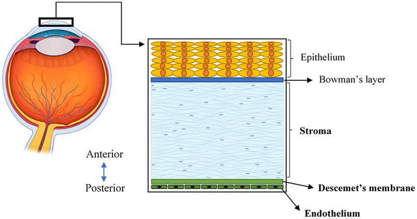
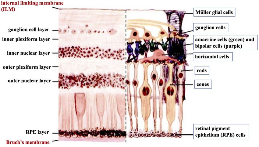
Song et al.                                                                                                                       Collagen in Ocular Bioengineering

 FIGURE 2 | A schematic illustration of lens anatomy and the matrix composition of the lens capsule in the enlarged area.

laminin networks bound together by nidogen and perlecan (5)                           signals to the brain to provide vision (6). It contains multiple
(Figure 2). The lens capsule acts as a supporting matrix for lens                     layers with various cell types (Figure 3). Retinal ganglion cells
epithelial cells anteriorly and fiber cells posteriorly. As a result                  (RGC) represent a type of neuron located at the inner surface
of this structure encapsulating all lens cells, it also protects them                 of the retina. The RGCs receive visual information from the
from infection. In younger eyes, the lens capsule also has a                          photoreceptors (rods and cones) via intermediate neuron types
role in determining the force required for lens accommodation,                        including bipolar cells, amacrine cells, and horizontal cells.
a process where the lens changes shape to alter our field of                          Rods and cones are responsible for sensing light. Bipolar cells
focus (23).                                                                           transfer visual information from photoreceptor cells to amacrine
   The human retina is the sensory tissue that lines the inner                        cells. Amacrine cells are interneurons in the retina that have
surface of the back of the eye, which senses light and sends                          short neurotic processes to connect to adjacent neurons and to

Frontiers in Surgery | www.frontiersin.org                                        3                                         August 2021 | Volume 8 | Article 639500
Song et al.                                                                                                                               Collagen in Ocular Bioengineering

 FIGURE 3 | A cross-sectional histological image of the retina, with its different layers (left) and the corresponding diagrammatic image depicting the different cell types
 of the retinal neural layers (right).

transfer neuronal signals. Horizontal cells, which are the laterally                     the passage of nutrients and metabolites between the RPE and
interconnecting neurons, have cell bodies in the inner nuclear                           underlying choriocapillaris (6). Bruch’s membrane also offers a
layer of the retina. They help integrate and regulate the input                          solid base and attachment site for RPE cells, acting as a part of
from multiple photoreceptors (6). Retinal pigmented epithelial                           the blood-retinal barrier (26). Bruch’s membrane may also be
(RPE) cells make up a single layer of the postmitotic cells. This                        involved in RPE differentiation (27) and wound healing (28, 29).
epithelia functions as a natural barrier and a regulator of the                          Type IV collagen is present on both sides of Bruch’s membrane
overlying photoreceptors (24). The final type of retinal-specific                        in a sandwich style with the middle layer containing elastic fiber-
cells is the Müller glial cells. These cells span the entire retina                      like bands, and can also be detected in the extracellular matrix
and connect with all other cell types via cellular processes that                        surrounding human RPE cells (30).
reach out to wrap around the neurons and the synapses. They                                 The ILM is not a true membrane, resulting from the fusion
also reach out to blood vessels, so as to act as an intermediary                         of the foot processes of the glia-like Müller cells. It forms a
between neurons and the circulatory system, thus regulating the                          physical barrier that protects the retina from toxins and from
flow of nutrients to the retina. Müller glia plays a critical role                       traction from the vitreous as the eye moves. Col-IV is the
in maintaining neuronal health and supporting visual function                            predominant extracellular matrix (ECM) protein in human ILM
(6). When light first enters the retina, it passes through the                           and accounts for ∼60% of its total proteins (31). Col-IV has
ganglion cell layer (GCL), then the inner plexiform layer (IPL),                         been detected throughout the entire thickness of ILM and is
inner nuclear layer (INL), outer plexiform layer (OPL), and outer                        likely to be secreted by retinal Müller cells (32). Several studies
nuclear layer (ONL) (6). All these neural layers comprising eight                        have identified that Col-IV is critical not only for the structural
types of retinal cells are located between the ILM and Bruch’s                           integrity of the basement membrane but also for neuron survival
membrane (Figure 3).                                                                     and angiogenesis (33, 34). Higher expression of Col-VI has been
   Basement membranes are specialized structures of the                                  found on the posterior side of the retina compared to its anterior
extracellular matrix that play an essential role in tissue                               side (35).
development and maintenance. Type IV collagens are abundant
components of all basement membranes (25). Bruch’s membrane                              The Structure of Col-I and -IV
and ILM represent two significant basement membranes within                              Collagens make up a supra-family of ECM proteins possessing
the human retina and are located at the inner and outer retina,                          a distinct triple-helical region formed from three polypeptide
respectively (Figure 3) (6). Bruch’s membrane primarily regulates                        chains (36). Currently, 28 genetically distinct collagen types

Frontiers in Surgery | www.frontiersin.org                                           4                                              August 2021 | Volume 8 | Article 639500
Song et al.                                                                                                        Collagen in Ocular Bioengineering

have been identified and described in the literature (37, 38).              opposed in different tropocollagen trimers. Formed elongated
Within this group, Col-I is classified as fibril-forming, while             fibrils can be 500 µm or more in length with a width of 500 nm
Col-IV is defined as network-forming due to their unique                    (36, 38). These fibrils also have a specific 3-dimensional packing
supramolecular organization. Due to its predominance in body                arrangement involving lateral associations between fibrils as they
tissues, Col-I biosynthesis has been more extensively explored              are staggered by about one-fourth of a molecular length. This
and will be outlined in this review; however, there is a notable            staggering also provides Col-I fibrils with a striated organization
lack of focus on the differences between Col-I and Col-IV                   where bands appear every 67 nm (50). Fibrillar organizations of
biosynthesis, which can be predicted on the basis of their differing        Col-I show a degree of crystallinity; however, this organization
supramolecular structures.                                                  varies throughout different tissues. Col-I aligns into straight
    At its most basic level, collagen biosynthesis involves                 parallel fibrillar arrangements in tendons, while in the human
the processing and aggregation of collagen monomers into                    corneal stroma, Col-I fibrils are arranged in 300 orthogonally
functional structures. Synthesis begins within the nucleus in               arranged sheets (51, 52).
generating relevant mRNAs, followed by the transcription of                    Following the spontaneous molecular arrangement,
mRNA molecules into a different α-chain (38). While Col-I                   additional stabilization of collagen is provided through
only possesses two types of α-chains (α1 and α2), Col-IV has                crosslinking. Lysyl oxidase (LO) facilitates crosslink formation
6 α-chains (α1–6) that form different network configurations                both in the head-to-tail alignment between adjacent telopeptide
to provide basement membrane specificity (39, 40). Col-I and                regions and in adjacent helical regions laterally (53, 54). LO
Col-IV are also considered heterotrimeric. This classification              initiates the formation of aldehydes from previously modified
results from the number of α-chain genes associated with each               amino acid residues, lysine, and hydroxylysine (38). Aldehydes
subtype; for example, Col-I trimers are composed of two α1                  formed in each region are then able to trigger aldol reactions with
chains and one α2 chain (39). Col-IV heterotrimers have greater             lysine residues in adjacent molecules, resulting in the formation
complexity as they can organize into three different isoforms:              of aldimine crosslinks. Intermolecular crosslinks following
α1α1α2, α3α4α5, and α5α5α6. The α1α1α2 Col-IV heterotrimer                  spontaneous molecular organization provide the required
is predominant throughout the basement membranes of the                     mechanical strength and stability for collagen organization. In
body (40); however, the α3α4α5 network has been identified                  comparison, Col-IV supramolecular assembly aims to create a
within specific tissues, including basement membranes within                mesh-like network structure. Within its polypeptide structure,
the eye. The adult human lens capsule contains only α3α4α5                  Col-IV chains have an N-terminal collagenous 7S domain, and a
network (41), whereas only α1α1α2 was found in the retinal ILM              C-terminal non-collagenous/globular domain (NC1), in addition
(42). Both α1α1α2 and α3α4α5 collagen IV networks co-exist in               to the central triple helix (40). In the creation of a network,
Bruch’s membrane (43), and all of six isoforms have been detected           varying arrangements of Col-IV trimers are created. Two
in adult Descemet’s membrane (3).                                           Col-IV trimers can covalently interact via their NC1 domain to
    Within the primary structure of collagen is a high proportion           form dimers while four 7S domains are able to crosslink into
of the repeating triplet sequence, Gly-X-Y. X, and Y in                     tetramers allowing for the creation of a strong and stable network
this sequence are predominantly occupied by proline (Pro)                   (40, 47, 55). More specifically, the 7S domains bond through the
and hydroxyproline (Hyp), respectively (38). High proportions               formation of disulphide bridges and covalent bonding of lysine
of Hyp are essential as this amino acid has a critical                      and hydroxylysine residues (47). This is a unique feature seen
role in stabilizing the triple helix through the formation of               in the Col-IV quaternary structure as the 7S domain contains
intramolecular hydrogen bonds. Accordingly, Xu et al. (2019)                cysteine and lysine residues. LO also plays a role in Col-IV
found hydrogen bond energy within helical regions to positively             crosslink formation as it again facilitates oxidative deamination
correlate with greater thermal stability (44). The formation of the         of lysine and hydroxylysine allowing for the formation of
triple helix in the procollagen molecule is likely a shared process         aldimine links, as seen in Col-I (56).
for Col-I and Col-IV. While uninterrupted triple helical domains
are the dominant structure of Col-I and have been found to have
a defined length of 300 nm (45, 46), Col-IV instead contains
21–26 interruptions within the Gly-X-Y sequence of the triple               CURRENT DEVELOPMENT OF TISSUE
helix, leading to greater intramolecular flexibility, more suited for       ENGINEERING IN TREATING OCULAR
network formation (47, 48).                                                 DISEASES
    Within the extracellular space, self-assembly is initiated, and
due to the significant differences in the resulting matrices, Col-          The ability to manufacture bioengineered tissue that mimics
I and Col-IV deviate at this stage. Released Col-I tropocollagen            existing intact tissue that can act as a means of repairing damage
molecules undergo a spontaneous but organized aggregation                   or to replace diseased layers presents obvious benefits in disease
process (38); albeit this spontaneous process has also been                 treatments. Using native matrix protein as the base material
found to be dependent on temperature, pH, ionic strength                    is a plausible direction and collagen, in particular Col-I, has
of the solution, and the concentration of collagenous and                   been widely investigated as a suitable candidate biomaterial. The
non-collagenous components (38, 49). Molecular assembly of                  current landscape of tissue engineering in cornea, lens, and retina
Col-I involves a linear alignment, with N- and C-terminal ends              is detailed in Section Cornea.

Frontiers in Surgery | www.frontiersin.org                              5                                    August 2021 | Volume 8 | Article 639500
Song et al. Collagen in Ocular Bioengineering
Cornea produced by vitrification, called “Collagen vitrigel,” can support
Corneal blindness is a worldwide problem that affects at least 10 re-epithelialisation on its surface, and stop epithelial cells from
million people (57–59). Corneal transplantation is an effective migrating into the cornea stroma (68). An attempt to increase
way to treat corneal blindness; however, there are still several its optical and mechanical properties was achieved by mixing
significant barriers to this procedure including shortage of donor the collagen solution with β-cyclodextrin to regulate collagen
tissue and graft rejection. Currently, only one cornea is available fibers to better mimic the normal corneal stromal structure
for every 70 patients worldwide (60). The lack of fully functional (69). This resulted in comparable mechanical properties to
eye bank facilities in third world countries, usually accompanied the native cornea; however, only with moderate transparency
by other limitations, such as the lack of staff training, equipment, (60–80% light transmission in visible light range), representing
and public awareness of corneal donation, impact the access and a relative disadvantage of this process. It was also found
ability to complete this vision rehabilitative procedure (61). High that the optical properties of the collagen vitrigel can be
tissue graft rejection rates have also been reported potentially improved by replacing β-cyclodextrin with α-cyclodextrin,
leading to reduced visual acuity. One study found that 10% and inducing further chemical crosslinking with 1-ethyl-
of grafts are rejected within the 1st year, increasing in up to 3-(3-dimethylaminopropyl)carbodiimide hydrochloride (EDC),
50% of patients who have had multiple prior graft procedures. suggesting further improvements may still be possible (70).
Each rejection episode represents a risk of total graft failure Chemical crosslinking is widely used in fabricating
and permanent blindness (62). Donor viability may be further bioengineered corneal implants. The most common chemical
impacted by the presence of transmissible diseases like hepatitis crosslinker is EDC and N-hydroxysuccinimide (NHS). Animal
A and HIV. Similarly, donor numbers may be impacted by collagen (porcine collagen) and recombinant human collagen
the increasing popularity of corneal laser refractive surgery (type I and type III) have been crosslinked with EDC and
which represents a relative contraindication for use in corneal NHS to fabricate an artificial cornea (71–73). These collagen
transplant procedures (58). structures have shown good mechanical properties (with
Corneal tissue engineering has become increasingly popular to up to 260 KPa tensile strength), optical properties (with
treat severe corneal injuries and is used in two main applications: up to 92.5% light transmittance), and show compatibility
constructing a bioengineered tissue to replace donor tissue and with corneal cells (71, 73). Other materials, such as silk
serving as a filler/implant to fill/replace partial damaged tissue. fibroin, may be added into the chemically crosslinked collagen
Due to its abundance within the cornea, Col-I is one of the hydrogel prior to crosslinking, to enhance certain mechanical
main natural polymers studied in this area. Researchers have properties, such as maximum tensile strain without affecting the
developed and used plastically compressed collagen (PC) to biocompatibility (74).
construct bioengineered corneal grafts (63). PC refers to collagen Electro-compacted (EC) collagen gels are produced by
gel from rat-tail collagen I being self-crosslinked at 37◦ C, and compacting collagen using a pH gradient created by electrodes.
then compressed and dehydrated to provide strength and corneal This can improve the packing density of the collagen gel. Kishore
shape. During this process, keratocytes (corneal cells located in et al. developed a collagen matrix using this method (75). The
the stroma) may be seeded into the structure. It is reported that collagen matrix was further crosslinked by EDC and NHS to
minimal cell death was induced by the compression of the gel, enhance its strength. The results show that although chemical
where the collagen fibers were dense and homogeneous, similar crosslinking reduced the visible light transmission (from 79–93%
to that of the intact corneal stroma (64). Its strength and optical to 67–89%), it dramatically increased the tensile modulus of the
properties can be further improved by introducing electrospun collagen gel (from 16 kPa to 1.8 MPa) (75). The structure is
poly(lactic-co-glycolic acid) (PLGA) mats and using a laser to also shown to be compatible with primary keratocytes. Another
create micro-holes in the matrix resulting in increased (15 times bioengineered corneal stroma layer fabricated by electro-
higher) light transmittance than the previous model (65). compaction and stacking collagen film has been developed by
There are other physical methods to make collagen- Chen et al. (76). The EC-compacted collagen solution showed
based corneal implants, including centrifugal ultrafiltration and a 5-fold of increase of storage modulus compared to non-
vitrification (66, 67). The process of centrifugal ultrafiltration EC compacted collagen and remains capable of promoting
involves the concentration of a collagen solution (from 5 to 125 the proliferation of human keratocytes. These collagen layers,
mg/ml) with 30 h of centrifugation, followed by rehydration in with aligned collagen fibers and human corneal stromal cells
water with an additional 10 h of centrifugation. The final collagen cultured on them, can be stacked and integrated by weighting
solution was neutralized and molded into a corneal shape. The down, to form a layered microstructure that closely mimics
Young’s modulus of the structure was 4.83 MPa, which was the corneal stromal structure. No further chemical crosslinking
between the strength of anterior corneal stroma (9.72 MPa) and processes are introduced in the weighting down process, with
posterior corneal stroma (2.04 MPa) (66). Further crosslinking the stacked structure having a much lower Young’s modulus
including photocrosslinking or chemical crosslinking were also than the native cornea (0.23 kPa compared to 23.05 kPa) (76).
tested, displaying a mean light transmittance rate of over 85% Alternatively, magnetic fields have been used to align the collagen
and higher Young’s modulus (34.89 MPa), compared to the fibers during the self-assembling process of the collagen gel
native corneal stroma (66). This was also compatible with (77). The magnetic field-generated collagen gel had a similar
keratocytes and corneal epithelial cells, further representing the arrangement of collagen fibers to the EC collagen gel, and it also
ability to closely mimic natural tissue (66). Collagen structures supported keratocyte growth (77). Proteoglycans extracted from
Frontiers in Surgery | www.frontiersin.org 6 August 2021 | Volume 8 | Article 639500Song et al. Collagen in Ocular Bioengineering
TABLE 2 | Main advantages and disadvantages of Collagen-crosslinking method within the implant structure. The implants mentioned above
in corneal bioengineering. adopt traditional manufacturing processes, such as casting that
Collagen-crosslinking Main advantages in Main disadvantages in
lack flexibility compared to modern fabrication processes, such
method corneal bio engineering corneal bio engineering as 3-D printing.
Physical-crosslinking High mechanical strength Lower optical properties Lens
(65) (65, 70) Although the cornea represents the primary structure responsible
Chemical-crosslinking High mechanical strength Potential cell toxicity (80) for light refraction, the lens remains important for fine-tuning
(71)
High optical
and precisely focusing the light that passes through it to
properties (73) the retina. A cloudy lens will prevent light transmission and
Photo-crosslinking Higher biocompatibility Slow crosslinking process can therefore lead to reduced visual acuity, and if significant,
(81) (66) blindness. Cataract, a condition of irreversible clouding of the
Electro-compaction Organized collagen fiber Lower mechanical natural lens, is the leading cause of blindness affecting ∼20
(76) property (76) million individuals globally (82–84). Cataract surgery is currently
the only method for treating cataract, with 28 million operations
performed annually (85). Cataract surgery requires the removal
of the clouded lens material and insertion of a prosthetic
porcine corneal tissues (35% of decorin and 65% of lumican, intraocular lens (IOL). IOLs have varied biomaterial composition
keratocan, and osteoglycin) have also been incorporated during and design in order to produce the best visual acuity outcomes
the fabrication of the collagen gel to improve the transparency; and prevent surgical complications; the most common of which
however, the details of the transmittance was not reported (77). is posterior capsular opacification (PCO). PCO results from
While there are many studies on collagen-based corneal remaining LECs post-surgery that are attached to the damaged
implants in development, the development of collagen-like anterior lens capsule. These cells can undergo an epithelial-to-
material-based corneal fillers/sealants are more recent. Collagen- mesenchymal transition (EMT) as they migrate to the posterior
like material-based filler can be used to seal corneal perforations capsule (86, 87). These trans-differentiated cells are contractile
and has been made and tested by Samarawickrama et al. and deposit excessive extracellular matrix, including Col-I and
(78). The collagen-like material-based filler was based on a Col-III that are not normally found within the normal adult
modified collagen peptide conjugated to polyethylene glycol lens (86, 88). These activities cause lens capsular wrinkling
(CLP-PEG). After its application to the wound site, the filler and opacification, correlated with loss of vision. Historically,
was further crosslinked by 4-(4,6-dimethoxy-1,3,5-triazin-2-yl)- PCO rates were as high as 20–40% of patients at 2–5 years
4-methylmorpholinium chloride (DMTMM) to form a structure follow-up after surgery (89), albeit more recent figures suggest a
that adheres to and seals the perforation. DMTMM was tested significantly decreased incidence. Neodymium: YAG (Nd:YAG)
individually with human epithelial and endothelial cell lines and laser capsulotomy is an effective procedure used within the clinic
was shown to have no cell toxicity. The performance of this filler to treat PCO. It involves the disruption of the central posterior
was compared to cyanoacrylate glue, a currently used treatment capsule by the laser to clear the visual axis, thereby improving
in clinics, with the results showing that the CLP-PEG filler glue, visual acuity (90). It uses a solid-state laser with a wavelength of
with an internal collagen patch, generated a much smoother 1,064 nm that can deliver high energy to ocular tissue resulting
surface than the cyanoacrylate glue. One disadvantage was that in tissue disruption without physically touching the tissue (91).
the bursting pressure of the CLP-PEG filler was much lower Although successful, Nd:YAG laser treatment is not without risk,
than the cyanoacrylate glue (86.6 mm Hg compared to 325.9). with damage to the IOL and retinal detachment noted in some
According to Islam et al., the CLP-PEG hydrogel is significantly cases (90, 92, 93).
weaker than normal human cornea due to its higher water Clinical and laboratory-based studies identified two key
content (90% compared to 78%) (79). While in vivo safety has factors affecting associated PCO incidence: IOL material and
been proven after 5-week long animal experiments done by design. Historically, most IOLs have been made up of three
implanting the gel into the cornea of guinea pigs (78), its weaker standard synthetic materials: polymethyl methacrylate (PMMA),
mechanical properties raise the concern of whether the CLP-PEG silicone, and acrylic polymers. PMMA was the first IOL material;
hydrogel may be stable under constant internal pressure for an however, PMMA IOLs were rigid and inflexible requiring a
extended period of time. The main advantages and disadvantages larger incision in surgery for appropriate insertion (>5 mm).
of the collagen-crosslinking method in corneal bioengineering This has previously been correlated with an increased risk of
are summarized in Table 2. PCO due to the disruption of the blood-aqueous barrier and
In conclusion, collagen corneal implants have already lens capsule (94, 95). In comparison, foldable IOLs (silicone
achieved good mechanical properties, optical properties, and and acrylic polymers) requiring only small incisions (Song et al. Collagen in Ocular Bioengineering
comparative studies have presented mixed results concerning utilized a biodegradable hyaluronic acid scaffold and found
PCO risk in silicone and acrylic IOLs. It has been found lenses of greater optical clarity and normal fiber arrangement
that instead of the material itself defining biocompatibility, the regenerated (117). It is believed these scaffolds provide the
material’s hydrophobicity/hydrophilicity may be the defining necessary mechanical support to the remaining lens capsule and
factor for PCO incidence. IOLs with a hydrophobic character LECs, mimicking the support previously provided by the natural
produce significantly lower rates of PCO in comparison to fiber mass (118). Furthermore, this then encourages normal
hydrophilic IOLs (100). In addition, when comparing PMMA, regeneration as opposed to aberrant proliferation and migration
silicone, hydrophobic acrylic, and hydrophilic acrylic IOLs, of LECs (i.e., PCO also linked to a sudden disruption of contact
the hydrophobic acrylic IOL produced significantly less PCO inhibition) (119, 120). With positive results for Col-IV-coated
compared to other materials (101). It is widely accepted that IOLs previously observed, Col-IV could also be a suitable scaffold
this hydrophobicity increases adhesion to the Col-IV of the candidate to be incorporated in future tissue engineering-based
lens capsule, and therefore creates closer apposition of the approaches to address both lens regeneration and PCO concerns.
IOL and remaining posterior capsule following surgery (102, This type of approach has yet to be trialed in humans and there
103). This close adherence provides a barrier to the migrating are limiting factors to consider. For example, older patients in
transdifferentiating lens epithelial cells (LECs). In comparison, whom the majority of cataract surgeries are performed (121),
IOLs with hydrophilic character have PCO rates not dissimilar have hard cataracts that may require more significant intraocular
to PMMA IOLs, as they have been described to promote aberrant surgical manipulation. This can lead to a significant loss of crucial
LEC proliferation and migration (102, 104). LECs and possible damage to the supporting lens capsule, both of
Extracellular matrix molecules including Col-IV, fibronectin, which are essential for LEC-mediated regeneration. Adult lenses
and laminin have been evaluated as potential adhesive coatings also have larger capsules “stretched” from years of continuous
or materials for IOLs and have been found to mimic the effects lens growth (118). This impacts on the mechanical environment
of hydrophobic IOLs, producing minimal PCO (105–107). Past present and makes it unconducive to regeneration. Therefore,
studies have noted that IOLs, either made from Col-IV or with a tissue-engineered scaffolds should consider these elements that
Col-IV coating produced significantly less PCO (108). In these may impact an outcome following scaffold implantation. For
studies, Col-IV was found to assist in stabilizing damage to example, if Col-IV-based scaffolds were developed, they may
the blood-aqueous barrier and preventing EMT transformations require specific dimensions or design to appropriately stretch
that normally initiate fibrotic PCO. The incorporation of native a “looser” capsule. Col-IV as a biomaterial could also be cast
lens capsules containing ECM molecules, in particular, Col-IV, into a “patch” and utilized to substitute the lens capsule lost
therefore, appears promising but requires more development and during surgery and hence minimize LEC disruption to maximize
further research. regeneration potential. The use of collagen biomaterials or
Another significant factor determining PCO incidence is IOL scaffolds in this field is not widely seen and the previous study
edge design. This factor holds significance as these edges also on the benefits of a Col-IV-based IOL is outdated (105–108).
have the potential to form a physical barrier to LEC movement. Therefore, moving forward, a tissue-engineering approach,
Studies comparing PCO outcomes between hydrophobic acrylic utilizing collagen-based scaffolds to encourage lens regeneration
IOLs and silicone IOLs both made with sharp edges found no is a potential and promising path that still requires a significant
significant differences after 3 years of observation (109–111). amount of work.
Sharp edges in combination with other IOL design factors, like
uninterrupted edges and appropriately angled haptics, have all Retina
been found to contribute to the reduction in PCO (112–114). Tissue engineering in the retina has previously been investigated,
Presently no treatment, surgical technique, or IOL with studies using a range of materials including decellularised
design/material arising from engineering this artificial lens natural tissues, such as amniotic membrane, lens capsule, Bruch’s
replacement has been found to eliminate PCO completely. membrane (BM), collagen I as well as synthetic materials
Therefore, moving forward, researchers have begun to look at (122); however, to date, there are no reports of Col-IV usage
options to create/regenerate natural lens structures, a process in in such retinal bioprinting studies. It is unclear why Col-
which tissue engineering may play a significant role. Mammals IV was not used as a main biomaterial for retinal tissue
have been found to possess lens regenerative abilities contingent engineering. Col-IV is an important protein for ocular health.
upon the remaining LECs being relatively undisrupted and Alport syndrome is the most typical Col-IV-related pathology
on an intact anterior and posterior lens capsule (115). This in the eyes. In 1990, a role for Col-IV in an inherited
method of lens regeneration is driven by these LECs and is genetic disease was subsequently discovered when mutations
called “LEC-mediated regeneration.” Studies in both rabbits in Col-IV a5, and later Col-IV a3, and Col-IV a4, were
and macaques found that upon fiber cell mass removal, a found to underlie X-linked and autosomal recessive forms of
whole lens structure was able to regenerate on the remaining Alportsyndrome, respectively (123). Ophthalmologic findings
lens capsule within 7 weeks and 5 months, respectively (116); include anterior lenticonus characterized by a thin, fragile lens
however, these regenerated lenses showed irregular fiber cell capsule (124), dot-and-fleck retinopathy (125), and temporal
growth that led to the development of opacities. To address this retinal thinning (126).
problem, other studies have inserted tissue-engineered scaffolds Despite no reports for collagen IV as a bioink, it has been
following fiber mass removal. For example, Gwon and Gruber used in retinal gluing. The aim of gluing is to achieve a strong
Frontiers in Surgery | www.frontiersin.org 8 August 2021 | Volume 8 | Article 639500Song et al. Collagen in Ocular Bioengineering
TABLE 3 | A summary of bioprinting methods.
Printing methods Advantages Disadvantages
Inkjet bioprinting SJI Fast, cost-friendly Requires low viscosity material
DOD Thermo
Piezoelectric
Electrostatic
Laser-assisted bioprinting Fast, more controllable Limited printable structure, high cell death
Stereolithography (SLA) Good cell viability Selective in the material of the bioink
Extrusion bioprinting Simple, flexible, and low-cost Requires shear-thinning material
and immediate adhesion between the retina and retinal pigment Bioprinting
epithelium (RPE). In 1989, researchers tried to apply “Matrigel” Unlike materials used in traditional 3D printing such as plastics,
that contained Col-IV and laminin, to study the effects of the materials used in bioprinting refer to biomaterials, usually
successful adhesives on retinal cells in vitro, and to investigate organic materials, such as collagen, gelatine and alginate, or
the potential biocompatibility of substrates. They found that bioink with a cell-laden ability (130). The most frequently
the “Matrigel” preparation stimulates the proliferation of bovine used methods in 3D bioprinting are inkjet bioprinting, laser-
retinal glial cells around retinal breaks. Pre-treatment with assisted bioprinting, and extrusion-based bioprinting (Table 3).
fibronectin supported the growth of retinal cells after sealing Additional technologies like vat photopolymerisation may also
(127); however, there are little to no further studies on Col- be used in bioprinting.
IV as a retinal sealant, with most studies conducted prior to Inkjet bioprinting, which is similar to the conventional inkjet
the 1990’s. This may be due to advancements in vitreoretinal printer, prints the structure by precisely depositing micro-drops
surgery to treat retinal diseases. More recently, gluing associated of bioinks to a substrate. The inkjet printing technique can be
with retinal tissue engineering appears to represent a renewed divided into two categories, continuous inkjet printing (SIJ),
focus in retinal surgery-related developments (122), with further which means continuously printing a stream of drops whilst
potential application in clinics. Tyagi and Basu performed glue- selecting the drops that are needed to be printed to the substrate,
assisted retinopexy for rhegmatogenous retinal detachments and drop on demand inkjet printing (DOD), where the ink
(GuARD) in patients, which allowed early visual recovery drop is only ejected out of the nozzle as needed (132, 133). The
while avoiding the problems of gas or oil tamponade and DOD technique can be further divided by the method used to
obviating the need for postoperative positioning that represents form the micro-droplets; thermo-inkjet printing, piezoelectric
a significant practical limitation for patients in the early inkjet printing, and electrostatic inkjet printing (134). The DOD
postoperative period (128). Ophthalmologists also found fibrin technique has been applied to fabricating a corneal-like structure
glue provided a superior adhesive for sealing retinal breaks, incorporated with corneal stromal cells, thus achieving good
while showing no additional adverse effects in patients (129). cell viability (up to 7 days) (135). Inkjet bioprinting has been
With the early successes reported in 1989, Col-IV may be a investigated in its potential of fabricating other tissues, including
valuable biomaterial that is to be used in gluing applications in bone and cartilage tissues (136, 137), blood vessels (138), and
clinical surgery. retinal layers (139). Inkjet printing is fast and cost-effective
compared to other bioprinting methods (140) but is limited by
the requirement of low-viscosity material to prevent clogging
during printing (141).
COLLAGEN-I AND -IV IN BIOPRINTING Laser-assisted bioprinting during the printing process
OCULAR TISSUES involves a pulse laser that is applied to a laser absorption layer,
with bioink-containing cells covering its lower surface not
Bioprinting belongs to 3D printing and is classified as additive directly exposed to the laser. This causes thermal expansion
manufacturing. The fundamental mechanism adopted here is that ejects micro-droplets of the bioink onto the substrate (142).
by stacking materials layer by layer to form a scaffold/structure This method can precisely control the type and density of the
based on computational images (130). Compared to classic cells during printing (143); hence, is often used to manufacture
molding methods, bioprinting has been in the spotlight in scaffold-free cell structures (144). A human corneal-like stroma
recent years, with its advantages and capability to generate with high cell viability has been produced with this technique,
customized structures based on recorded images, as well as the using a Col-I based bioink and human stem cells (145). Further
reproducibility of cell printing. In recent years, publications developments have improved the strength of the laser-assisted
about in situ printing, directly printing biomaterials/cells to bioprinted structure, as evidenced by this technique that is
injured sites using hand-held printers to reconstruct the wound used to successfully print mesenchymal stromal cells for bone
and promote healing, gave us a glimpse of what future surgeries regeneration, with the aid of pre-printed nHA-collagen disks
may be like (131). (146). Laser-assisted bioprinting does not carry the risk of
Frontiers in Surgery | www.frontiersin.org 9 August 2021 | Volume 8 | Article 639500Song et al. Collagen in Ocular Bioengineering blocking the printing nozzles and remains a relatively fast printed in a supporting material to obtain higher resolution and process. The main current limitation of this technique is the high structural support for printing low-viscosity bioinks that have rates of cell death during the printing process that may impact difficulty maintaining the printed shape during printing. This the long-term survival of the tissue (142, 147). technique has already been used to print a range of human Photopolymerisation or photocrosslinking is a tissue including artificial human corneal stroma and heart tissues further technique used in 3D bioprinting, known as (158, 159). stereolithography (SLA). For this technology, a laser beam is applied directly to the printing material to initiate Collagen-I Based Bioink photopolymerisation/photocrosslinking in a selected area Since Col-I is the major component of the human corneal stroma, of bioink and the 3D structure is printed layer-by-layer. The most of the bioink under current investigation for use in corneal bioink used in this printing process is usually required to be applications contains this as its major constituent; however, this photopolymerisable or to contain a photoinitiator to be able to is usually combined with other materials in order to gain enough crosslink. As an example, methacrylate gelatine (GelMa) and printability or postprinting mechanical properties. These bioinks eosin-Y combined with visible light are common materials and can be printed with the earlier mentioned bioprinting methods. photoinitiators used for this cell printing process (148). This The bioink used in 3D printing of human cornea using FRESH combination has been successfully used to print human corneal- printing by Isaacson et al. was a combination of up to 8 mg/ml like stroma and shows good cell compatibility post-printing methacrylated bovine Col-I and sodium alginate (158). The (149). Lithium phenyl-2,4,6-trimethylbenzoylphosphinate (LAP) authors reported that with increased concentration of collagen in is another photoinitiator used with GelMa and UV. Other the bioink, and the addition of sodium alginate, the printability studies printing different tissue or organs, including artificial and transparency were improved. A formulation of the bioink cartilage and liver using this same combination, also have good that contained 2.66 mg/ml Col-I and 2% of sodium alginate was cell viability (150, 151). Stereolithography has been adopted reported as their choice of best overall properties (158). Another in making artificial blood vessels with a photopolymerisable example of combining bovine Col-I with sodium alginate to make polyacrylate material (152). As the bioink used in printing is the collagen-based bioink for corneal bioprinting was developed required to be photo-cross linkable, this remains a relative by Kutlehria et al. (160). In this study, they used the SLA limitation of the technique. technique to fabricate the supporting structure and then used Extrusion bioprinting is the most common bioprinting used in extrusion printing to print corneal stroma-like tissue onto the current applications (153). During extrusion printing processes, structure. The collagen in use was an acid-soluble bovine Col- the shear-thinning bioink is extruded from a syringe by the I. The sodium alginate used acted to assist the solidification pressure created by either air, a piston, or screw. The extrusion 3D of the printed structure with the use of calcium chloride as a printer can be a single syringe, a multi-syringe, or joint syringes, crosslinking agent. Gelatine, incorporated with the bioink, was with coaxial printing tips to meet different needs. Extrusion added to enhance its printability. The optimized concentrations printing is widely used in tissue engineering with a variety of the components of the bioink include 4% gelatin, 3.25% of bioinks. Multi-syringe extrusion printing has been used in alginate, and 5 mg/ml collagen (160). A similar formulation of printing human skin with two different layers, both dermis and bioink was used by Wu et al., who also combined collagen with epidermis (154). An example of coaxial extrusion bioprinting has other natural polymers, including gelatine and sodium alginate been published in a study that used alginate as the shell that was (161); however, they used normal extrusion-based bioprinting immediately crosslinked by the calcium ion that was contained techniques, and Col-I sourced from rat tail. As extrusion printing in the mixture of GelMa and calcium chloride during the requires the bioink to have high printability, this bioink has initial mixing process, binding the GelMa together before further high gelatine content at 10% weight per volume, to improve crosslinking (155). Extrusion bioprinting has great potential in printability. The collagen concentration was low at 0.83 mg/ml surgery. In addition to printing the entire structure, it can also be and the bioink also contained 1% alginate. The printed structure used in in situ printing. O’Connell et al. developed an extrusion was immersed with calcium chloride to further strengthen the printing-based hand-held device called a “biopen” for treating structure. The printed structure was found to be transparent cartilage injuries (131, 156). As a hand-held device, it increases and cell compatible; however, the authors also reported that surgical dexterity and portability. It adopts the coaxial extrusion the alginate structure could not be degraded by cells, therefore printing mechanism and integrates a UV-curing attachment to potentially inhibiting cell proliferation (161). solidify the material during, and post-printing. Another device, Other methods to facilitate the liquid to gel transition developed by Hakimi et al., is also a handheld 3D printer based of collagen-based bioink included using temperature-sensitive on double syringe extrusion printing (157). Targeting a range biomaterials and other natural cross-linkers. Duarte Campos of tissues, this dispensing method uses a cartridge that applies et al. used low gelling temperature agarose as a component in a crosslinker on the top of the material while printing (157). their bovine Col-I based bioink (135). The composition included In the above-reviewed extrusion methods, a viscous bioink is 2 mg/ml Col-I and 5 mg/ml agarose. The bioink was held in usually required to maintain the shape of the printed structure the printer with a temperature above the gelation point and the during the printing process. For low viscous bioinks, a method printed structure was held at room temperature for the gelling called “freeform reversible embedding of suspended hydrogels of agarose, then at 37◦ C for gelling the collagen. The structure (FRESH)” has been developed. In this method, the bioink was printed by this bioink can show letters of text placed under it Frontiers in Surgery | www.frontiersin.org 10 August 2021 | Volume 8 | Article 639500
Song et al.                                                                                                         Collagen in Ocular Bioengineering

without distortion, and had good cell compatibility (over 95%               concern that the photoinitiator and the light-curing process may
cell viability); however, it had a lower mechanical strength than           be cytotoxic. Sorkio et al. suggested that photocrosslinking can be
cornea (135). Sorkio et al. have used a bioink with 1.2 mg/ml               important not only to further enhance the mechanical properties
human collagen in combination with human plasma, thrombin,                  of the printed structure but also expressed concern for its impact
and hyaluronic acid to print the cell-loaded corneal-like structure         on cell viability (145). Diamantides et al., reported that the cell
(145). Thrombin served as a crosslinker to assist bioink gelation.          viability of the chondrocytein, the collagen bioink decreased to
Stem cells were printed by laser-assisted bioprinting in parallel           76%, with 10 s of 1.2 W/cm2 blue light, and 0.5 mm riboflavin
with the main structure, forming a corneal-like structure, with             photo-crosslinking (164); however, Ibusuki et al., have shown
cells surrounded by collagen fibers. The structure showed                   that with 40 s of photocrosslinking using 0.5 W/cm2 blue light
good cell compatibility; however, it required non-transparent               and 0.5 mM riboflavin, the cell viability of chondrocytesin, the
supporting material during printing, resulting in a translucent             collagen solution, was still over 90% (81). The cytotoxic effect
final structure, with no report of its mechanical properties (145).         of photocrosslinking could be reduced by lowering the strength
    A bioink primarily incorporating decellularised cornea was              of the curing light, and therefore, it is possible to introduce
used to print a corneal model using an extrusion printing                   photocrosslinking into the development of cell encapsulating
technique (162). The decellularised cornea was dissolved in                 collagen-based bioinks to enhance methods on bioengineering
acetic acid and pepsin with a concentration of 20 mg/ml and                 a cornea.
later neutralized by NaOH to make the bioink. The printed
corneal model had over 75% light transmittance in the visible               Collagen-IV Based Bioink
light spectrum and was compatible with human turbinate-                     To date, there is little to no exploration into the field of Col-IV
derived mesenchymal stem cells (hTMSCs) (162). The collagen                 bioprinting, and the utilization of Col-IV as a versatile bioink.
content of the decellularised cornea solution was suggested to be           Hence, the current use of collagen in bioinks is essentially limited
∼86% (163); however, the detail of the collagen types was not               to Col-I as previously discussed. In comparison, the current
given (163).                                                                use of Col-IV emphasizes cell culturing, where it is utilized
    In conclusion, the printing method used in published studies            as a coating. A previous study tested a number of coatings of
is FRESH (158), extrusion printing (160–162), DoD (135), and                a polydimethylsiloxane substrate including Col-I and Col-IV
laser-assisted printing (145). For the crosslinking methods, the            (165). Substrates coated with Col-IV were found to produce the
chemical crosslinkers that are widely used in fabricating collagen-         most ideal phenotypic expression in bovine corneal endothelial
based corneal-like structures are not popular among the 3-D                 cells, with strong ZO-1 expression and minimal cytoskeletal α-
cell printing projects. As all these projects incorporated cells in         SMA, indicating no abnormal EMT. This was a predicted result
the bioink, more gentle crosslinking methods were used that                 as normal corneal endothelial cells have been found to secrete
include crosslinking using natural biomaterials, such as alginate-          Col-IV as their native collagen, but following an EMT, these
calcium, gelatin and thrombin, and low-temperature agarose.                 cells instead produced Col-I (166, 167). A follow-up study tested
Most of the reported corneal bioprinting research studies used              cultured primary human cells on coated Col-I gels and found that
lower concentrations of Col-I (0.82 to 5 mg/ml) to maintain                 only gels coated with Col-IV produced confluent monolayers of
transparency of printed structure but required additional gentle            high cell density suitable for transplant (168). A similar study
crosslinkers. The only bioink that appears to have a higher Col-            found that of the different ECM-coating proteins tested, only
I concentration used 20 mg/ml of the decellularised cornea with             Col-IV-coated silk fibroin films allowed for the formation of
86% being collagen (162). This higher concentration of collagen             confluent monolayers of primary human corneal endothelial cells
has sufficient printability for extrusion printing without the need         that maintained apolygonal morphology (169).
to add gelatine (162); however, it would be challenging to define              For future applications of bioprinting, including the
the composition of the material obtained from decellularised                construction of a full-thickness corneal substitute, Col-IV
corneas that contain many different proteins. This uncertainty              printing, with or without cells, could hold significant promise.
could be a significant potential limitation to the broader                  Col-IV bioprinting of layers extends beyond the printing of layers
applicability and use of the bioink. The collagen sources are either        in a potential biomimetic corneal substitute, as this collagen
animal-based, such as bovine (135, 158, 160) and rat (161), or              is ubiquitous in the basement membranes of the body. Hence,
from human tissue (145, 162). The current studies of 3-D printing           the development of Col-IV inks and bioinks is essential for the
of corneal tissue remain primarily proof-of-concept studies that            recreation of 3D scaffolds for research, and clinical applications
still have a notable period before their actual clinical use. The           in which the cultured cells require a basement membrane to
printed structures are mainly focused on corneal stromal layers             support their physiological function.
and cells, except the study done by Wu et al. that explored the cell
viability of encapsulated epithelial cells (161). Although a number         CONCLUSION AND FUTURE TRENDS
of tissues represent potential alternatives to natural tissue, none
of the above-mentioned printed structures have reported similar             We have reviewed the application of tissue engineering in the
or exceeded the mechanical properties compared to the intact                cornea, the lens, and the retina with a focus on Col-I and Col-
human cornea. It is also notable that none of the above-                    IV. Compared to the lens and the retina, tissue engineering of
mentioned projects used photo-crosslinking, either during the               corneal structures is heavily studied. This may be due to its
printing process or post-printing. This may be because of the               relatively simple-layered structure and a strong practical need to

Frontiers in Surgery | www.frontiersin.org                             11                                     August 2021 | Volume 8 | Article 639500
You can also read