ANALYSIS OF INFECTIOUS MONONUCLEOSIS CLINICAL CASES IN VARNA REGION 2010- Journal of IMAB
←
→
Page content transcription
If your browser does not render page correctly, please read the page content below
https://doi.org/10.5272/jimab.2019251.2332
Journal of IMAB
                                              Journal of IMAB - Annual Proceeding (Scientific Papers). 2019 Jan-Mar;25(1)
ISSN: 1312-773X
https://www.journal-imab-bg.org
                                                    Original article

                                           ANALYSIS OF INFECTIOUS MONONUCLEOSIS
                                           CLINICAL CASES IN VARNA REGION (2010-
                                           2016)
                                           Tsvetelina Kostadinova 1 , Liliya Ivanova 2,3 , Zhivka Stoykova 2,3 , Tatina
                                           Todorova4, Denitsa Tsaneva2
                                           1) Section Medical Lab Technicians, Medical College, Medical University,
                                           Varna, Bulgaria.
                                           2) Department of Microbiology and Virology-Medical University, Varna,
                                           Bulgaria.
                                           3) Laboratory of Clinical Virology-University hospital “St. Marina”, Varna,
                                           Bulgaria
                                           4) Department of Preclinical and Clinical Sciences, Faculty of Pharmacy,
                                           Medical University, Varna, Bulgaria.

        ABSTRACT                                                 liva, lack of specific antibodies prior to clinical symptoms,
        Purpose: Infectious mononucleosis (IM) is an acute       and the appearance of anti-VCA IgM at the onset of symp-
and self-limiting lymphoproliferative disease, and accord-       toms [1]. It is found to occur in more than 50% of cases (in
ing to literature data in 90% of cases is associated with pri-   different studies the range is 25% to 77%) when the pri-
mary Epstein-Barr (EBV) viral infection. However, the            mary EBV infection is in adolescence and post-adolescence
mononucleosis-like syndrome is also caused by a number           [2]. This is the pattern of infection in developed societies.
of other pathogens such as HHV-6, CMV, HIV, adeno-                       IM varies from region to region. In the United States,
viruses, etc., which makes the laboratory diagnosis neces-       500 cases per 100,000 population are reported annually,
sary to identify the etiology of the disease. We compared        with a higher prevalence amongst 15-24-year-olds [3]. A
clinical with serological IM data and defined serological        lower annual average - 130 cases per 100,000 population
profiles requiring further laboratory investigation.             was established in Israel [4].
        Materials/ Methods: We investigated 746 patients                 However, the mononucleosis-like syndrome is also
with clinical symptoms for IM who had been tested in a           caused by a number of other pathogens, such as HHV-6,
virological laboratory at the „St.Marina“ University Hos-        CMV, HIV, adenoviruses, and others [5,6], which makes the
pital in the period 2010-2016, 57.6% (95% CI: 54.0% -            laboratory diagnosis necessary to identify the etiology of
61.2%, n = 430) were men. The average age of the subjects        the disease. Laboratory confirmation of IM is performed by
was 9.56 years (SD ± 8.26), lower in the boys. We used an        serological methods. Mass tests used to detect heterophile
indirect ELISA to detect anti-VCA IgM/IgG (Euroimmun,            antibodies (Paul and Bunnell test) are non-specific and can
Germany).                                                        be found positive in other conditions. They are probably
        Conclusion: The serological markers used in our          due to polyclonal activation and are not directed against
laboratories (anti-VCA IgM/IgG) in combination with              specific viral antigens [7]. These tests have proven low
symptoms and other laboratory results are in most cases suf-     specificity in young children under 5 years of age. In about
ficient to confirm primary EBV infection, but there are cases    5% of adults, they can give a false positive and in a 10% -
where additional studies are needed to accurately deter-         20% false negative result [8]. The specific tests, the main
mine the stage of the infection. Serological profiles requir-    markers of which are viral antigens (VCA and EBNA), have
ing ongoing studies are the isolated anti-VCA IgM and            much better informative value. The main method of diagno-
anti-VCA IgG models.                                             sis is ELISA, although the immunofluorescence method is
                                                                 the gold standard. In order to detect a primary infection in
       Keywords: Infectious mononucleosis, Epstein-Barr          the ELISA, antibodies against IgM and IgG viral capsid an-
virus, ELISA, anti-VCA IgM/IgG,                                  tigen (VCA) are determined, the first occurring in the begin-
                                                                 ning, and the second remaining persist for life. In the world
       INTRODUCTION                                              literature, there is also a third marker to use – anti-EBNA 1
       According to the literature, 90% of cases of IM are       IgG that is lacking in primary infection and appears later.
due to primary EBV infection. Viral etiology is determined       The combination of the three antibodies may in most cases
by the high levels of EBV replication in the oropharynx          determine the stage of the infection. In the diagnosis of pri-
in subjects with clinical manifestations of the disease,         mary EBV infection, the variability in the appearance of
which results in high levels of infectious virus in the sa-      these antibodies should be considered, and additional mark-

2332                                            https://www.journal-imab-bg.org               J of IMAB. 2019 Jan-Mar;25(1)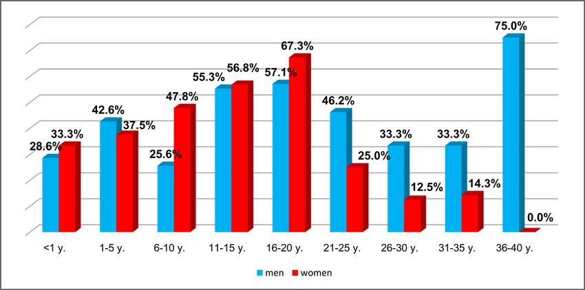
ers and methods should be used [6,9]. This study aims to               2. Statistical methods - The results were processed
compare clinical with serological data (anti-VCA IgM / IgG)     with the statistical program SPSS, vs 23. The average age
for infectious mononucleosis and to define serological pro-     of the sample, the relative proportions and the confidence
files that require further laboratory investigation.            intervals were determined. We used the chi-square test for
                                                                correlation with p < 0.05 as statistically significant.
        MATERIALS AND METHODS
        We investigated 746 samples of patients with clini-            RESULTS
cal symptoms on IM who had been tested in the virological              Data on acute infection based on ELISA studies for
laboratory at the University Hospital “St. Marina” in the       anti-VCA IgM, alone or in combination with anti-VCA IgG,
period 2010-2016. The male patients were 57.6% (95% CI:         were found in 43.2% (95% CI: 39.6% -46.8%, n = 322) of
54.0% -61.2%, n = 430) and 42.4% (95% CI: 38.8% -46.0%,         the patients – 41.9% (95% CI: 37.2% - 46.7%, n = 180) of
n = 316) were women. The average age of males was 8.87          the males and 44.9% (95% CI: 39.4% -50.6%, n = 142) of
(SD ± 8.19) and was lower than that of female subjects          the females.
10.49 (SD ± 8.27). For the purpose of our analysis, patients           The highest proportion of the laboratory confirmed
were divided into 10 age groups at regular intervals of 5       cases with IM was registered in the age group 16-20 years
years except for first and last group.                          - 62.2% (95% CI: 52.5% - 71.2%, n = 69), followed by age
        We used:                                                group 11-15 - 56.0% (95% CI: 44.1% - 67.7%, n = 42). Al-
        1. Serological methods - indirect ELISA for detec-      though according to our analysis, the clinical diagnosis of
tion of anti-VCA IgM/IgG (Euroimmun, Germany). Samples          IM was more frequent in early childhood and early school
were tested according to the standard manufacturer’s instruc-   age, the laboratory confirmed cases were in the period of
tions.                                                          adolescence (Table1).

       Table 1. Number and proportion of the patients with diagnosis IM and patients positive in ELISA anti-VCA IgM

  Age                 N*           Proportion (%,95%CI)                      N**         Proportion ( %, 95%CI)
  < 1 y.               10          1.3% (0.6-2.5)                             3          30.0% (6.7-65.2)
  1-5 y.              322          43.2% (39.6-46.8)                         131         40.7% (35.3-46.3)
  6-10 y.             157          21.0% (18.2-24.1)                          55         35.0% (27.6-43.0)
  11-15 y.            75           10.1% (8.0-12.4)                           42         56.0% (44.1-67.7)
  16-20 y.            111          14.9% (12.4-17.6)                         69          62.2% (52.5-71.2)
  21-25 y.            29           3.9% (2.6-5.5)                             10         34.5% (17.9-54.3)
  26-30 y.            20           2.7% (1.6-4.1)                             5          25.0% (8.7-49.1)
  31-35 y.            13           1.7% (0.9-3.0)                             3          23.1% (5.0-53.0)
  36-40 y.             6           0.8% (0.3-1.7)                              3         50% (11.8-88.2)
  41 +                 3           0.4% (0.1-1.2)                             1          33.3% (0.6-2.5)
  All                 746          100%                                      322         43.2%(39.6-46.8)

       N*- patients with diagnosis IM; N**- patients positive in ELISA anti-VCA IgM

                                                                       The average age of anti-VCA IgM positive patients
                                                                diagnosed with IM was 9.99 (SD ± 7.78), lower in men (9.7
                                                                years (SD ± 8.57)). Statistically significant differences (p
Fig. 1. Proportion of anti-VCA IgM-positive by age and sex
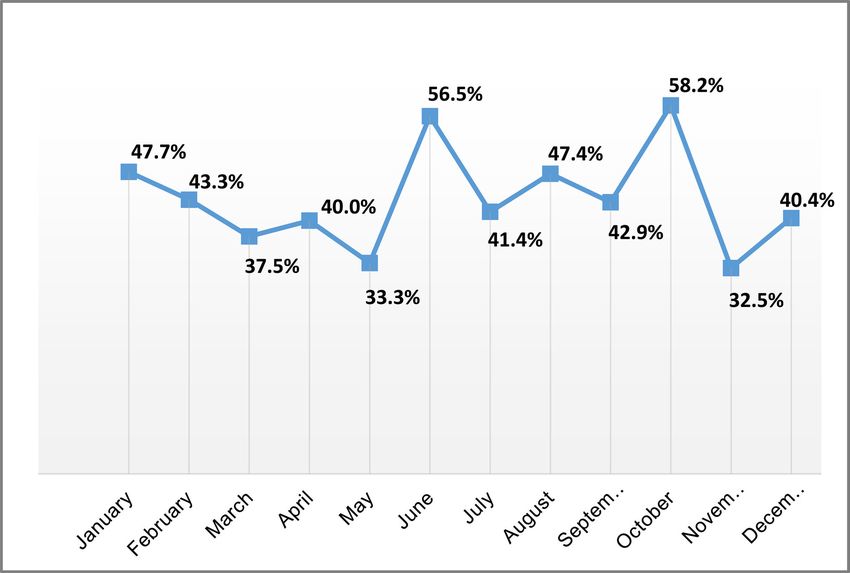
When analyzing anti-VCA IgM negative subjects other respiratory and influenza viruses were circulating. The
with clinical diagnosis of IM, 33.9% (95% CI: 54.8% - highest was the incidence in December - 16.8% and in No-
64.4%, n = 253) were positive in anti-VCA IgG. This was vember - 15.4% and at least in August (8.5%) and Septem-
one of the groups that require the use of additional meth- ber (7.7%). Laboratory confirmed cases (anti-VCA IgM
ods or markers to identify isolated IgG models. positive) were more frequent in October and June, with no
Our data showed that the most common diagnosis distinct seasonality (Figure 2).
of IM was during the colder months of the year when many
Fig. 2. Laboratory confirmed cases of IM by months
2334 https://www.journal-imab-bg.org J of IMAB. 2019 Jan-Mar;25(1)DISCUSSION called “deep kissing”, whereby a greater amount of virus
When analyzing ELISA results for the presence of is acquired. This leads to rapid colonization of B lympho-
anti-VCA IgM, fewer than half of the patients showed a cytes and consequent induction of a more potent T cell im-
positive result. A lower proportion was found in a Pleven mune response [2,14]. The development of clinical symp-
region study of 37 hospitalized patients in the 2008-2012 toms of IM is considered largely determined by the high
period where EBV serological evidence was found in 38% viral load and the high number of NK and CD8 + T cells.
[10]. In a study of 330 patients in the age group 18- 23, Several studies have found a correlation between viral load
other authors found a positive result in 55.9% of those sur- and symptom development [14,16]. Our data showed more
veyed [4]. Possible causes are an inaccurate or guideline frequent diagnosis at earlier ages, but serological confir-
diagnosis or the presence of a profile with isolated anti- mation was obtained in adolescent patients.
VCA IgG, which is observed in about 7% according to lit- There is currently no recognized seasonal pattern in
erature data [11]. A high degree of variability in the sero- the manifestation of IM. According to a 12-year observa-
logic response to EBV has been identified, particularly in tion in one university, it peaked in October, and in a study
the positivity of IgM antibodies against VCA. This requires conducted in Israel during the summer months [4]. We also
careful interpretation in cases only positive for anti-VCA cannot claim seasonality in the disease.
IgG with clinical data for primary infection where additional
serological tests such as the IgG antibody avidity test CONCLUSION:
should be used [6]. Although IM is a self-limiting disease, 1. Detection of anti-VCA IgM/IgG in most cases is
various studies indicate an increased risk of developing HL sufficient to confirm primary EBV infection. This is most
[7,12]. More recent data indicate a link with some NHL likely due to the presence of both classes of antibodies
subtypes [13]. IM is more common in primary infection in 2. No antibodies to EBV were found in 22.9% of IM
the teenage period. Upon monitoring of seronegative stu- patients tested in our study. In these cases, the role of other
dents (n = 510) from Edinburgh, Scotland, 110 of them were viruses involved in the etiology of the disease may be pre-
seroconverted within the university stay, and 27 (25%) de- sumed.
veloped IM symptoms [2]. In another study among seron- 3. In 33.9% of people diagnosed with IM, we found
egative students in Minnesota, United States, observed for only anti-VCA IgG, and this is a group that has to be tested
seroconversion during their university stay, 77% of the pri- for other markers to be confirmed in future laboratory prac-
mary infected have developed IM symptoms [14]. In a ret- tice.
rospective analysis for our region, we found that 1/3 of the 4. Patients with isolated anti-VCA IgM models
primary infections were in an age range of 10 years to 20 should also be eligible for further studies, given the possi-
years, which we associate with adolescence [15]. The in- bility of false positive results. In our study, this was ap-
creasing proportion of cases of IM during adolescence is proximately 11.0%, predominant in younger ages.
explained by the beginning of sexual contacts and the so-
REFERENCES:
1. Niedobitek G, Agathanggelou entation of EBV positive infectious 8. De Paschale M, Clerici P. Sero-
A, Herbst H, Whitehead L, Wright mononucleosis in young adults. logical diagnosis of Epstein-Barr virus
DH, Young LS. Epstein-Barr virus Epidemiol Infect. 2003 Aug;131(1): infection: Problems and solutions.
(EBV) infection in infectious mononu- 683-689. World J Virol. 2012 Feb 12;1(1):31-43.
cleosis: Virus latency, replicationand 5. Morris MC, Edmunds WJ, The [PubMed] [Crossref]
phenotype of EBV-infected cells. J changing epidemiology of infectious 9. Klutts JS, Ford BA, Perez
Pathol. 1997 Jun;182(2):151-9. mononucleosis? J Infect. 2002 Aug; NR, Gronowski AM. Evidence-based
2. Crawford DH, Macsween KF, 45(2):107-109. approach for interpretation of Epstein-
Higgins CD, Thomas R, McAulay 6. Hess RD. Routine Epstein-Barr Barr virus serological patterns. J Clin
K, Williams H, et al. A cohort study Virus Diagnostics from the Laboratory Microbiol. 2009 Oct;47(10):3204-10.
among university students: identifica- Perspective: Still Challenging after 35 [PubMed] [CrossRef]
tion of risk factors for Epstein-Barr vi- Years. J Clin Microbiol. 2004 Aug; 10. Gancheva G, Doychinova Ts,
rus seroconversion and infectious mo- 42(8):3381-7. [PubMed] [Crossref] Lukanov Ts, Tsvetanova H, Gancheva
nonucleosis. Clin Infect Dis. 2006 Aug 7. Evans AS, Niederman JC, S, Hristov H. [Infectious mononucle-
1;43(3):276-82. [PubMed] [Crossref] Cenabre LC, West B, Richards VA. A osis in the Pleven region (2008-2012)
3. Hellwig T, Jude K, Meyer B. prospective evaluation of heterophile - Clinical, laboratory and immunologi-
Management Options for Infectious and Epstein-Barr virus-specific IgM cal aspects.] [in Bulgarian] MedInfo.
Mononucleosis. US Pharm. 2013 antibody tests in clinical and subclini- 2013 Oct;10;1-6.
May;38(5):38-41. cal infectious mononucleosis: speci- 11. De Paschale M, Cagnin D,
4. Grotto I, Mimouni D, Huerta ficity and sensitivity of the tests and Cerulli T, Manco MT, Agrappi C, Mirri
M, Mimouni M, Cohen D, Robin persistence of antibody. J Infect Dis. P, et al. Search for Anti-EA(D) Anti-
G, et al. Clinical and laboratory pres- 1975 Nov;132(5):546-54. [PubMed] bodies in Subjects with an “Isolated
J of IMAB. 2019 Jan-Mar;25(1) https://www.journal-imab-bg.org 2335VCA IgG” Pattern. Int J Microbiol. tory of infections and the risk of non- Raykov T, Stojkova Z, Tsankova G.
2010; 2010: 695104. [PubMed] Hodgkin lymphoma: an InterLymph Seroprevalence of Epstein-Barr Virus in
[Crossref] pooled analysis. Int J Cancer. 2012 North-Eastern Bulgaria. Acta
12. Ambinder R. Disease and Nov 15;131(10):2342-8. [PubMed] Microbiol Bulg. 2016 Sep;32(3):33-
Pathogenesis Associated with Epstein- [Crossref] 38.
Barr Virus. In: Human Cancer Viruses. 14. Balfour HH, Odumade OA, 16. Balfour HH, Holman CJ,
Principles of Transformation and Schmeling DO, Mullan BD, Ed JA, Hokanson KM, Lelonek MM,
Pathogenesis. Editors: Nicholas J. Knight JA,et al. Behavioral, virologic, Giesbrecht JE, White DR, et al. A pro-
Jeang KT, Wu TC. Transl Res Biomed. and immunologic factors associated spective clinical study of Epstein-Barr
Basel, Karger. 2008, vol 1, pp 137- with acquisition and severity of pri- virus and host interactions during
149. [Crossref] mary Epstein-Barr virus infection in acute infectious mononucleosis. J In-
13. Becker N, Falster MO, Vajdic university students. J Infect Dis. 2013 fect Dis. 2005 Nov 1;192(9):1505-12.
CM, de Sanjose S, Martínez-Maza Jan;207(1):80-8. [PubMed] [Crossref] [PubMed] [Crossref].
O, Bracci PM, et al. Self-reported his- 15. Kostadinova Ts, Ivanova L,
Please cite this article as: Kostadinova T, Ivanova L, Stoykova Zh, Todorova T, Tsaneva D. Analysis of infectious mono-
nucleosis clinical cases in Varna region (2010-2016). J of IMAB. 2019 Jan-Mar;25(1):2332-2336.
DOI: https://doi.org/10.5272/jimab.2019251.2332
Received: 09/06/2018; Published online: 22/01/2019
Address for correspondence:
Tsvetelina Kostadinova Popova
Section Medical Lab Technicians, Medical College, Medical University, Varna,
84 Tsar Osvoboditel Blvd., Varna, Bulgaria
E-mail: ckostadinova@abv.bg
2336 https://www.journal-imab-bg.org J of IMAB. 2019 Jan-Mar;25(1)You can also read