Annals of Case Reports - Gavin Publishers
←
→
Page content transcription
If your browser does not render page correctly, please read the page content below
Annals of Case Reports
Nikitha M, et al. Ann Case Report 6: 586
Case Report DOI: 10.29011/2574-7754.100586
A Rare Case of Adult Ileocolic Intussusception Due to a Vaneks Tumour
Nikitha M, Selvam A*, Ashwath S, Sudarshan P B, Kumaran K
Department of Surgery, Saveetha Medical College and hospital, Saveetha Institute of Medical and Technical Sciences (SIMATS),
Kuthambakkam, Tamil Nadu, India.
*
Corresponding author: Dr Agil Selvam, Department of Surgery, Saveetha Medical College and hospital, Saveetha Institute of
Medical and Technical Sciences (SIMATS), Kuthambakkam, Tamil Nadu, India.
Citation: Nikitha M, Selvam A, Ashwath S, Sudarshan P B, Kumaran K (2021) A Rare Case of Adult Ileocolic Intussusception Due
to a Vaneks Tumour. Ann Case Report 6: 586. DOI: 10.29011/2574-7754.100586
Received Date: March 19, 2021; Accepted Date: March 24, 2021; Published Date: March 29, 2021
Abstract
Intussusception occurs when a more proximal portion of bowel invaginates into the more distal bowel. Intussusception
in adult is a rare condition, its presentation is acute with clinically vague signs. Incidence of vaneks tumour presenting as
intussusception is 8.6%. Vaneks tumour is a least common benign small bowel neoplasm. We present to you a case of a 32
year old female presenting with an acute abdomen, which was later diagnosed to be an ileocolic intussusception. The diagnosis
was made based on Computed Tomographic images of the abdomen showing bowel within a bowel appearance. A classical
right Hemicolectomy with ileotransverse anastamosis was successfully performed. Post-operative histopathological picture
confirmed lead point causing the intussusception as inflammatory fibroid polyp or Vaneks tumour.
Intussusception in adults is a rare condition, its presentation is acute with clinically vague signs, making initial diagnosis
difficult and tricky. Once diagnosis is confirmed intervention should be prompt and appropriate as it is lifesaving. We share
this case report for the rarity of this condition and to reinforce the knowledge of this atypical presentation of intussusception.
Introduction normal delivery, sterilised. On clinical examination, pallor present.
Intussusception occurs when a more proximal portion Patient was afebrile with stable vital signs. On per abdominal
of the bowel (intussusceptum) invaginates into the more distal examination, abdomen was found to be soft with mild distension.
bowel (intussuscipiens). Intussusception is common in children. Right hypochondrial tenderness was present. No guarding/
Only 5%-16% of intussusception occurs in adults. Most of adult rigidity. Bowel sounds on auscultation were sluggish. Our clinical
intussusception has a lead point. Vaneks tumour is one such lead diagnosis included cholecystitis. Routine blood investigations
point. Vaneks tumour, or inflammatory fibroid polyp, is one of were within normal limits. On ultrasound, few dilated bowel loops
the least common benign small bowel tumours. Its Peak incidence were seen, otherwise normal. Abdomen x-ray erect and chest x-ray
occurs in sixth and seventh decades of life, with a slight male were normal.
preponderance. It’s incidence in GIT is 0.3-0.5%. Incidence of On CECT abdomen, right hypochondrium and right lumbar
Vaneks tumour in terminal ileum is 18-20%. A incidence of Vaneks region showed dilated ileum, caecum and proximal ascending
tumour presenting as intussusception is 8.6%. We herein present a colon seen invaginating into the distal ascending colon, hepatic
rare case of a 32 year old female with ileocolic intussusceptions flexure and seen up to the proximal transverse colon. Mesenteric
due to IFP. fat and mesenteric vessels also seen invaginating along with
the above mentioned bowel loops. All these features were
Case Report representative of an ileocolic intussusception at ileo-caecal
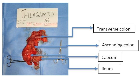
A 32 year old female who was apparently normal two junction “Target sign- positive”atient was taken for laparotomy
weeks back, presented with a three day history of colicky upper and proceed. Intra-operatively, an ileocolic intussusception was
abdominal pain, predominantly of the right hypochondrium. identified as a huge mass in the ascending colon. Appendix was in
History of multiple episodes of loose stools in the past 2 weeks. right iliac fossa, identified at the point of ileum invaginating into
History of (2-3) episodes of bilious vomiting since the past 1 the caecum. 10cms of ileum has intussuscepted into right colon &
week. History of weight loss present. No history of fever, loss of presented as a huge mass in the ascending colon. Intussuscepiens
appetite. No history of blood in stools or melena. Patient does not (large bowel) and intussusceptum (small bowel) (Figure 1). A
have any co-morbid disease. No history of previous surgery. P2L2 brief trial of reduction was done , but was unsuccessful. Viability
1 Volume 6; Issue 02
Ann Case Rep, an open access journal
ISSN: 2574-7754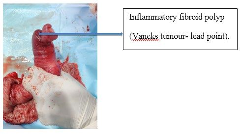
Citation: Nikitha M, Selvam A, Ashwath S, Sudarshan P B, Kumaran K (2021) A Rare Case of Adult Ileocolic Intussusception Due to a Vaneks Tu-
mour. Ann Case Report 6: 586. DOI: 10.29011/2574-7754.100586

of the bowel was good. A classical right hemicolectomy & end           D, E    - From polypoidal lesion- shows a polypoidal lesion
to side ileo-transverse anastomosis with hand sewn anastomosis         covered with colonic and ileal mucosa with focal ulceration
& mesentery rent was closed. On cutting open the specimen, a           covered with neutrophilic exudates.
11cm long intussusceptum and the returning segment formed by
                                                                             The submucosa is expanded and shows myxomatous stroma
ileum both along with the intussuscepiens component formed by
                                                                       with benign spindle shaped cells (Figure 3), prominent thick-
the ascending colon appeared as a bulky mass, with a huge 5x4
                                                                       walled vessels and many proliferating capillaries. There was
cm ulcero proliferative polyp found in the apex as the lead point
                                                                       scattered inflammatory cell infiltrate composed was plasma cells,
(Figure 2). Post op period was uneventful.
                                                                       lymphocytes and eosinophils. No evidence of dysplasia/malignancy
                                                                       seen. IHC markers were positive for CD30, negative for SMA and
                                                                       S100. Features were suggestive of INFLAMMATORY FIBROID
                                                                       POLYP (IFP).

Figure 1: Showing a segment of terminal ileum measuring 7.5cm,
partially cut open large intestine (along taenia coli) measuring
22cm, appendix measuring 9.5cm. Intussusceptum measuring
21cm.
                                                                       Figure 3: (H&E, 10x) The polyp composed of benign spindle
                                                                       shaped cells, thick walled blood vessels, proliferating capillaries,
                                                                       inflammatory cell infiltrate of plasma cells, eosinophils and few
                                                                       lymphocytes.

                                                                       Discussion
                                                                              In 1674, Barbette first described intussusception. Later
                                                                       in1789, Hunter described about intussusception. First surgeon
                                                                       to operate on a child diagnosed with intussusception was Sir
                                                                       Jonathan Hutchinson in 1871(4). In adults intussusception is
                                                                       very rare & its incidence being 2-3 per 10,00,000 per year (1).
                                                                       Its defined as invagination of proximal part of small intestine
                                                                       along with its mesentery into the adjacent segment of bowel
Figure 2: Intussusceptum showing a pedunculated polyp                  & leads to various complications like obstruction, impaired
measuring 4x3x2cm. Stalk measuring 1cm, with a region of               peristalsis & gangrene (4). Any lesion in bowel wall/ irritant
ulceration and slough.                                                 in the lumen is the leading point for intussusception & initiates
                                                                       invagination (4). Ingested food and subsequent peristaltic activity
Histopathological examination of the specimen-
                                                                       of the bowel produces an area of constriction above the stimulus
A       - Proximal resected margin- section showed wall and            and relaxation below, thus telescoping the lead point through
mucosa of small intestine which appears viable.                        the distal bowel lumen (4). Freely moving part of bowel &
                                                                       retroperitoneally or adhesively fixed parts are the most common
B       - Distal resected margin- showed colonic wall and mucosa
                                                                       locations for intussusception (2). Intussusceptions have been
which appears viable with no specific pathology.
                                                                       classified according to location into three major categories, i.e,
C       - Tip of appendix- shows wall of appendix with mucosal         enteroenteric, ileocolic or ileocecal, and colocolic (4). In ileocolic
lymphoid hyperplasia.                                                  intussusception, the ileum invaginates through the ileocecal valve

2                                                                                                                            Volume 6; Issue 02
Ann Case Rep, an open access journal
ISSN: 2574-7754
Citation: Nikitha M, Selvam A, Ashwath S, Sudarshan P B, Kumaran K (2021) A Rare Case of Adult Ileocolic Intussusception Due to a Vaneks Tu-
mour. Ann Case Report 6: 586. DOI: 10.29011/2574-7754.100586

(4). Adult intussusception occurs more frequently in small bowel         intussusception presents as chronic condition but with non specific
(50%-88%) than in the large bowel (12%-50%) (4). In adults, the          symptoms suggestive of intestinal obstruction (1,2,15) Abdominal
aetiology, presentation & management is very different from that         pain, nausea, vomiting, diarrhoea and bleeding per rectum are the
of children (1). Idiopathic or secondary viral illness are the most      common symptoms (11,13,14) Acute intestinal obstruction is a
common causes in children. In adults, various causes are present. In     very rare presentation of intussusception (1). Classical triad of
90% cases, a lead point is found to cause intussusceptum (1). Most       abdominal pain, sausage shaped palpable mass in per abdomen
lead points in gastrointestinal tract involve primary or metastatic      examination & passage of red current jelly stools is very rarely seen
malignancy, lipomas, leiomyomas, adenomas, neurofibromas,                in adults (1). Only in 24-42% of patients, palpable abdomen mass
postoperative adhesions, meckels diverticulum, foreign bodies,           is felt. Intussusception is suggestive when a shifting abdominal
vascular anomalies, lymphoid hyperplasia, trauma, celiac disease,        mass or mass that’s only palpable when symptoms arises (2).
cytomegalovirus colitis, lymphoid hyperplasia secondary to lupus,
                                                                                Various radiological methods are used to describe
henoch-schonlein purpura, wiskott-aldrich syndrome, appendiceal
                                                                         intussusception: x-ray abdomen erect, ultrasound abdomen,
stump, or inflammatory fibroid polyps (IFP) (4). Benign lesions
                                                                         barium studies, angiography & radionucleotide studies (2). Plain
account for almost 25% cases of intussusception in adults (1). The
                                                                         abdominal x-ray may shows signs of intestinal obstruction if its
commonest benign lesion is a lipoma in the colon (1).
                                                                         present (2). On ultrasonography, a classical “target/ doughnut
       In our case, the lead point was found to be a Vanek’s             sign” on transverse view and the “pseudokidney sign” in
tumour. Vanek’s tumour/ Inflammatory fibroid polyp (IFP) are rare        longitudinal view is identified. The major disadvantage being, gas
clinically benign mesenchymal tumours originating in submucosa           filled bowel loops (2). Abdominal CT is the most useful technique
of GIT. Incidence being unknown (3). It’s incidence in GIT is 0.3-       in diagnosing intussusception with accuracy 58%-100%. Signs of
0.5%. Incidence of Vanek’s tumour in terminal ileum is 18-20%. A         target/ sausage, mesenteric fat & vessels are identified. Metastasis
Vanek’s tumour presenting as intussusception is 8.6% (5). In 1949,       & lymphadenopathy can also be viewed.
Vanek first described this as “gastric submucosal granuloma(s) with
                                                                               In the adult population, once intussusceptions is diagnosed,
eosinophilic infiltration”. These lesions were found throughout the
                                                                         prompt surgical intervention is warranted to avoid complications
GIT (3). A few identified events of IFP’s are reactive inflammatory
                                                                         of ischemia, necrosis, and perforation (3). Traditionally, surgical
process with trauma, allergic reaction, and bacterial, physical,
                                                                         resection is the treatment of choice for symptomatic IFPs.
chemical or metabolic stimuli (3). Recently it was identified that
                                                                         Resection is curative, and only one case of polyp recurrence is
reports of familial occurrence & recognition of activating platelet-
                                                                         found in the literature (3). The appropriate management of adult
derived growth factor receptor alpha (PDGFRA) mutations in
                                                                         intussusceptions remains controversial, with the debate focusing
these tumours suggest that IFPs represent true neoplasms (3).
                                                                         mostly on the issue of primary en bloc resection vs initial reduction
GIST & IFP share a common oncogenetic pathway since they have
                                                                         followed by more limited resection (4). Reduction by surgery
similar PDGFRA gene mutations (3,12) Immunohistochemically,
                                                                         before resection may theoretically permit more limited resection;
IFPs are negative for CD117 and variably positive for CD34 (3).
                                                                         however, the risk of potential intraluminal seeding or venous
In contrast, GISTs have characteristically positive CD117 and
                                                                         tumour dissemination during the manipulation of a malignant
CD34 immunostaining (3). The most common site is the gastric
                                                                         lesion should also be taken into consideration (4). The incidence of
antrum (60-70%), followed by small bowel (18-20%), colorectum
                                                                         malignancy as the cause of small intestinal intussusceptions ranges
(4-7%), and far less commonly (1%) in oesophagus, duodenum,
                                                                         from 1% to 47%, and the majority of lesions are metastatic (4).
gallbladder, and appendix (3,6,8) The polyps are typically solitary,
                                                                         Therefore, recent reports have recommended initial reduction of
but rare metachronus lesions have been reported in familial cases
                                                                         externally viable small bowel prior to resection (4).
(3). Most IFPs grow intraluminally and are smaller than 4cm (3).
In this case the polyp measured 4x3x2cm in dimensions.                   Conclusion
      Clinical manifestations depend largely on tumour location                Intussusception in adults is a rare occurrence and when a adult
and size. Often IFPs are asymptomatic and are identified                 patient presents with a slightly chronic symptoms of abdomen pain
incidentally either during endoscopic or surgical procedures (3).        with diarrhoea and a tender spot in a quadrant of abdomen, surgeon
When present in small intestine they are more likely to present          should think about intussusception as a differential diagnosis. CT
with chronic colicky abdominal pain, small bowel obstruction,            scan is the diagnostic imaging of choice. And surgery should not be
intussusception, and weight loss (6,9) GI bleeding is a rare             delayed in order to prevent bowel ischemia and gangrene. Chances
presenting symptom, and if present, it may indicate significant          of malignant lesion as a lead point should be thought about and a
ulceration or ischemia (3).                                              proper resection of bowel with anastomosis will be an ideal form
       The     clinical    presentation,   most   often   in   adults,   of surgical treatment.

3                                                                                                                             Volume 6; Issue 02
Ann Case Rep, an open access journal
ISSN: 2574-7754
Citation: Nikitha M, Selvam A, Ashwath S, Sudarshan P B, Kumaran K (2021) A Rare Case of Adult Ileocolic Intussusception Due to a Vaneks Tu-
mour. Ann Case Report 6: 586. DOI: 10.29011/2574-7754.100586
The debate focuses mostly on the issue of primary en-bloc 7. Z Jukic, Z Ferencic, P Radulovic, A Mijic, and A Fucic (2014) “Estrogen
resection Vs initial reduction followed by more limited resection. and androgen receptors in inflammatory fibroid polyp (Vanek’s tumor):
case report,” Anticancer Research. 34: 7203-7206.
Reduction by surgery before resection may theoretically permit
more limited resection; however, the risk of potential intraluminal 8. H Neishaboori, I Maleki, and O Emadian (2013) “Jejunal intussusception
caused by huge Vanek’s tumor: a case report,” Gastroenterology and
seeding or venous tumour dissemination during the manipulation Hepatology from Bed to Bench. 6: 210-213.
of a malignant lesion should also be taken into consideration. In
our patient, due to failed attempt at reduction, primary en-bloc 9. R Nonose, JS Valenciano, CM da Silva, CA de Souza, and CA
Martinez (2011) “Ileal intussusception caused by Vanek’s tumor: a
resection was carried out. case report,” Case Reports in Gastroenterology. 5: 110-116.
References 10. S Siminas, E Qasem, R Shukla, and R Turnock (2014) “Inflammatory
fibroid polyp: a rare benign tumor of the alimentary tract in
1. Inflammatory fibroid polyp: an unusual cause of ileoileal children presenting as intussusception-case report and review of
intussusception- Haley S Adams, Brian Bergstorm, Bret Haines, literature,” European Journal of Pediatric Surgery Reports. 2: 6-19.
Nathan Roberts. Department of general surgery, Oklahama state
University, Tulsa, OK, USA. 11. C Zhang, M Cui, J Xing, Y Shi, and X Su (2014) “Massive gastrointestinal
bleeding caused by a giant gastric inflammatory fibroid polyp: a case
2. World Journal of Emergency surgery. Ileocolic intussusception- A report,” International Journal of Surgery Case Reports. 5: 571-573.
rare cause of acute intestinal Obstruction in adults; Case report
and literature review. Muhammad Njam Khan, Avi Agarwal and Paul 12. H U Schildhaus, T Cavlar, E Binot, R Büttner, E Wardelman, et al.
Strauss. Department of general surgery, Royal Hospital, Gosport, UK (2008) Inflammatory fibroid polyps harbor mutations in the platelet-
and department of general surgery, Darnet Valley Hospital, Dartford, derived growth factor receptor alpha (PDGFRA) gene, Journal of
Kent, UK. Pathology. 216: 176-182.
3. Intussusception due to inflammatory fibroid polyp: A case report and 13. T Liu, M Lin, E A Montgomery, A D Singhi (2013) “Inflammatory fibroid
comprehensive literature review. World journal of gastroenterology. polyps of the gastrointestinal tract: spectrum of clinical, morphological,
Sami Akbulut, department of surgery, Diyarbakir education and and immunohistochemistry features,” American Journal of Surgical
research hospital, turkey. Pathology. 37: 586-592.
4. Adult ileo-ileo-caecal intussusception: Case report and literature 14. T Miyata, H Yamamoto, H Kita et al. (2004) “A case of inflammatory
review. Sanjeev Singhal et al, 2012. fibroid polyp causing small-bowel intussusception in which retrograde
double-balloon enteroscopy was useful for the preoperative
5. World Journal of gastroenterology. Vanek’s tumor of the small bowel diagnosis,” Endoscopy. 36: 344-347.
in adults. Bassam Abboud, department of general surgery, hotel Dieu
de France hospital, faculty of medicine, saint joseph University, Beirut, 15. S Yakan, C Caliskan, O Makay, AG Denecli, and MA Korkut (2009)
Lebanon, 2015. “Intussusception in adults: clinical characteristics, diagnosis and
operative strategies,” World Journal of Gastroenterology. 15: 1985-
6. J Vanek (1949) “Gastric submucosal granuloma with eosinophilic 1989.
infiltration,” American Journal of Pathology. 25: 397-411.
4 Volume 6; Issue 02
Ann Case Rep, an open access journal
ISSN: 2574-7754You can also read