Antegrade and Retrograde Crossing of Chronic Total Occlusions Using the Outback Re-entry Device
←
→
Page content transcription
If your browser does not render page correctly, please read the page content below

Original Contribution

Antegrade and Retrograde Crossing
of Chronic Total Occlusions Using the
Outback Re-entry Device
  Timothy W.I. Clark, MD, MSc1; Ansar Z. Vance, MD1; Mark P. Mantell, MD2;
  Shilpa Reddy, MD1; Christopher Shackles, DO, MPH1

                                                                                                                ns
                                                                                                              io
Abstract

                                              y at
Purpose. The Outback device (Cordis) enables true lumen re-entry during subintimal recanalization of chronic total occlusions

                                            nl ic
(CTOs). This study compared outcomes of patients who underwent subintimal recanalization of lower-extremity arterial CTOs

                                           O un
utilizing the Outback device via antegrade and retrograde approaches. Methods. A retrospective analysis identified 39 patients
with Rutherford 3 (n = 13), 4 (n = 13), and 5 disease (n = 13) where the Outback device was utilized (19 antegrade crossing

                                         se m
femoropopliteal CTOs, 20 retrograde [17/20 transpedal access crossing femoropopliteal/tibioperoneal CTOs, 3/20 femoral
                                      l U om
access crossing iliac CTOs]) after conventional techniques failed. Mean age was 70.5 years and 67% were men. Most patients
had multifocal and/or long-segment occlusions, with 41% having combined above- and below-knee disease. Results. Overall
technical success was 90% (95% antegrade and 85% retrograde cohort; P=.15). There were no major complications and 4 minor
                                    na C

complications (prolonged bleeding, femoral pseudoaneurysm requiring thrombin injection, and 2 small access-site hematomas).
Fifteen percent of the retrograde cohort subsequently underwent distal bypass, compared with 0% in the antegrade cohort
                                  so P

(P=.23). A single amputation occurred, in the antegrade group. Twelve-month target-vessel unassisted primary patency was
                                er HM

higher with antegrade use (76% in the antegrade group vs 48% in the retrograde group; P=.03), but 12-month assisted primary
patency was similar (85% in the antegrade group vs 79% in the retrograde group; P=.85). Conclusion. The Outback can be used
safely and effectively from both antegrade and retrograde approaches during recanalization of CTOs. Lower target-vessel
                            r P 21

unassisted primary patency using the retrograde transpedal approach indicates the need for closer surveillance to achieve
high rates of limb salvage.
                         Fo 20

J CRIT LIMB ISCHEM 2021;1(1):E11-E16.
Key words: chronic total occlusions, lower-extremity occlusions, retrograde transpedal approach
                             ht
                          ig
                   yr
            op

Peripheral arterial disease affects nearly 20% of the population                  The Outback re-entry catheter (Cordis) is one of several
and more than 200 million people worldwide.1 Chronic total                    devices that has been developed to overcome this challenge of
       C

occlusions (CTOs) of lower-extremity vessels are present in                   true lumen re-entry. The Outback catheter has a hollow, curved
more than 40% of patients with symptomatic peripheral vascular                needle attached to the distal end of the catheter. This needle can
disease.2 Successfully traversing these lesions with endovascular             be properly aligned and advanced through a side port under
techniques can be challenging secondary to a variety of lesion-re-            fluoroscopic guidance to achieve re-entry and 0.014˝ diameter
lated characteristics, including a resistant fibrous cap, severe              guidewire passage into the true lumen of the target vessel.6
calcification, and long lesion length.3 While the development of                  The Outback device is intended for crossing of lower-ex-
subintimal and rendezvous techniques has increased the success                tremity CTOs from an antegrade direction. However, there are
rate in traversing CTOs, re-entering the true lumen of the target             circumstances in which the Outback device may be advantageous
vessel remains a challenge.4,5 Failure to gain re-entry into the              from a retrograde access when spontaneous re-entry fails. For
true lumen after attempting subintimal recanalization can lead                example, when attempting antegrade crossing of a CTO from a
to significant increases in procedure-related complications sec-              contralateral femoral approach, advancement of the Outback
ondary to compromise of collateral vasculature and may result                 device over a steep aortic bifurcation may be challenging. Advanc-
in subsequent amputation.3,5                                                  ing the Outback device antegrade through the subintimal space

Vol. 1 · no. 1 March 2021                                                                                                                   E11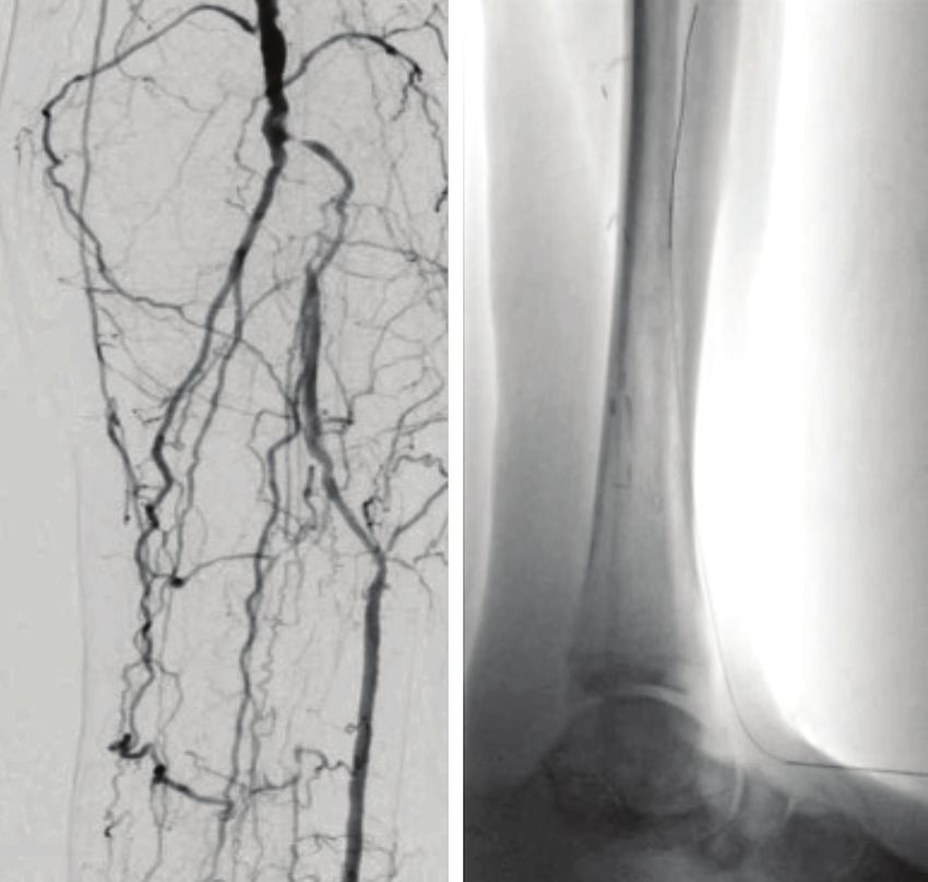
Crossing CTOs Using the Outback Re-entry Device                                                                                  CLARK, et al.

 Table 1. Patient demographics.
                                                                             with symptomatic lower-extremity peripheral arterial disease
                                                                             treated via endovascular intervention with the Outback device
 Patient Demographics                                    Patients (n = 39)   over a 9-year period ending in 2020 were identified. Three of these
 Men                                                          26 (67%)       patients were lost to follow-up, leaving 39 patients included in
                                                                             the study. Patients in whom the Outback device was used in the
 Age (years)
                                                                             aorta (for example, during aortic dissection fenestration) were
  Mean                                                        70.5 ± 9.6
                                                                             excluded from the study.
  Range                                                        44-90

 Diabetes mellitus                                                           Patient population. Patient demographics and comorbidities
                                                                             are detailed in Table 1. The mean patient age was 70.5 ± 9.6 years

                                                                                                            ns
  Type I                                                      12 (31%)
                                                                             and 67% were men. Rutherford classification of patients in the
  Type II                                                     13 (33%)       study cohort is also summarized in Table 1. The Outback device

                                                                                                          io
  Insulin dependent                                           17 (44%)       was used in conventional antegrade technique in 19 cases, all of

                                                y at
                                                              39 (100%)      which were femoropopliteal CTOs, and in retrograde technique
 Hypertension
                                                                             in 20 cases after conventional endovascular crossing techniques

                                              nl ic
 Dyslipidemia                                                 36 (92%)
                                                                             had failed. In 17/20 retrograde cases (85%), the Outback device

                                             O un
 Chronic kidney disease                                       16 (41%)       was introduced via transpedal access to cross femoropopliteal
  Hemodialysis                                                8 (20.5%)      or tibioperoneal CTOs and in 3/20 cases (15%), the device was

                                           se m
                                                                             introduced via a common femoral access to traverse iliac CTOs.
 Coronary artery disease                l U om                36 (92%)
                                                                             The majority of patients had multifocal and/or long-segment
 Ambulatory                                                   38 (97%)       (>10 cm) occlusions, with 49% involving the femoropopliteal
 Smoking                                                                     segments and 41% with combined above- and below-knee disease.
                                      na C

  Never                                                        9 (24%)
                                                                             Endovascular treatment. Endovascular treatment was per-
                                    so P

  Prior                                                       20 (54%)
                                                                             formed at the discretion of the performing provider, as described
                                  er HM

  Active                                                       8 (22%)       briefly. Initial access of the common femoral artery was gained
 Rutherford class                                                            in antegrade or retrograde fashion, based on the location of the
                                                                             diseased arterial segment. When treating distal disease without
  3                                                           13 (33%)
                              r P 21

                                                                             a more proximal lesion, the common femoral artery was ac-
  4                                                           13 (33%)
                                                                             cessed in an antegrade fashion whenever possible. Access was
                           Fo 20

  5                                                           13 (33%)       performed under direct ultrasound guidance and using standard
  6                                                            0 (0%)        micropuncture technique with subsequent placement of a 5-7
                                                                             Fr vascular sheath. After performing diagnostic angiograms,
                               ht

 Prior amputation                                              4 (10%)
                                                                             the patient was systemically heparinized using a weight-based
                            ig

 Prior bypass                                                  5 (13%)       bolus of heparin (70 U/kg) and intermittent boluses to maintain
                                                                             activated clotting times > 250 seconds during the procedure.
                    yr

 Data presented as mean ± standard deviation or number (%).
                                                                             We then attempted to cross the diseased arterial segment in
            op

                                                                             antegrade fashion, while remaining intraluminal using a 4
may also be difficult due to cumulative friction from proximal               Fr catheter system and a 0.035˝ hydrophilic wire. When in-
       C

calcified plaque. Finally, retrograde use of the Outback device may          traluminal attempts to cross the lesion were unsuccessful, an
be helpful when crossing tibial vessel CTOs. However, there are              intentional subintimal access was achieved utilizing established
limited data on the use of the Outback device from a retrograde              techniques.5 If conventional attempts to re-enter the true lumen
approach. This study compares clinical outcomes of patients who              were unsuccessful, retrograde transpedal access was obtained with
underwent subintimal recanalization of lower-extremity arterial              placement of only a 3 Fr inner micropuncture initially (Figure 1).
lesions utilizing the Outback re-entry device via antegrade and              Sequential escalation of transpedal access size was performed as
retrograde approaches.                                                       needed for intended devices. True-lumen re-entry was typically
                                                                             first attempted utilizing traditional catheters, snares, controlled
Methods                                                                      antegrade and retrograde tracking (CART) technique, or re-
                                                                             verse CART technique, per operator preference.7 If true-lumen
    A retrospective, single-center study was performed at a ter-             re-entry was still unsuccessful from rendezvous techniques,
tiary referral center and was approved by the institutional review           sharp re-entry with the Outback device was attempted via
board. Using a prospectively maintained database, 42 patients                the antegrade access or with placement of a 6 Fr thin-walled

E12                                                                                                 Journal of Critical Limb Ischemia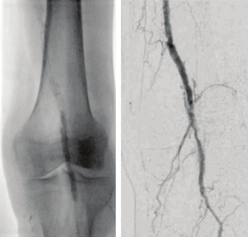
Crossing CTOs Using the Outback Re-entry Device                                                                                  CLARK, et al.

 A                               B                                   C                                     D

                                                                                                        ns
                                                                                                      io
                                           y at
                                         nl ic
                                        O un
                                      se m
E                                F
                                   l U om
                                 na C
                               so P
                             er HM

                                                                     Figure 1. Elderly male with history of a chronic non-healing left foot ulcer.
                                                                     (A) Initial digital subtraction angiography (DSA) shows heavily calcified
                                                                     chronic total occlusion of the popliteal artery. (B) After unsuccessful at-
                         r P 21

                                                                     tempts to cross occlusion (CTO) in antegrade fashion, retrograde access
                                                                     was achieved via the dorsalis pedis artery. Initial attempts to regain true
                      Fo 20

                                                                     lumen access were unsuccessful. (C) The Outback re-entry device was ad-
                                                                     vanced in retrograde fashion via dorsalis pedis access and used to achieve
                                                                     re-entry into the popliteal artery proximal to the chronic occlusion. (D)
                          ht

                                                                     After successful re-entry, the snare was advanced from antegrade femo-
                                                                     ral access and a .014˝ wire was captured. (E) The wire was subsequently
                       ig

                                                                     externalized through common femoral access and the subintimal tract/
                                                                     true lumen re-entry site was dilated with a 5 mm low-profile balloon.
                 yr

                                                                     (F) The lesion was then successfully stented with a 6 mm x 5 cm Viabahn
          op

                                                                     (Gore) with completion DSA showing markedly improved flow through the
                                                                     previously occluded segment.
      C

sheath (Terumo) via retrograde access. The preference was            appropriately aligning the integrated fluoroscopic markers.6 After
for antegrade Outback re-entry when possible to minimize             withdrawing the 0.014˝ wire back into the Outback catheter, the
pedal access size. After reaching the distal end of the target       curved nitinol needle of the device was advanced into the vessel
lesion, re-entry was performed utilizing the Outback catheter        lumen. The 0.014˝ wire was advanced into the true lumen of the
as close to re-established true lumen as possible (Figure 1). In     vessel, and the Outback catheter was subsequently exchanged
patients with issues deterring conventional femoral access           for a low-profile balloon, which was used to predilate the site
(hostile anatomy from prior surgical bypass/revision or con-         of re-entry to allow for 0.035˝ wire exchange and ultimately
comitant common femoral disease), a primary pedal approach           definitive treatment (Figure 1).
was undertaken.                                                          The diseased arterial segments were then treated at the dis-
    The Outback catheter was advanced over an 0.014˝ guidewire.      cretion of the operator utilizing balloons, atherectomy devices,
The orientation of the needle tip toward the intraluminal re-entry   or stents. A completion angiogram was performed to ascertain
site was established utilizing 2 orthogonal fluoroscopic views and   the presence of a persistent flow-limiting dissection, rupture, or

Vol. 1 · no. 1 March 2021                                                                                                                    E13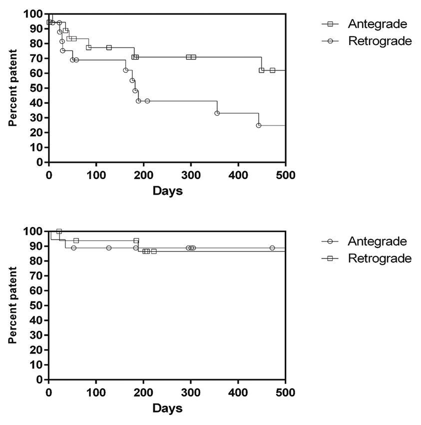
Crossing CTOs Using the Outback Re-entry Device                                                                                          CLARK, et al.

 Table 2. Lesion characteristics and interventions performed.            Table 3. Comparison of retrograde vs antegrade Outback
                                                                         utilization cohorts.
 Characteristic                                          Patients
                                                          (n = 39)                                           Retrograde      Antegrade       P-Value
                                                                                                              (n = 20)        (n = 19)
 Retrograde Outback use                                 19 (45.2%)
                                                                         Technical success                     17 (85%)      18 (95%)          .15a
 Antegrade Outback use                                  20 (47.7%)
                                                                         Technical failure                         3 (15%)    1 (5%)
 Lost to follow-up                                        3 (7.1%)
                                                                         Subsequent amputation                     0 (0%)     1 (5%)           .29a
 Lesion location
                                                                         Mean time to amputation                    n/a       5 days
  Aortoiliac                                              3 (7.7%)

                                                                                                               ns
                                                                         Type of amputation
  Femoropopliteal                                       19 (48.7%)
                                                                          Ray                                      0 (0%)     0 (0%)

                                                                                                             io
  Infrapopliteal                                         1 (2.6%)
                                                                          Transmetatarsal                          0 (0%)     0 (0%)

                                               y at
  Femoropopliteal + infrapopliteal                       16 (41%)
                                                                          Below knee                               0 (0%)     1 (5%)           .29a
 Outcomes

                                             nl ic
                                                                          Above knee                               0 (0%)     0 (0%)
  Technical success                                     35 (89.7%)

                                            O un
                                                                         Limb salvage rate                    20 (100%)      18 (95%)          .29a
  Technical failure                                      4 (10.3%)
                                                                         All-cause mortality                       5 (25%)    7 (37%)          .25a

                                          se m
  Major complications                                     0 (0%)
                                       l U om                            Subsequent bypass                         3 (15%)    0 (0%)           .01a
  Minor complications                                    4 (10.3%)
                                                                         Follow-up procedures (n)
 Interventions performed
                                                                          Mean                                 0.9 ± 1.1     0.4 ± 0.7         .08b
                                     na C

  PTA alone                                              8 (22.9%)
                                                                          Range                                     0-4         0-2
  Stent placement                                       27 (77.1%)
                                   so P

                                                                         Data presented as number (%).
      Above knee                                        22 (62.3%)
                                 er HM

                                                                         a
                                                                          Fisher’s exact test; bUnpaired t-test.

      Below knee                                          0 (0%)

      Above and below knee                               5 (14.2%)      restenosis/occlusive disease and suboptimal evaluation with ABI,
                             r P 21

  Drug-eluting balloon                                   8 (22.9%)      PVR/SVP, or Doppler ultrasound.
  Drug-eluting stent                                     9 (25.7%)
                          Fo 20

                                                                        Statistical analysis. Demographic data were used to tabulate
 Rheolytic mechanical thrombectomy                       4 (11.4%)
                                                                        the characteristics of the included patients as reported in
 Atherectomy                                             2 (5.7%)       the hospital clinical database. Complications were defined in
                              ht

 Data presented as number (%).                                          accordance with consensus reporting guidelines.8 Statistical
                           ig

                                                                        evaluations were conducted to investigate associations/differ-
                                                                        ences in technical success, subsequent amputation, all-cause
                    yr

distal embolus. Tibial run-off vessels were described as patent         mortality, complications, primary/assisted primary/secondary
            op

if they provided in-line flow to the foot or occluded if they con-      patency, and reintervention frequency. All statistical analyses
tained proximal or mid-level occlusions with or without distal          were conducted using STATA software, version 11 (STATA SE).
        C

reconstitution.                                                         A P-value of ≤.05 was considered statistically significant. Sub-
                                                                        group Kaplan-Meier analyses were constructed using Prism
Post procedure and follow-up. Patients were started on clopido-         (GraphPad Software).
grel with initial 300 mg oral loading dose followed by 75 mg daily
for a minimum of 3 months if stents were placed. Patients were          Results
maintained on aspirin 81 mg oral daily for life. Postprocedural
follow-up comprised clinic visits at 1, 3, 6, and 12 months after the   Procedure outcomes. Technical success in crossing the CTO
procedure and yearly thereafter. At each visit, clinical evaluation     was achieved in 35 of 39 total cases (90%) (Table 2). Technical
included non-invasive studies (ankle-brachial indices [ABIs] ±          success was 95% in the antegrade group (18/19 cases) and 85% in
pulse volume recordings [PVRs]/segmental limb pressures [SLPs]          the retrograde group (17/20 cases), which was not statistically
and Doppler ultrasound). Cross-sectional imaging (computed              significant (P=.15) (Table 3). Of note, even in the failure cohort,
tomographic angiography and magnetic resonance angiography)             there were no instances in which the Outback device could not
was reserved for patients with significant clinical concerns for        be advanced in retrograde fashion to engage the target lesion.

E14                                                                                                   Journal of Critical Limb Ischemia
Crossing CTOs Using the Outback Re-entry Device CLARK, et al.
There was no significant difference in all-cause mortality
between groups (P=.50). A total of 3 patients (15%) in the retro-
grade cohort subsequently underwent successful distal bypass,
compared with 0% in the antegrade cohort (P=.23). Almost half of
all patients (18/39) required reintervention, with a mean number
of 0.4 reinterventions in the antegrade group (range, 0-2) and
0.9 reinterventions in the retrograde group (range, 0-4). While
there was a trend toward an increased reintervention rate in the
retrograde group, this did not reach statistical significance (P=.08).
Subgroup Kaplan-Meier analysis of antegrade vs retrograde
ns
transpedal cohorts among technically successful Outback utili-
zations showed superior 12-month target-vessel primary unas-
io
sisted patency with antegrade use (76% in the antegrade cohort
y at
vs 48% in the retrograde cohort; P=.03), but similar 12-month
assisted primary patency (85% in the antegrade cohort vs 79%
nl ic
in the retrograde cohort; P=.85) (Figure 2).
O un
Discussion
se m
l U om Endovascular recanalization of CTOs can be achieved either
via intraluminal or subintimal approaches, with similar clinical
Figure 2. Kaplan-Meier estimates of (A) primary unassisted patency and outcomes between both approaches.9-11 Intraluminal crossing
na C
(B) primary assisted patency between antegrade and retrograde use of cannot always be achieved and remains particularly challenging
the Outback re-entry catheter. There was superior 12-month target-vessel in the setting of long-segment or heavily calcified CTOs. The
so P
primary unassisted patency with antegrade use (76% antegrade vs 48% development of subintimal crossing techniques has increased
er HM
retrograde; P=.03), but similarly high 12-month assisted primary patency
the ability to traverse these occlusions; however, true-lumen
(85% antegrade vs 79% retrograde; P=.85).
re-entry can remain challenging.4,5,9 Prior studies have demon-
strated the feasibility, safety, and efficacy of subintimal flossing
r P 21
The majority of patients (77%) required stent placement with antegrade and retrograde intervention (SAFARI) to enable
secondary to insufficient luminal gain after angioplasty, with true-lumen re-entry.12-16 While SAFARI technique is associated
Fo 20
the majority of the stents (62%) placed above the knee. Although with high technical success, advanced techniques may be nec-
almost half of the study cohort (44%) suffered from a component essary if re-entry still cannot be achieved. Re-entry devices,
of infrapopliteal disease, only a small fraction of patients (14%) such as the Outback re-entry catheter, have been developed to
ht
required both above- and below-knee stent placement for com- address this need.
ig
bined disease (Table 2). Recent studies have shown high success rates of true-lumen
There were no major procedure-related complications and 4 re-entry utilizing the Outback catheter, with 96% reported tech-
yr
minor complications reported (2 in the antegrade group and 2 in nical success rate and 2% major complication rate.17,18 However,
op
the retrograde group). The minor complications were prolonged limited data are available regarding outcomes and complication
bleeding in 1 patient, femoral pseudoaneurysm requiring throm- rates of Outback re-entry device use in retrograde fashion. The
C
bin injection in 1 patient, and small access-site hematomas in 2 present study aims to report institutional experience with retro-
patients. The difference in incidence of complications between the grade Outback re-entry device use and compare outcomes with
antegrade and retrograde groups was not statistically significant antegrade use. The results showed no statistically significant
(Fisher’s exact test P=.96). difference in technical success, overall survival, or complication
rates when the Outback re-entry catheter was utilized in ante-
Follow-up outcomes. A single amputation occurred in the an- grade or retrograde directions. Notably, no complications directly
tegrade group on postprocedure day 5, while none occurred in related to retrograde use of the Outback device, such as retrograde
the retrograde group. The single patient requiring amputation access-site occlusion/dissection or tibial vessel perforation, were
initially presented with acute critical limb ischemia secondary observed. While antegrade Outback device use showed superior
to a thrombosed stent complex. While this lesion was able to 12-month target-vessel unassisted primary patency as compared
be successfully recanalized utilizing the Outback device in with retrograde use (76% in the antegrade cohort vs 48% in the
the traditional antegrade fashion, the foot remained cool and retrograde cohort; P=.03), 12-month assisted primary patency
mottled, requiring eventual amputation. was similar in both groups (85% in the antegrade cohort vs 79%
Vol. 1 · no. 1 March 2021 E15Crossing CTOs Using the Outback Re-entry Device CLARK, et al.
4. Schanzer A, Conte MS. Critical limb ischemia. Curr Treat Options Cardiovasc Med.
in the retrograde cohort; P=.85). There was a trend toward an 2010;12:214-229.
increased reintervention rate in the retrograde group; however,
5. Bolia A, Miles KA, Brennan J, et al. Percutaneous transluminal angioplasty of oc-
this did not reach statistical significance (P=.08). It is possible clusions of the femoral and popliteal arteries by subintimal dissection. Cardiovasc
that this observation is influenced by a larger percentage of Intervent Radiol. 1990;13:357-363.
the retrograde cohort comprising patients with infrapopliteal 6. Hausegger KA, Georgieva B, Portugaller H, et al. The OUTBACK® catheter: a new
disease (antegrade 36% vs retrograde 50%). The smaller vessel device for true lumen reentry after dissection during recanalization of arterial
caliber and often concomitant above-knee disease represent a occlusions. Cardiovasc Intervent Radiol. 2004;27:26-30.
more advanced disease presentation, which may require closer 7. Michael TT, Papayannis AC, Banerjee S, Brilakis ES. Subintimal dissection/reentry
attention during follow-up. strategies in coronary chronic total occlusion interventions. Circ Cardiovasc Interv.
2012; 5:729-738.
ns
Study limitations. This study has several limitations. The sample 8. Khalilzadeh O, Baerlocher M, Shyn P, et al. Proposal of a new adverse event
classification by the Society of Interventional Radiology Standards of Practice
size was small and data were collected retrospectively. Use of the
io
Committee. J Vasc Intervent Radiol. 2017;28:1432-1437.
Outback re-entry device and decision to obtain retrograde access
y at
9. Jacobs DL, Motaganahalli RL, Cox DE, et al. True lumen reentry devices facilitate
was left to the operator, thus introducing significant selection
subintimal angioplasty and stenting of total chronic occlusions: initial report. J
bias. Furthermore, treatment of the target lesion was determined
nl ic
Vasc Surg. 2006;43:1291-1296.
by the operator and subject to heterogeneity within the cohort.
O un
10. Thukkani AK, Kinlay S. Endovascular intervention for peripheral artery disease.
Patients did not undergo routine surveillance cross-sectional Circ Res. 2015;116:1599-1613.
imaging of the distal access site; therefore, delayed access-site
se m
11. Clark TWI, Watts M, Kwan T. Percutaneous femoropopliteal recanalization using a
occlusions or stenoses may have been under-reported. Given completely transpedal/transtibial approach. Cardiovasc Interv Radiol. 2016;39:1750-
l U om
the smaller size of pedal vessels, potential loss or compromise 1758.
of the distal access site due to the need for a sheath when using 12. Spinosa DJ, Leung DA, Matsumoto AH, et al. Percutaneous intentional extralu-
Outback device in retrograde fashion should be considered. We minal recanalization in patients with chronic critical limb ischemia. Radiology.
na C
also recognize the potential for inadvertent vessel injury resulting 2004;232:499-507.
in arterial extravasation and possible compartment syndrome 13. Spinosa DJ, Harthun NL, Bissonette EA, et al. Subintimal arterial flossing with
so P
when performing Outback-facilitated re-entry. Although this antegrade-retrograde intervention (SAFARI) for subintimal recanalization to treat
er HM
chronic critical limb ischemia. J Vasc Interv Radiol. 2005;16:37-44.
complication was not observed in the present series, it is imper-
ative to keep appropriate stent-grafts on hand in case vascular 14. Kinlay S. Management of critical limb ischemia. Circ Cardiovasc Interv. 2016;9:e001946.
re-entry results in vessel rupture. 15. Khanolkar UB, Ephrem B. Endovascular reconstruction of popliteal and infrapop-
r P 21
liteal arteries for limb salvage and wound healing in patients with critical limb
Conclusion ischemia - a retrospective analysis. Indian Heart J. 2016;68:77-82.
Fo 20
16. Adam DJ, Beard JD, Cleveland T, et al. Bypass versus angioplasty in severe ischemia of
the leg (BASIL): multicentre, randomized controlled trial. Lancet. 2005;366:1925-1934.
Use of the Outback re-entry catheter was associated with
similarly high rates of technical success when comparing ante- 17. Ramjas G, Thurley P, Habib S. The use of a reentry catheter in recanalization of
ht
chronic inflow occlusions of the common iliac artery. Cardiovasc Intervent Radiol.
grade and retrograde use, even when the latter approach required
2008;31:650-654.
ig
tibioperoneal access and Outback device advancement via tibial
18. Aslam MS, Allaqaband S, Haddadian B, et al. Subintimal angioplasty with a true
arteries. Higher primary-assisted patency was observed in the
yr
reentry device for treatment of chronic occlusion of the arteries of the lower
antegrade group, although assisted patency rates were similar. extremity. Catheter Cardiovasc Interv. 2013;82:701-706.
op
The use of this technology for these applications appears safe
and effective and has the potential to further increase the spec-
C
From the 1Section of Interventional Radiology, Department of Radiology, University of Penn-
trum of patients who can benefit from successful endovascular sylvania Perelman School of Medicine, Philadelphia, Pennsylvania; and 2Division of Vascular
recanalization. Surgery, Department of Surgery, Penn Presbyterian Medical Center, University of Pennsylvania,
Philadelphia, Pennsylvania.
Disclosure: The authors have completed and returned the ICMJE Form for Disclosure of Potential
References Conflicts of Interest. Dr Clark reports royalties from Teleflex and Merit Medical; consultant
income from Teleflex, Becton Dickinson, Forge Medical, and B. Braun; shareholder in Forge
1. Fowkes FG, Rudan D, Rudan I, et al. Comparison of global estimates of prevalence Medical and Verix Health. The remaining authors report no conflicts of interest regarding
and risk factors for peripheral artery disease in 2000 and 2010: a systematic review the content herein.
and analysis. Lancet. 2013;382:1329-1340.
The authors report that patient consent was provided for publication of the images used herein.
2. Gallagher KA, Meltzer AJ, Ravin RA, et al. Endovascular management as first
therapy for chronic total occlusion of the lower extremity arteries: comparison Manuscript accepted February 12, 2021.
of balloon angioplasty, stenting, and directional atherectomy. Endovasc Ther.
Address for correspondence: Timothy W.I. Clark, MD, Professor of Clinical Radiology and
2011;18:624-637. Surgery, University of Pennsylvania Perelman School of Medicine, Division of Interventional
Radiology, Penn Presbyterian Medical Center, 51 N 39th St., Philadelphia, PA 19104. Email:
3. Lipsitz EC, Ohki T, Veith FJ, et al. Does subintimal angioplasty have a role in the Timothy.Clark@pennmedicine.upenn.edu
treatment of severe lower extremity ischemia? J Vasc Surg. 2003;37:386-391.
E16 Journal of Critical Limb IschemiaYou can also read