ARTICLE Building Your Own Neuroscience Equipment: A Precision Micromanipulator and an Epi-fluorescence Microscope for Calcium Imaging
←
→
Page content transcription
If your browser does not render page correctly, please read the page content below
The Journal of Undergraduate Neuroscience Education (JUNE), Fall 2020, 19(1):A134-A140

ARTICLE
Building Your Own Neuroscience Equipment: A Precision Micromanipulator and
an Epi-fluorescence Microscope for Calcium Imaging
James Ryan1, Bruce R. Johnson2, and David Deitcher2
1
 Biology Department, Hobart and William Smith Colleges, Geneva, NY 14456; 2Department of Neurobiology and
Behavior, Cornell University, Ithaca, NY 14853.

A faculty member’s ability to develop meaningful research-             calcium imaging of neuronal activity in living Drosophila
oriented laboratories in neurobiology is often hampered by             brains. This later technique uses transgenic flies with a
the rapid pace of new technologies and the increasing cost             genetically encoded calcium indicator, GCaMP, linked to
of equipment. To help undergraduate neuroscience faculty               green fluorescent protein (GFP). During an action potential,
meet these challenges, we introduce two important                      calcium ions (Ca2+) enter neurons and are observed as an
neuroscience research tools we designed and built. The first           increase in fluorescence intensity from a series of video
is a precision micromanipulator for neurophysiology                    images. These neuronal firing patterns can be assessed
applications costing less than $40 USD. We compare data                qualitatively and quantitatively to understand neural circuits
generated using the DIY manipulator with commercial                    leading to specific behaviors. We plan to develop curricula
micromanipulators costing over $1000. The second tool is               around the use of the epi-fluorescence microscope for
our newly designed 3D printed epi-fluorescence                         calcium imaging in the next year, and to provide detailed
microscope. Commercial fluorescence imaging devices                    parts sources and construction guides for the student and
often cost over $20,000, but our 3D printed version is                 faculty DIY experience.
constructed for less than $1200. This epi-fluorescence
microscope uses interchangeable LED light sources and                     Key words: micromanipulator; neurophysiology; epsp;
filter sets to image static fluorescence in prepared slides and        epi-fluorescence; GFP; mCherry; GCaMP; Calcium imaging

This report is based on our presentation at the 2020 FUN               can be constructed by faculty or students for about $40.00
Virtual Meeting where we introduced two new teaching and               in parts. Fluorescence microscopes with imaging cameras
research tools for undergraduate neuroscience faculty. The             are expensive, often costing over $20,000 each. This cost
rapid pace of new technologies and the increasing cost of              often puts fluorescence microscopes out of reach for many
equipment can restrict the development of meaningful                   undergraduate neuroscience courses and faculty/student
research-oriented laboratories in neuroscience. However,               research. Our imaging microscope can be constructed
the present availability of inexpensive industrial parts,              using about $1200 in parts. A major goal for our DIY work
efficient supply chains for parts distribution, 3-D printing and       is to increase the toolbox for neuroscience teaching,
open source software development can facilitate faculty                learning and research for students and faculty, especially at
development of do-it-yourself (DIY) laboratory teaching and            national and foreign institutions with limited financial
research tools to overcome curricular and research financial           resources.
limitations.
    Previously the Cornell Hoy Neurobiology group and                  DIY EQUIPMENT
collaborators have designed a variety of inexpensive or free           Precision Micromanipulator Design
tools for neuroscience teaching and research laboratories.             We designed an inexpensive micromanipulator that student
These include an extracellular amplifier (Land et al., 2001),          lab teams could assemble from a kit and use for any
suction electrodes (Land et al., 2001; Johnson et al., 2007),          laboratory exercise or research that requires fine probe
a temperature control device (Krans and Hoy, 2005),                    placement. The precision micromanipulator can be built in
micromanipulators (Krans et al., 2006), physiological                  approximately one hour. It consists of two modified x-y
stimulators (Land et al., 2004), LED control boxes for                 microscope stages, several 3D printed parts, and various
optogenetic stimulation (Pulver et al., 2010; Vilinsky et al.,         nuts and bolts (Figure 1). The two x-y mechanical stages
2018), and electrophysiological data acquisition and                   can be purchased online (Amazon, eBay) for under $20USD
analysis software (Lott et al., 2009). Here we describe our            each. These stages have movement range of 60mm x
recent       progress     developing        an      inexpensive        30mm and an accuracy of 0.1mm. The manipulator is
micromanipulator for student neurobiology teaching labs                constructed by first removing the slide holders from both x-
and research, and an inexpensive fluorescence imaging                  y stages and then removing the y-axis from the x-axis on
device for dynamic and static fluorescence visualization.              one of the two stages. The single x-axis is joined to the other
    Precision micromanipulators from commercial sources                x-y stage using an aluminum L-bracket with M3 screws and
cost ~$500 to over $1000 USD. The one described below                  nuts. Finally, the completed x-y-z stage is epoxied and

                          JUNE is a publication of Faculty for Undergraduate Neuroscience (FUN) www.funjournal.org
Ryan et al.   DIY Neuroscience Equipment   A135

                                                                different muscle fibers using the two manipulators. Both
                                                                muscle fibers were easily penetrated, and both recordings
                                                                were stable and of similar fidelity. Thus, the DIY manipulator
                                                                has the precision and stability to record intracellular
                                                                potentials.    The EPSP amplitudes recorded with the
                                                                Narishige M333 micromanipulator (blue trace) have higher
                                                                amplitudes than those recorded with our DIY manipulator
                                                                (red trace), probably because the electrode of the blue
                                                                recording was closer to a synaptic site. These muscles do
                                                                not fire action potentials, and EPSP amplitude decays with
                                                                distance from the synaptic site (Hoyle, 1983). The red and
                                                                blue recordings in Figure 3 are not the same EPSPs
                                                                because the SF muscles are poly-neuronally and multi-
                                                                terminally innervated by different, spontaneously active
                                                                motor neurons (Atwood, 2008).

                                                                Epi-fluorescence Microscope Design
                                                                The goal here was to design an affordable epi-fluorescence
                                                                microscope that small student teams could also assemble
                                                                from a kit and use to bring cutting edge research into the
                                                                undergraduate neuroscience curriculum. To meet this goal,
                                                                we designed and built an epifluorescence microscope for
Figure 1. The components for assembling the DIY precision
micromanipulator. There are 2 x-y mechanical stages, an         approximately $1200 USD. The 3D printed epi-fluorescence
aluminum rod and L-bracket, a set of 3D printed parts, and      microscope described here is suitable for many
miscellaneous nuts and bolts for assembly.                      neuroscience applications including calcium imaging in
                                                                living neurons.
screwed into the 3D printed manipulator clamp. The 3D
printed parts include a manipulator clamp, a thumb screw
housing, and a set of 4 probe holder clamps (Figure 2).
These parts are printed from .stl files using PLA, ABS, or
similar filaments at 80% infill for added strength. The .stl
files can be sent to a local 3D printer or uploaded to an
online printing service such as CraftCloud. The 3D printed
thumb screw is both threaded into the M6 hex cap and
epoxied in place. The completed thumb screw is then
threaded into a nut housed inside the 3D printed manipulator
clamp. We are preparing parts lists, construction manuals
and videos for future dissemination.

DIY Micromanipulator Performance
We compared the stability and fine movement accuracy of
our DIY manipulator with a much more expensive (~$1300)
Narishige M333 micromanipulator that is normally used for
student electrophysiological recordings in the “Principles of
Neurophysiology“ laboratory class at Cornell University
(BioNB4910).       A conventional electrophysiological rig
(Wyttenbach et al., 2018) with 2 A-M Systems Model 1600
intracellular amplifiers, an ADInstruments Power Lab for
data acquisition with LabChart software, and glass
intracellular microelectrodes filled with 3M KCl (resistances
15-20 MOhm) were used to record and display the
membrane potentials from crayfish muscle fibers. We
recorded intracellular excitatory synaptic potentials (EPSPs)
from two different crayfish superficial flexor (SF) muscle
fibers with electrodes and amplifier headstages clamped to
our DIY and the Narishige manipulators. Details of the
saline composition, dissection and recording protocols for
crayfish muscle are found in the Crawdad lab manual
(Wyttenbach et al., 2014)                                       Figure 2. The fully assembled micromanipulator with intracellular
                                                                amplifier head-stage clamped in place.
    Figure 3 shows EPSPs recorded simultaneously from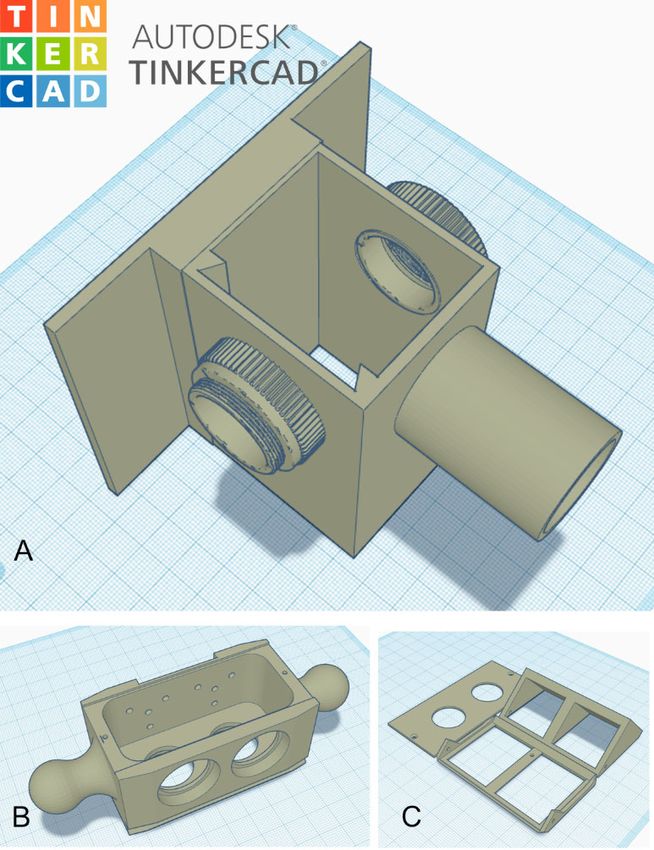
The Journal of Undergraduate Neuroscience Education (JUNE), Fall 2020, 19(1):A134-A140 A136
4). The wiring diagram for the LED controller has been
described previously (Pulver et al., 2011 Vilinsky et al.,
2018). For GFP a Cree XLamp XP-E2 high power blue
(470nm peak, $4USD, LED Supply.com) LED is coupled to
a BuckPuck DC LED driver ($20USD, LEDSupply.com).
The LED and a 20mm narrow spot LED lens are housed in
a 1 inch diameter aluminum heat sink ($15USD,
LEDSupply.com). The LED and its heat sink housing are
inserted into the 3D printed LED port. This design allows
rapid exchange of different colored LEDs.
Figure 3. Simultaneous intracellular recordings of excitatory The 3D printed parts were designed using the free, online
postsynaptic potential (EPSPs) from 2 different crayfish muscle CAD program TinkerCad (AutoDesk). TinkerCad allows you
fibers. Top red trace DIY manipulator; resting potential, -84 mV; to build complex parts from a library of basic shapes or to
calibration bars, 1 mV. Bottom blue trace, Narishige M3330 import and modify existing .stl files. Finished parts are
manipulator; resting potential, -75 mV; calibration bar, 2 mV. Note exported as .stl files for 3D printing (Figure 5). The .stl files
higher gain in red trace. Horizontal calibration bar, 500 ms.
can be printed on any 3D printer. However, printing these
parts using an online print company such as CraftCloud
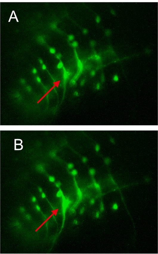
The scope consists of several off the shelf components allow greater choice of print materials. The optical housing
coupled with a set of 3D printed parts for the optical housing described here was printed using MJF Nylon or resin. These
(Figure 4). The base of the scope is an aluminum optical materials are stronger and provide higher detail than that of
breadboard drilled for M6 metric threads. Attached to this PLA or ABS plastics.
base is a 2x2 inch extruded aluminum column with t-slots Finally, the microscope is modular in design. Removing
which carries the 3D printed optical components and a the fluorescence filters and adding an LED light source from
precision lab jack for raising and lowering the specimen below the specimen converts the scope into a conventional
stage. light microscope. Screw a post and micromanipulator into
Attached to the top of the precision lab jack is a clear the base and it becomes a neurophysiology rig.
plexiglass plate (130 x 190 mm) that holds the specimen
slide or dish. Positioning of the specimen in the x-y planes
is achieved by a standard x-y mechanical microscope stage
mounted to the upper surface of the plexiglass stage. The
lab jack allows the entire stage assembly to be lowered
easily to replace specimens or add fluids.
On the vertical column, the microscope uses a linear
stage with micrometer adjustment for precision focusing the
objective lens. Attached to the linear stage is the 3D printed
optical housing. The main housing contains the
fluorescence filters and has a male c-mount on top for
attaching the CMOS camera and a female c-mount on the
bottom for the 20x long working distance objective lens.
The monochrome global shutter CMOS camera (~$180
from Basler, model da1280-54um/uc) is capable of acquiring
images in low light at up to 54 frames per second. This
camera has a resolution of 1280x960 pixels and uses a 1/3”
Aptina AR0134 sensor. However, any camera with a c-
mount can be attached to this port. Inside the main housing
is a double filter slider that accepts two sets of fluorescence
filters. The excitation and emission filters are 25mm
diameter and the dichroic mirror (beamsplitter) is
25.5x36mm. The calcium imaging described below used a
single band filter set tailored for green fluorescent protein
(GFP) from Iridian Spectral Technologies: 469-35nm
excitation, 497nm dichroic mirror, and 525-39nm emission
filter ($450 USD). A second filter set for mCherry or Alexa
Fluor 595 can be added to the filter slider if desired. In
addition, these filter sliders are very inexpensive to 3D print,
allowing the user to have several sliders and filter
combinations ready to swap out in seconds.
The light source for the scope is an interchangeable high-
power LED with heat sink connected to an LED controller
box powered by either AC or DC and capable of being driven Figure 4. The epi-fluorescence microscope with LED controller.
by a computer stimulus generator via a BNC cable (Figure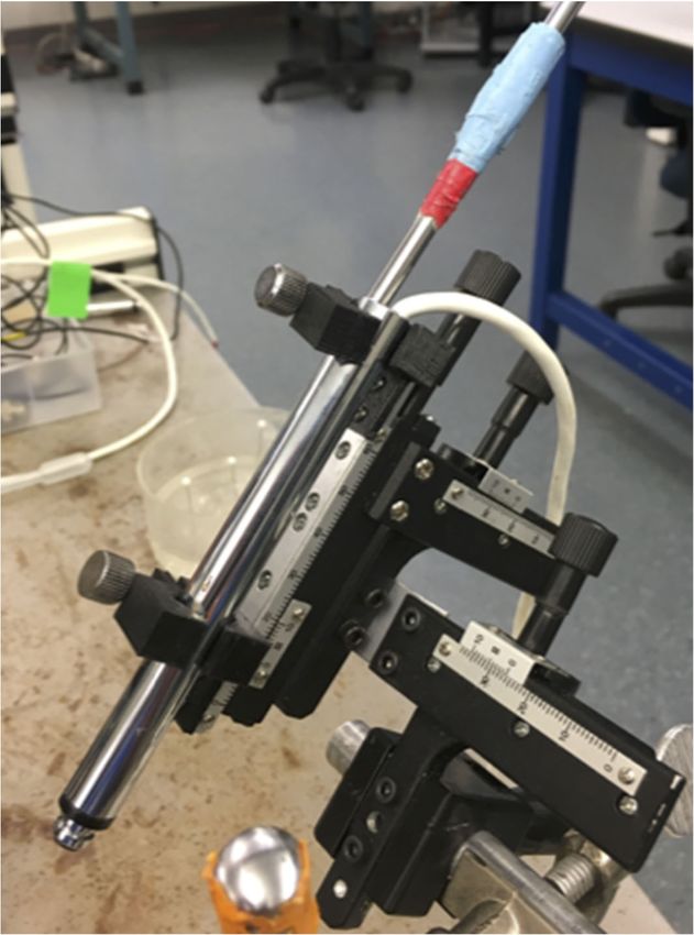
Ryan et al.   DIY Neuroscience Equipment      A137

                                                                     autoexposure to locate the preparation, then manually
                                                                     adjusted to reveal the GCaMP7b labeled motoneurons.

                                                                     Flourescent Imaging
                                                                     To test the epifluorescence microscope, we first attempted
                                                                     to image neurons expressing GFP in the ventral ganglion of
                                                                     the larva. Figure 6 shows static GFP imaging of a ventral
                                                                     ganglion of a third instar larva expressing the membrane-
                                                                     bound GFP fusion, CD8-GFP in selected neurons driven by
                                                                     the GAL4/UAS binary expression system. CD8-GFP is a
                                                                     useful label to highlight the morphology of neurons.
                                                                          Next, to show the utility of the microscope we imaged a
                                                                     ventral ganglion expressing the calcium-sensitive protein
                                                                     GCaMP7b in a pair of motor neurons in each segment.
                                                                     Images were sent at 10 fps for 300 seconds from the camera
                                                                     to a lap top computer, using camera supplied software, and
                                                                     converted into an AVI format with Fiji (Image J) software.
                                                                     Figure 7 shows two frames of dynamic imaging of actively
                                                                     firing motor neurons in the abdominal ganglia of a fruit fly
                                                                     larvae from the video link (https://youtu.be/zB55QNQ_IrM).
                                                                     For a short while after the larval fly’s CNS is removed, CNS
                                                                     motor networks will produce spontaneous neural activity that
                                                                     is correlated with larval crawling and turning (fictive
                                                                     locomotion, Pulver et al., 2015). Action potential-induced
                                                                     depolarization        results        in     calcium     entry

Figure 5. 3D printable microscope parts shown in TinkerCad prior
to export as .stl files. A) Main optical housing, B) Double filter
slider, and C) the filter holder components.

Fly Stocks for Imaging Preps
Fluorescent Drosophila lines were raised on standard
cornmeal-molasses food. The stocks used were UAS
GCaMP7b; P{GMR94G06-GAL4}attP2 for calcium imaging
from the Bloomington Stock Center (#80907and #40701)
and jus-GAL4 UAS-CD8-GFP (available from D. Deitcher)
for static fluorescent imaging of membrane-bound GFP.

Larval and CNS Preparation
Wandering third instar larvae were selected with forceps,
placed in a Sylgard petri dish and rinsed with water, then
pinned at the mouth hooks and posterior end with fine insect
pins with the dorsal (trachea) side oriented up. Room
temperature saline (135 mM NaCl, 5 mM KCl, 2 mM CaCl2,
4 mM MgCl2, 5 mM BES, pH 7.5) for calcium imaging was
added to the preparation. A superficial incision is made from
the tail to the mouth hook with micro-scissors and the “guts”
are carefully removed to reveal the CNS composed of brain
lobes and ventral ganglion. Additional pins are placed to
flatten the cuticle flaps into a “filet” (Pulver et al., 2011).
    For static fluorescent imaging, the Sylgard disc was
adhered to a glass slide with double-sided tape and then
imaged using appropriate camera settings. For dynamic
calcium imaging, the CNS was excised from the larval filet
and gently placed on a Sylgard-coated glass depression               Figure 6. Expression of membrane-bound GFP in ventral
slide and covered with saline. The slide was placed on the           ganglion neurons that express the neural gene, julius seizure
microscope stage.       Initially the camera was set to              (jus).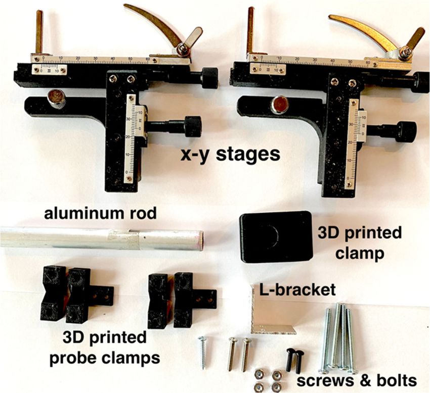
The Journal of Undergraduate Neuroscience Education (JUNE), Fall 2020, 19(1):A134-A140   A138

which is detected by the genetically encoded calcium
indicator, GCaMP that is expressed in selective
motoneurons. Thus, calcium-induced fluorescence during
neural activity is a proxy for identifying actively firing neurons
that normally drive muscles to execute larval locomotion.
These neuronal firing patterns can be assessed qualitatively
and quantitatively to understand neural circuits leading to
specific behaviors.

DISCUSSION
We present two new teaching and research tools as “proof
of concept” of our recent DIY efforts to increase the faculty
toolbox for neuroscience teaching, learning and research.
Our DIY manipulator performed well compared to the
commercial Narishige M333 when recording synaptic
potentials. The DIY manipulator could be used for any
student lab exercise or research that requires fine probe
placement, such as for cell injection of dyes or genetic
material. Although students and faculty have not yet
formally tested and assessed it, initial and limited feedback
for manipulator assembly and use was positive with
constructive criticisms from faculty participating in the
January 2020 CrawFly workshop in San Antonio. A
manipulator made by one of the 2020 CrawFly participants
was used to record miniature end-plate potentials from fruit
fly larval muscle during the workshop (data not saved). The
inexpensive manipulator could expand the range of
electrophysiological exercises possible in a student/faculty
lab.     For example, faculty could add conventional
intracellular and whole cell patch recording to student
electrophysiology exercises. Our students will construct and
use the DIY manipulator for electrophysiological student lab
exercises.       Learning outcomes, specific student skill
objectives, and data analysis suggestions for our
electrophysiology exercises are found in the Crawdad lab
manual (Wyttenbach et al., 2014).
     Our initial results of imaging static GFP fluorescence in
fixed tissue and dynamic GFP fluorescence from living                 Figure 7. GCaMP7b fluorescence in select motoneurons of the
neurons during fictive larval locomotion (Pulver et al., 2015)        ventral ganglion. Two single frames taken from series illustrate
show that our fluorescence microscope is operationally                waves of neural activity during fictive locomotion. A. Earlier fame
ready to be brought into the student laboratory classroom             shows cell at red arrow with low light intensity. B. Later frame
and into faculty research labs. Any type of fluorescence              shows higher intensity light emission from the same neuron,
                                                                      indicating greater electrical excitability. See short video link
tissue could be imaged with the appropriate excitation and
                                                                      (https://youtu.be/zB55QNQ_IrM) for entire series of images.
emission filters. Our fluorescence microscope facilitates
unique and high powered research experiences for students             microscope and dynamic imaging project will promote
and faculty with limited financial resources. We are not the          interdisciplinary thinking and skills in our neuroscience
first to propose inexpensive fluorescence imaging for the             students. For example, they will study the excitable
student neuroscience lab or for research. Our imaging                 properties of neurons though calcium imaging as a proxy for
microscope design was inspired by previous efforts                    signal transmission by action potentials, and the advantages
including from the “FlyPi” group (Chagas et al., 2017), the           of imaging for exploring neuronal circuit activity (Yang and
miniscope produced by Zhang et al. (2019) and an imaging              Yuste, 2017). Students will examine the properties of light
system recently adapted to a classroom compound                       and optics, and understand principles of fluorescence
microscope (Sane et al., 2020- this JUNE issue). For an               imaging (Cox, 2012) as they build and use their imaging
imaging microscope design that fit our needs, we searched             microscopes to measure fluorescence in living preparations
for the least expensive, yet high quality options for the             expressing fictive locomotion. They will learn introductory
microscope imaging components, light filters, camera,                 coding to control camera software and analyze images, and
microscope and mounting stage parts, and JR designed the              practice engineering design and construction by building the
3-D printed optical housing, filter holders, and slider.              microscope. This includes 3D printing, soldering, fine scale
     Our student learning objectives and skills for the imaging       assembly, and troubleshooting. Finally, students will perfect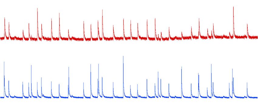
Ryan et al.   DIY Neuroscience Equipment     A139

their dissection and dexterity skills by preparing Drosophila      CrawFly
larval nervous systems for imaging.                                (https://www.adinstruments.com/training/education/applicat
    We are still preparing imaging analysis protocols              ion-workshops?field_event_type_tid=2977).
appropriate for our undergraduate student use. μManager
software can be used to gather the imaging movies from the         REFERENCES
camera, and image J to process the images and select               Atwood HL (2008) Parallel ‘phasic’ and ‘tonic’ motor systems of the
“regions of interest”. Data analysis programs such as                crayfish abdomen. J Exp Biol 211:2193-2195.
Dataview          (Heitler,        2009;         https://www.st-   Basu,A, Mondoux MA, Whitt JL, Isaacs AK, Narita T (2017)
andrews.ac.uk/~wjh/dataview/) can be used to visualize and           Integrative approach to STEM concepts in an introductory
measure fluorescent amplitude and activity patterns (see             neuroscience course: Gains in interdisciplinary awareness. J
Sane et al., 2020, this JUNE issue). Our imaging software            Undergrad Neurosci Educ 16(1):A102-A111.
protocols, in prep, will be designed for student interaction to     Bennett ML, Gadlin H (2012) Collaboration and team science:
modify Python code at various phases of image acquisition,           From theory to practice. JIM 60:768-775.
                                                                   Chagas AM, Prieto-Godino LL, Arrenberg AB, Baden B (2017) The
analysis and visualization.                                          €100 lab: A 3D- printable open-source platform for fluorescence
    We hope that our goals for the imaging microscope                microscopy, optogenetics, and accurate temperature control
project in particular will be broadly applicable at other            during behaviour of zebrafish, Drosophila, and Caenorhabditis
institutions.    The National Research Council (2003)                elegans.            PLoS           Biol         15(7):e2002702.
emphasizes that future science research progress will                https://doi.org/10.1371/journal. pbio.2002702.
require greater STEM interdisciplinary training. We plan to        Cox G (2012) Fluorescence and fluorescence microscopy. In:
create an interdisciplinary classroom where students from            Optical imaging techniques in cell biology pp 35-48. Boca Raton,
different STEM studies work together to develop the                  FL: CRC Press LLC.
confidence to build and understand equipment and solve             Crisp KM, Muir GM (2012) Assessing development of an
                                                                     interdisciplinary perspective in an undergraduate neuroscience
biological problems. Interdisciplinary science education is          course. J Undergrad Neurosci Educ 10(2):A88-95.
considered critical for the future recruitment of talented         Fromherz S, Whitaker-Fornek J, Andrew A. Sharp AA (2018)
scientists (Kazar and Elrod, 2012). Our imaging project              Classroom-based research experiences to support underserved
promotes interdisciplinary learning with engineering design,         STEM student success: From introductory inquiry to
physics (optics and light principles), biology (genetics and         optogenetics in the embryonic chicken. J Undergrad Neurosci
neuroscience), and computer science (introductory coding             Educ 17(1):A97-A110.
to control camera software and analyze images). Team-              Glover EM, Lauzon O (2018) Using a contrast illusion to teach
based learning skills required for research (Bennett and             principles of neural processing. J Undergrad Neurosci Educ
Gadlin, 2012) will be fostered as student teams build, test,         17(1):A81-A88.
                                                                   Heitler WJ (2009) Practical tools for analysing rhythmic neural
and trouble shoot their own instrumentation, and gather and          activity.     J    Neurosci     Methods      185:151-64.      doi:
analyze data to answer biological questions. Too often our           10.1016/j.jneumeth.2009.09.009
students treat instrumentation as a “black box”, with little       Hoyle G (1983) Muscles and their neural control pp 483-525. New
understanding of how scientific equipment works. We are in           York, NY: John Wiley and Sons.
the process of developing written and video guides for             Johnson BR, Hauptman SA, Bonow RH (2007) Construction of a
construction and use of the manipulator and the imaging              simple suction electrode for extracellular recording and
microscope, along with laboratory modules to guide inquiry           Stimulation. J Undergrad Neurosci Educ 6(1):A21-A26.
driven research experiences for students (Fromherz et al.,         Kazar A, Elrod S (2012) Facilitating interdisciplinary learning:
2018).                                                               Lessons from Project Kalioedoscope. Change 44(1): 16-25.
                                                                   Krans JL, Hoy RR (2005) Tools for physiology labs: An inexpensive
    Our plans for a Spring 2020 rollout of student team              means of temperature control. J Undergrad Neurosci Educ
construction and use of the manipulator and imaging                  4(1):A22-A26.
microscope in the laboratory classroom were derailed by the        Krans J, Gilbert C, Hoy R (2006) Teaching insect retinal physiology
Covid-19 pandemic, but we will continue our curriculum               with newly designed, inexpensive micromanipulators. Adv
development for both Cornell and Hobart and William Smith            Physiol Educ 30:254-261.
students this Spring 2021.          We will follow inclusive       Land BR, Wyttenbach RA, Johnson BR (2001) Tools for the
classroom practices of clearly structured assignments and            student physiology lab: An inexpensive high-performance
expectations, and emphasize that all ideas are welcome,              amplifier and suction electrode for extracellular recording. J
respected and open for discussion (Penner, 2018). We will            Neurosci Meth 106:47-55.
                                                                   Land BR, Johnson BR, Wyttenbach, RA, Hoy RR (2004) Tools for
assess student interdisciplinary awareness (Basu et al.,             Physiology Labs: Inexpensive equipment for physiological
2017; Crisp and Muir, 2012) before and after imaging                 stimulation. J Undergrad Neurosci Educ 3:A30-A35.
microscope use, and student perceived experience and               Lott GK III, Johnson BR, Bonow R H, Land BR, Hoy RR (2009) g-
confidence in building and using the imaging microscope              PRIME: A free, windows based data acquisition and event
(Glover and Luzon, 2018). Our student learning outcomes              analysis software package for physiology in classrooms and
will be examined by grading student mastery of microscope            research labs. J Undergrad Neurosci Educ 8(1):A50-A54.
construction, designing and testing hypotheses of neuronal         National Research Council (2003) BIO2010: Transforming
network activity through data gathering and interpretation,          undergraduate education for future research biologists.
student written journal-like articles and oral presentations on      Available       at  https://www.nap.edu/catalog/10497/bio2010-
                                                                     transforming-undergraduate-education-for-future-research-
their work. Our project results will be disseminated through         biologists.
additional journal articles and faculty workshops such as          Penner MR (2018) Building an inclusive classroom. J Undergrad
The Journal of Undergraduate Neuroscience Education (JUNE), Fall 2020, 19(1):A134-A140 A140
Neurosci Educ 16(3):A268-A272. Neurosci Educ 16 (3):A277-A281.Yang W, Yuste R (2017) In
Pulver SR, Hornstein NJ, Land BL, Johnson BR (2011) vivo imaging of neural activity. Nat
Optogenetics in the teaching laboratory: Using Methods 14(4):349-359. doi: 10.1038/nmeth.4230.
channelrhodopsin-2 to study the neural basis of behavior and Zhang L, Bo Liang B, Barbera G,1, Hawes S, Zhang Y, Stump K,
synaptic physiology in Drosophila. Adv Physiol Educ 35(1):82- Baum I, Yang Y, Li Y, Lin D-T (2019) Miniscope GRIN lens
91. system for calcium imaging of neuronal activity from deep brain
Pulver SR, Bayley TG, Taylor AL, Berni J, Hedwig B (2015) structures in behaving animals. Curr Protocols Neurosci 86:e56.
Imaging fictive locomotor patterns in larval Drosophila. J doi: 10.1002/cpns.56.
Neurophysiol 114(5):2564-77.
Booth JRH, Sane V, Gather M, Pulver SR (2020) Inexpensive
Methods for Live Imaging of Central Pattern Generator Activity Received September 22, 2020; accepted October 22, 2020.
in the Drosophila Larval Locomotor System. J Undergrad
Neurosci Educ 19(1):A124-A132. This work was funded by a grant from the Cornell Center for Teaching
Vilinsky.I, Hibbard KL, Johnson BR, Deitcher DL (2018) Probing Innovation (DD and BRJ), and support from Hobart-William Smith Colleges
synaptic transmission and behavior in Drosophila with (JR). We thank Dr. Stefan Pulver of the University of St. Andrews in
Scotland for DIY inspiration and advice.
optogenetics: A laboratory exercise. J Undergrad Neurosci Educ
16 (3):A289-A295. Address correspondence to: Dr. James Ryan, Biology Department, Hobart
Wyttenbach RA, Johnson BR, Hoy RR. (2014) Crawdad: An Online and William Smith Colleges, Geneva, NY 14456. Email: ryan@hws.edu
Lab Manual for Neurophysiology Sinauer Associates, Inc.,
Sunderland, MA. Copyright © 2020 Faculty for Undergraduate Neuroscience
Wyttenbach RA, Johnson BR, Hoy RR (2018) Reducing the cost of
electrophysiology in the teaching laboratory. J Undergrad www.funjournal.orgYou can also read