High-speed multiplane structured illumination microscopy of living cells using an image-splitting prism - Uni Bielefeld
←
→
Page content transcription
If your browser does not render page correctly, please read the page content below
Nanophotonics 2020; 9(1): 143–148

Research article

Adrien Desclouxa, Marcel Müllera, Vytautas Navikas, Andreas Markwirth,
Robin van den Eynde, Tomas Lukes, Wolfgang Hübner, Theo Lasser, Aleksandra Radenovic*,
Peter Dedecker* and Thomas Huser*

High-speed multiplane structured illumination
microscopy of living cells using an image-splitting
prism
https://doi.org/10.1515/nanoph-2019-0346                                      of a second camera to acquire a laterally highly resolved
Received September 5, 2019; revised October 22, 2019; accepted                3D image stack. We demonstrate the performance of mul-
November 4, 2019                                                              tiplane SIM by applying this instrument to imaging the
                                                                              dynamics of mitochondria in living COS-7 cells.
Abstract: Super-resolution structured illumination micro-
scopy (SR-SIM) can be conducted at video-rate acquisition                     Keywords: super-resolution optical microscopy; PACS10:
speeds when combined with high-speed spatial light mod-                       42.30.Wb; multiplane image acquisition; structured illu-
ulators and sCMOS cameras, rendering it particularly suit-                    mination microscopy.
able for live-cell imaging. If, however, three-dimensional
(3D) information is desired, the sequential acquisition of
vertical image stacks employed by current setups signifi-
cantly slows down the acquisition process. In this work,
                                                                              1 Introduction
we present a multiplane approach to SR-SIM that over-
                                                                              Conventional fluorescence microscopy is inherently
comes this slowdown via the simultaneous acquisition of
                                                                              limited in its spatial resolution due to diffraction. Optical
multiple object planes, employing a recently introduced
                                                                              super-resolution imaging techniques allow us to over-
multiplane image splitting prism combined with high-
                                                                              come this limitation. For super-resolution structured illu-
speed SIM illumination. This strategy requires only the
                                                                              mination microscopy (SR-SIM), this is achieved by using
introduction of a single optical element and the addition
                                                                              high spatial illumination frequencies that down-modulate
                                                                              spatial frequencies beyond the cutoff into the passband of
a
 Adrien Descloux and Marcel Müller: These authors contributed                 the microscope [1–4].
equally to this work.                                                              Linear implementations of SR-SIM create a sinusoi-
*Corresponding authors: Aleksandra Radenovic, Laboratory of                   dal interference pattern in the sample plane, leading to
Nanoscale Biology, École Polytechnique Fédérale de Lausanne,                  a spatial resolution improvement of at best twofold over
1015 Lausanne, Switzerland, e-mail: aleksandra.radenovic@epfl.ch;
                                                                              wide-field microscopy and additional contrast enhance-
Peter Dedecker, Laboratory for Nanobiology, Department of
Chemistry, KU Leuven, 3000 Leuven, Belgium,
                                                                              ment for high spatial frequencies and strong suppres-
e-mail: peter.dedecker@kuleuven.be; and Thomas Huser,                         sion of out-of-focus light by filling the missing cone of the
Biomolecular Photonics, Department of Physics, Bielefeld                      instrument’s optical transfer function [3, 5, 6]. In return,
University, 33501 Bielefeld, Germany, e-mail: thomas.huser@                   only a small number of raw images with defined illumina-
physik.uni-bielefeld.ded. https://orcid.org/0000-0003-2348-7416               tion patterns [nine in the standard two-dimensional (2D)
Adrien Descloux, Vytautas Navikas and Tomas Lukes: Laboratory of
                                                                              implementation, less when using advanced algorithms
Nanoscale Biology, École Polytechnique Fédérale de Lausanne, 1015
Lausanne, Switzerland                                                         [7]] are needed for the image reconstruction process. The
Marcel Müller and Robin van den Eynde: Laboratory for                         excitation powers are comparable to conventional wide-
Nanobiology, Department of Chemistry, KU Leuven, 3000 Leuven,                 field imaging, so photo-damage can be minimized, and no
Belgium                                                                       special dyes (e.g. dyes that are photo-switchable or inher-
Andreas Markwirth and Wolfgang Hübner: Biomolecular Photonics,
                                                                              ently blinking) are required for the approximately twofold
Department of Physics, Bielefeld University, 33501 Bielefeld,
Germany
                                                                              resolution enhancement. The combination of these fea-
Theo Lasser: Max Planck Institute for Polymer Research, 55128                 tures makes SR-SIM a very fast super-resolution imaging
Mainz, Germany                                                                technique, with current instruments [8, 9] providing 2D
  Open Access. © 2019 Aleksandra Radenovic, Peter Dedecker, Thomas Huser et al., published by De Gruyter.         This work is licensed under the
Creative Commons Attribution 4.0 Public License.
                                                                                                                            Unauthenticated
                                                                                                            Download Date | 2/11/20 8:32 PM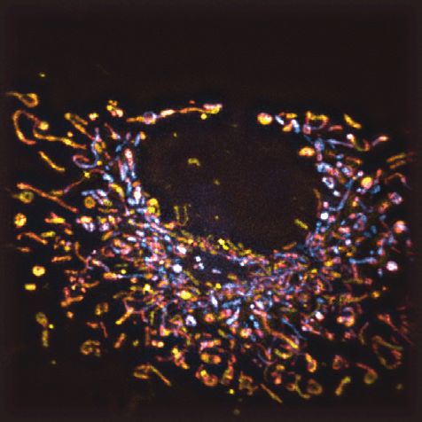
144          A. Descloux et al.: High-speed multiplane structured illumination microscopy of living cells

imaging with approximately 100-nm spatial resolution in                     and provided the first multiplane SR-SIM images in a pro-
the lateral direction at video-rate speed and even faster.                  totype system. Although this proved to be a great demon-
     Live-cell imaging is the primary domain for SR-SIM.                    stration of the future of SR-SIM imaging, these tests ended
Here, its high temporal resolution excels, especially when                  up being limited by the slow speed of the commercial
observing highly dynamic processes on subsecond time                        SR-SIM platform, which was not originally conceived for
scales. If, however, three-dimensional (3D) volumet-                        video-rate super-resolution imaging. Here, we present a
ric imaging is required, current state-of-the-art SR-SIM                    multiplane imaging approach to SR-SIM that circumvents
systems slow down the acquisition rate significantly,                       these issues and achieves high-speed imaging rates previ-
because of the need to physically move the object through                   ously available only to 2D SR-SIM.
the focal plane, e.g. by piezo-translation stages. The slow-
down arises not just from the repeated image acquisi-
tions, but also from the intermediate sample movement
that needs to be accounted for. Also, advanced control                      2 Multiplane SIM
electronics are required if stage movement, SR-SIM illu-
mination, and camera exposure are to be synchronized                        Our work is based upon an imaging strategy for multiplane
precisely.                                                                  detection that divides the detected fluorescence light into
     Such delays can be avoided if a full 3D volume of                      multiple image planes by introducing discrete optical path
the sample could be acquired at once. Two different                         length differences to the image path [14–16]. This path
approaches to multiplane image detection currently exist:                   length difference guarantees perfect object-image conju-
a diffractive element, based on a phase-shifting spatial                    gation for eight object planes equally displaced along the
light modulator or a lithographically-produced optical                      axial direction. As seen in Figure 1, the classical 8f setup of
element [10, 11], can be introduced into a conjugated                       the detection path allows us to easily realize a telecentric
image plane and create almost arbitrary multiplane detec-                   instrument ensuring identical scaling of all eight images
tion schemes. Chromatic aberrations can be corrected                        obtained from different axially displaced object planes.
using additional elements [12]. Recently, this approach                         While image splitting via subsequent changes in
was combined with a commercial SR-SIM microscope [13]                       detection path length is a robust concept, its typical

                                                      SIM illumination
                                                                           SMF
                                                  532 nm                          2x telescope              Multi-plane detection
                                                 1W laser                          periscope
                Sample
                                                                                                                 Camera 2
                                   M
                                                                                                     DMD
                                                                                                                             Camera 1

                                                                  Mask
                          150 mm                 150 mm                      300 mm   M

                 Objective
                60x \ 1.2 NA       DM     TL                IIP
                                        140 mm                           160 mm             200 mm

Figure 1: Optomechanical schematics of the high-speed multiplane SR-SIM imaging system.
The SR-SIM pattern is created by a 532- nm 1-W laser (Roithner) coupled into a single-mode fibre for mode cleaning. The light at the distal
end of the fibre is then collimated, expanded by a factor of 2 and directed onto a digital micromirror device (DMD), which is used as a fast,
electronically controlled optical grating. A segmented aperture mask removes spurious diffraction orders, arising due to the binary nature
of the DMD. A relay lens system focuses the light into the back focal plane of the objective lens (Olympus UPLSAPO 60× /1.2 NA water
immersion). Excitation and emission light is wavelength-separated by a dichroic mirror (DM). In the detection path, the tube lens (TL) forms
an intermediate image (image plane), where a field stop (IIP) sets the field-of-view size and prevents overlapping signals in the multiplane
detection system. A magnification of ×58.5 is achieved, which corresponds to a projected pixel size of 111 nm for the sCMOS cameras (ORCA
Flash 4.0, Hamamatsu). Lenses form a relay telescope into the main component, the precision-made multiplane prism [16]. It provides 2 × 4
copies of the image onto two scientific sCMOS cameras, where each copy has a defined path-length difference and thus defined axially
offset focal plane.

                                                                                                                      Unauthenticated
                                                                                                      Download Date | 2/11/20 8:32 PM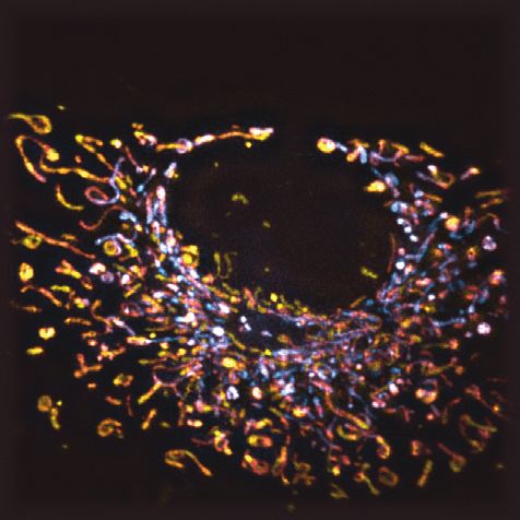
A. Descloux et al.: High-speed multiplane structured illumination microscopy of living cells   145

implementation with discrete optical elements is sensi-            in some of the lateral resolution enhancement for optical
tive to thermal drifts and vibration and rather difficult to       sectioning capability. In brief, using a slightly coarser SIM
align. A recent development [16] solved this problem by            pattern (frequency of 1.8 μm−1, or 555-nm periodicity, here)
integrating this approach into a single, stable, precision-        leads to an overlap of the shifted copies of the optical trans-
made optical element: a compound image-splitting prism.            fer function obtained by SIM, which, in turn, allows to fill
This device allows for the simultaneous detection of up            their missing cone and thus provides axial sectioning [5, 6].
to eight diffraction limited images obtained at equally            Additionally, using a coarser SIM pattern simplifies the
spaced vertical planes with only minimal alignment of the          aspect of polarization control. For optimal pattern contrast,
overall optical setup. Compared to diffraction-based mul-          the polarization of the interfering beams would have to be
tiplane microscopy [13], our approach has the advantage            rotated to match the three orientations of the SIM pattern.
that the prism consists of a single, compound refractive           However, when using a coarser pattern, and thus steeper
element, instead of two separate gratings and a refrac-            interference angles, the influence of polarization becomes
tive correction prism. This greatly improves the stability         less pronounced [17]. This allows us to use a fixed linear
and simplifies the alignment of the system. While the dif-         polarization while maintaining reasonable pattern modu-
fractive solution can in principle be tailored to a specific       lation contrast for all pattern orientations, which both sim-
imaging application, the method presented here is very             plifies the optical setup and avoids light loss in the filter
versatile and, through a careful selection of tube lenses,         elements that would otherwise occur. To summarize, we
can be used at any wavelength, allowing for example 3D             tuned the SIM system to a compromise of both optical sec-
correlative microscopy [16].                                       tioning and lateral resolution improvement, while operat-
     We combined our prism-based detection path with               ing at the minimum of nine raw SIM frames to achieve high
a coherent SIM excitation path based on a high-speed               imaging speeds.
digital mirror device (DMD-DLP7000, Vialux) and a high-
power laser (532 nm, 1 W, Roithner). By utilizing two syn-
chronized sCMOS cameras (Hamamatsu Orcaflash V4.0),
we achieved high-speed volumetric imaging with 50-ms               3 Results
exposure time. Taking camera readout and device syn-
chronization into account, this results in about 1.3 recon-        We applied our multiplane SIM system to the fast imaging
structed SIM data sets per second, to image a full volume of       of mitochondrial dynamics [18, 19]. Here, SIM is an ena-
about 40 × 40 × 2.45 μm3. Both the DMD-based spatial light         bling imaging method, as its increase in spatial resolu-
modulator and the cameras could, in principle, be tuned            tion and inherent background suppression allow us to
to provide even higher imaging speeds [8], if required by          visualize the mitochondria and their 3D morphology and
the application and if sufficiently bright fluorophores are        dynamic changes thereof, which cannot be observed with
used to compensate for the lower signal-to-noise ratio.            conventional wide-field resolution [20]. Furthermore, the
     In its current configuration, the SIM is operated in          rapid movement of mitochondria requires high imaging
coherent two-beam mode. Here, the ±1st diffraction orders          speed, while their 3D nature greatly benefits from acquir-
generated by the DMD grating pattern can interfere in the          ing signals from multiple z-planes. However, the struc-
sample plane to generate a lateral modulation of the exci-         tures are still rather “thin” compared to their length, and
tation pattern. The 0th order is blocked, so no axial mod-         a coverage of 2.45 μm (eight planes with an axial sampling
ulation of the SIM pattern is introduced. This approach            of 350 nm) as provided by our system is typically suffi-
sacrifices the axial resolution improvement possible in full       cient to capture the entire 3D mitochondrial structure in
three-beam SIM, but comes with a set of advantages. First,         a single-shot multiplane exposure. In this case, no axial
including SIM modulation along the axial direction would           movement of the sample is needed at all, which greatly
entail aligning each z-plane with the axial periodicity of the     simplifies data acquisition and decreases the acquisition
SIM pattern, which would take away some of the systems             time. The data processing and results are presented in
robustness and simplicity. Also, the acquisition of 15 instead     Figure 2, where our imaging was performed with 50-ms
of nine raw images is required for a direct SIM reconstruc-        exposure time per raw data frame. As mentioned earlier,
tion of three-beam SIM data, which impacts overall imaging         three angles and three phases are required for the 2D
speed, as well as sample photobleaching and phototoxic-            SIM image reconstruction process, thus reaching 770-ms
ity. Most importantly, the optical sectioning intrinsically        acquisition time for the full imaged volume.
provided by three-beam SIM can also be obtained in a two-              Figure 2A summarizes the flow of data. A key chal-
beam approach (albeit at lower axial resolution) by trading        lenge is that the eight planes are all slightly shifted with

                                                                                                                  Unauthenticated
                                                                                                  Download Date | 2/11/20 8:32 PM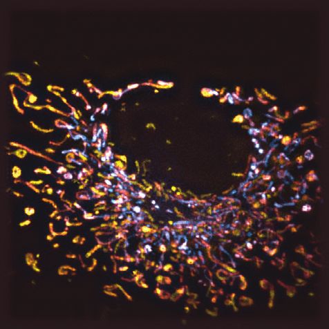
146 A. Descloux et al.: High-speed multiplane structured illumination microscopy of living cells
A B
Raw camera frame
e
m
Ti
F
Raw frames
W
coregistration
&
3D LR deconvolution
z = 0 µm z = 0.7 µm z = 1.4 µm z = 2.1 µm
SIM dec
Average raw SIM frames
pseudo 3D WF
]
µm
5
2.
Plane by plane
,
[0
z:
2D SIM
reconstruction
F
W
Estimation of
plane to plane
c
de
Transformation 2.5
F
W
matrix T(z)
(µm)
0
M
SIM dec
SI
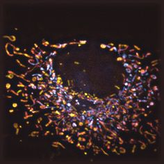
Figure 2: Data processing and SIM image reconstruction procedure.
(A) The raw images are first averaged to produce a pseudo wide-field image. This image is then used to estimate the interplane
transformation matrix T(z). This matrix is used to coregister the raw SIM images and reorder the data in x, y, z, t space. Then, the data are
either averaged (WF), deconvolved in 3D (WF dec), reconstructed (SIM), or deconvolved and reconstructed (SIM dec). (B) Colour-coded
maximum intensity z-projection of 3D images for all four modalities on a fixed COS-7 cells labelled with MitoTracker Orange. Inset shows a
zoomed in comparison between WF and deconvolved SIM. Scale bar 5 μm.
2.5
F
(µm)
W
c
c
de
0
de
M
F
W
SI
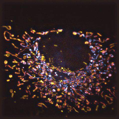
t=0s t = 10 s t = 20 s t = 30 s
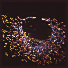
Figure 3: Time series of SR-SIM imaging data following mitochondrial dynamics (stained with MitoTracker Orange) in COS-7 cells, colour-
coded maximum intensity z-projection of the deconvolved and SIM reconstructed eight planes (Figure 2). The full series contains 74 time
points, each acquired with 50-millisecond SR-SIM acquisition time to allow for motion artefact–free imaging (see Supplementary Material:
Visualization 1). The series has been renormalized in brightness to compensate for photo bleaching, but no further processing beyond a
standard SIM reconstruction had to be performed. Insets (field of view 7 × 7 μm2) show deconvolved SIM exhibiting high spatial motility of
the mitochondria during the imaging. Scale bar 5 μm.
respect to each other, due to unavoidable limitations in plane (SIM dec). Results are shown in Figure 2B, where
the prism manufacturing. By cross-correlating two con- we encoded the depth information using a colour-coded
secutive planes, we are able to recover the prism transfor- maximum z-projection. Using the recently introduced
mation matrix T(z) and perform a subpixel coregistration image resolution estimation method [21], we measure
for all the raw SIM frames. We then average all the frames a resolution of 460 nm for WF and 434 nm for WF dec.
to form a pseudo–wide-field image (WF), deconvolve the This relatively high value is due to the presence of out-of-
3D WF using 10 iterations of Lucy–Richardson (LR) decon- focus light, which is partially removed by the 3D decon-
volution (deconvlucy, MATLAB 2017b), reconstruct the volution. For SIM, we measure 266 nm (263 nm for SIM
SIM image plane by plane (SIM), or deconvolve in 3D the dec), which corresponds to a resolution gain of about 1.7
raw SIM frames and reconstruct the SIM images plane by compared to WF. We note that for SIM the deconvolution
Unauthenticated
Download Date | 2/11/20 8:32 PM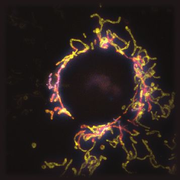
A. Descloux et al.: High-speed multiplane structured illumination microscopy of living cells   147

mostly improves the image contrast and does not change            for low signal-to-noise image reconstruction, we envi-
the resolution. We used a custom 2D SIM reconstruction            sion that 5- to 10-millisecond exposure times, and thus
algorithm implemented in MATLAB, following closely the            volumetric imaging within 50 to 100 milliseconds, will
work from Müller et al. [22]. For the 3D LR deconvolution,        be possible. Very recent development into advanced
we used an experimentally acquired 3D PSF of the setup,           denoising SR-SIM reconstruction algorithms [23] points
obtained by imaging, localizing, and averaging the 3D             to solutions that will allow for the use of significantly
image of 15 sparsely distributed subdiffraction (100-nm           lower signal levels in the image reconstructions and thus
diameter) fluorescent beads.                                      might allow us to push for even faster imaging speed.
    In Figure 3, the time series of a second data set is          Advanced fluorescent dyes will allow for even more
shown. With an exposure time of 50 milliseconds, we               extended observation times. Novel, smart data-driven
achieved about 1.3-fps volumetric SIM imaging speed. We           feedback loops should also be able to dynamically adapt
estimate for the resolution 520 nm for WF, 460 nm for WF          the imaging speed depending on the observed dynamics.
dec, 375 nm for SIM, and 265 nm for SIM dec. The small            These approaches will all complement multiplane video-
degradation of resolution for SIM and WF is due to una-           rate SR-SIM imaging quite well.
voidable sample movement. No further degradation of                    In its current state, image-splitting multiplane SR-SIM
resolution over time is observed. However, we find (see           technology provides an early demonstration of what this
Supplementary Material: Visualization 1) that this speed          technology will be able to achieve in more improved con-
allows us to capture the 3D dynamics of the mitochon-             figurations. As the approaches and their implementations
drial network. While the reconstructed sequence had to            evolve, we believe they will provide an important tool for
be bleach-corrected by normalizing the average of each            future high-speed super-resolution 3D imaging of living
frame, 74 time points (about 60 seconds) could easily             cells and organisms.
be acquired, without the need to resort to any advanced
image reconstruction algorithms.

                                                                  5 Sample preparation and staining

4 Conclusion                                                     No. 1.5 cover glass coverslips were cleaned with a piranha
                                                                  solution and coated with fibronectin (0.5 μm/ml).
                                                                  Cells were grown in Dulbecco modified eagle medium
The results presented here demonstrate the first proof of
                                                                  without phenol red medium, containing 10% of foetal
concept of high-speed two-beam multiplane SIM imaging
                                                                  bovine serum. Mitochondria were stained with 100 nm
of living cells using an image-splitting prism. They show
                                                                  MitoTracker Orange (Thermo Fisher) according to manu-
that the combination of multiplane image detection with
                                                                  facturer-provided staining protocol for 30 min. Then cells
fast two-beam SIM illumination indeed yields the desired
                                                                  were either fixed with 4% paraformaldehyde or used for
high-speed volumetric imaging. These results also indi-
                                                                  live-cell imaging. For live-cell imaging, cells were washed
cate that this system is well suited to image mitochondrial
                                                                  twice with grown medium and imaged in phosphate-buff-
dynamics, as both the necessary temporal and spatial
                                                                  ered saline, pH 7.4, which proved to reduce background
resolution is reached.
                                                                  fluorescence, and short-term imaging was performed in a
     Our work also provides several insights into current
                                                                  custom-built incubator at 37°C.
limitations and challenges. For example, the current
DMD implementation encounters significant losses of
the excitation light due to spurious diffraction, with a          Acknowledgments: The authors thank Kristin Grußmayer
maximum of about 3 mW reaching the sample (single-                for her advice on sample preparation and setup design. This
mode fibre coupling: 25%, DMD transmission: 6%, SIM               project has received funding from the European Union’s
mask: 30%, dichroic and objective lens: 70%). Newer               Horizon 2020 research and innovation programme under
liquid crystal on silicon–based designs will allow for a          the Marie Sklodowska-Curie grant agreement no. 752080
10× improvement in power management albeit at the                 and no. 766181. T. Lasser and A. Radenovic acknowl-
cost of more complex timing requirements. By utiliz-              edge support from the Horizon 2020 framework program
ing polarization control and higher pattern frequen-              of the European Union via grant 686271. R.v.d.E. thanks
cies, the lateral spatial resolution could also be pushed         the Research Foundation-Flanders for a doctoral fellow-
towards ~130 nm. In combination with more advanced                ship, GBM-D5931-SB. P.D. acknowledges support from the
algorithms taking full advantage of the 3D information            European Research Council via ERC Starting Grant 714688

                                                                                                                 Unauthenticated
                                                                                                 Download Date | 2/11/20 8:32 PM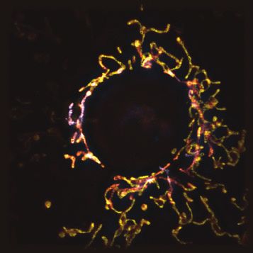
148 A. Descloux et al.: High-speed multiplane structured illumination microscopy of living cells
and from the Research Foundation-Flanders via grants [11] Abrahamsson S, Chen J, Hajj B, et al. Fast multicolor 3D imag-
G062616N, G0B8817N, G0A5817N, and VS.003.16N. ing using aberration-corrected multifocus microscopy. Nat
Methods 2013;10:60.
[12] Abrahamsson S, Ilic R, Wisniewski J, et al. Multifocus microscopy
Conflicts of interest: The authors declare that they have with precise color multi-phase diffractive optics applied in func-
no conflicts of interest related to this article. tional neuronal imaging. Biomed Opt Express 2016;7:855–69.
[13] Abrahamsson S, Blom H, Agostinho A, et al. Multifocus
structured illumination microscopy for fast volumetric super-
resolution imaging. Biomed Opt Express 2017;8:4135–40.
References [14] Juette MF, Gould TJ, Lessard MD, et al. Three-dimensional
sub–100 nm resolution fluorescence microscopy of thick
[1] Gustafsson MG. Surpassing the lateral resolution limit by a fac- samples. Nat Methods 2008;5:527.
tor of two using structured illumination microscopy. J Microsc [15] Geissbuehler S, Sharipov A, Godinat A, et al. Live-cell multi-
2000;198:82–7. plane three-dimensional super-resolution optical fluctuation
[2] Heintzmann R, Cremer CG. Laterally modulated excitation imaging. Nat Commun 2014;5:5830.
microscopy: improvement of resolution by using a diffraction [16] Descloux A, Grußmayer K, Bostan E, et al. Combined multi-
grating. Proc SPIE 1999;3568:85–196. plane phase retrieval and super-resolution optical fluctuation
[3] Gustafsson MG, Shao L, Carlton PM, et al. Three-dimensional imaging for 4D cell microscopy. Nat Photonics 2018;12:165.
resolution doubling in wide-field fluorescence microscopy by [17] O’Holleran K, Shaw M. Polarization effects on contrast in struc-
structured illumination. Biophys J 2008;94:4957–70. tured illumination microscopy. Opt Lett 2012;37:4603–5.
[4] Heintzmann R, Huser T. Super-resolution structured illumina- [18] Sison M, Chakrabortty S, Extermann J, et al. 3D time-lapse
tion microscopy. Chem Rev 2017;177:13890–908. imaging and quantification of mitochondrial dynamics. Sci Rep
[5] O’Holleran K, Shaw M. Optimized approaches for optical sec- 2017;7:43275.
tioning and resolution enhancement in 2D structured illumina- [19] Große L, Wurm CA, Brüser C, Neumann D, Jans DC, Jakobs S. Bax
tion microscopy. Biomed Opt Express 2014;5:2580–90. assembles into large ring-like structures remodeling the mito-
[6] Shaw M, Zajiczek L, O’Holleran K. High speed structured chondrial outer membrane in apoptosis. EMBO J 2016;35:402–13.
illumination microscopy in optically thick samples. Methods [20] Kner P, Chhun BB, Griffis ER, Winoto L, Gustafsson MG. Super-
2015;88:11–9. resolution video microscopy of live cells by structured illumina-
[7] Ströhl F, Kaminski CF. Speed limits of structured illumination tion. Nat Methods 2009;6:339–42.
microscopy. Opt Lett 2017;42:2511–4. [21] Descloux A, Grußmayer KS, Radenovic A. Parameter-free image
[8] Lu-Walther H-W, Kielhorn M, Förster R, Jost A, Wicker K, resolution estimation based on decorrelation analysis. Nat
Heintzmann R. Fastsim: a practical implementation of fast Methods 2019;16:918–24.
structured illumination microscopy. Methods Appl Fluores [22] Müller M, Mönkemöller V, Hennig S, Hübner W, Huser T.
2015;3:014001. Open-source image reconstruction of super-resolution struc-
[9] Song L, Lu-Walther H-W, Förster R, et al. Fast structured illu- tured illumination microscopy data in ImageJ. Nat Commun
mination microscopy using rolling shutter cameras. Meas Sci 2016;7:10980.
Technol 2016;27:055401. [23] Huang X, Fan J, Li L, et al. Fast, long-term, super-resolution
[10] Abrahamsson S, Usawa S, Gustafsson M. A new approach imaging with hessian structured illumination microscopy. Nat
to extended focus for high-speed high-resolution biological Biotechnol 2018;4:451–9.
microscopy. In: Three-Dimensional and Multidimensional
Microscopy: Image Acquisition and Processing XIII, vol. 6090.
International Society for Optics and Photonics; 2006. p. Supplementary Material: The online version of this article offers
60900N. 10.1117/12.647022. supplementary material (https://doi.org/10.1515/nanoph-2019-0346).
Unauthenticated
Download Date | 2/11/20 8:32 PM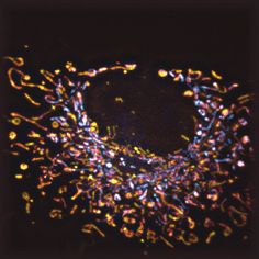

You can also read