Green Tea Extract Exerts Anabolic Effects on Extracellular Matrix of the Skin
←
→
Page content transcription
If your browser does not render page correctly, please read the page content below
www.ijpsonline.com
Green Tea Extract Exerts Anabolic Effects on
Extracellular Matrix of the Skin
M. TURKOGLU, E. PEKMEZCI1* AND S. KILIC
Biota Laboratories, R & D Center, Istanbul, 34785, 1Istanbul Medipol University, Department of Dermatology, Istanbul,
34214, Turkey
Turkoglu et al.: Anabolic Effects of Green Tea Extract on Skin
An in vitro trial was carried out to reveal the effects of an extract of Camellia sinensis (green tea extract)
on the major extracellular matrix components of human skin. After preparing the green tea extract its
phytochemical contents were analysed and its effects on gene expression of hyaluronan synthase 2, matrix
metalloproteinase-9 and elastase in a human dermal fibroblast cell line were determined. Cell proliferation
assay was performed using XTT reagent. Ribonucleic acid isolations were realized by using TRI reagent.
Expressions of the relevant enzymes and a control enzyme glyceraldehyde-3-phosphate dehydrogenase
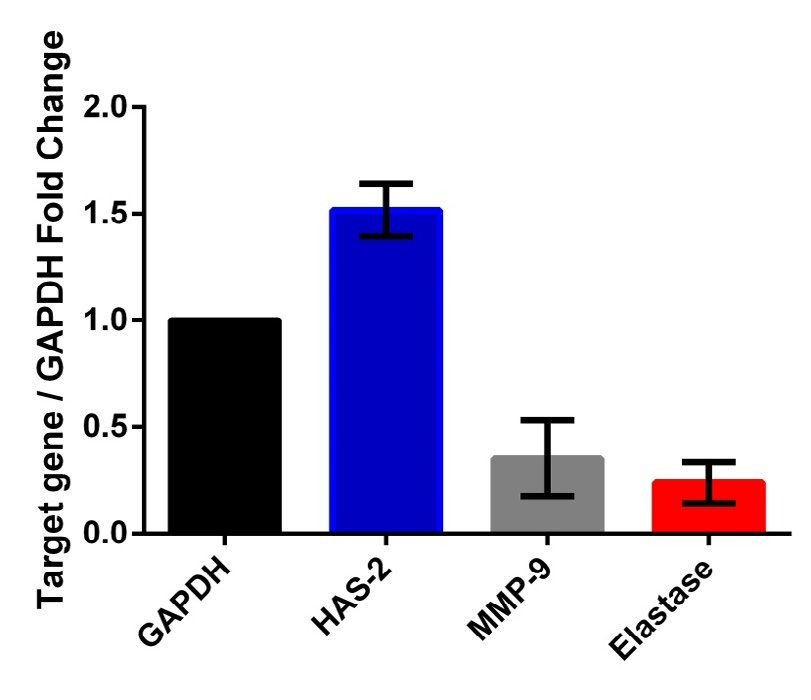
were determined using RT-qPCR analysis. Green tea extract caused statistically significant upregulation
of hyaluronan synthase-2 gene expression compared to untreated control cells. Also, as a positive outcome,
the treatment resulted in significant downregulations of matrix metalloproteinase-9 and elastase gene
expressions. Green tea extract was found to have substantial anabolic effects on hyaluronic acid, collagen
and elastin. The results obtained in this study might partially explain the molecular basis of the health
benefits and antiaging effects of Camellia sinensis on skin.
Key words: Camellia sinensis, green tea, hyaluronan synthase, matrix metalloproteinase, elastase
Tea plant, Camellia sinensis, is a member of the green tea extracts protected the skin from a direct UV
Theaceae family. Polyphenolic compounds called exposure preventing sunburn and erythema[4,5]. Tea
cathecins are thought to be responsible for the polyphenols alleviate the UVB-induced destructive
majority of health benefits associated with this plant. morphological changes in human keratinocyte
The potential health benefits of C. sinensis on skin (HaCaT) cell line via interacting with induced reactive
include, protection from the detrimental effects of oxygen species (ROS)[2]. Sunscreens containing
ionizing and ultraviolet (UV) radiation, improvement 2-3 % C. sinensis extract substantially protect against
in wound healing and cancer chemoprevention[1-3]. photoaging and photoimmunology related biological
Author’s previous studies reported that black or events such as cutaneous erythema and epidermal
March-April 2020 Indian Journal of Pharmaceutical Sciences 368thickening[6]. Cream with 10 % C. sinensis extract and Luna® Phenyl-Hexyl column with a solvent gradient of
300 mg twice daily green tea oral supplementation, 2 % acetic acid-acetonitrile with EDTA; acetonitrile
which were given to moderately photoaged women 9-32 % over 15 min at 278 nm and temperature was
revealed significant improvement in the elastic tissue kept at 35º.
contents of analysed specimens[7]. Topical application A solution of 2,2-diphenyl-1-picrylhydrazyl (DPPH,
of various C. sinensis extracts found effective in atopic 0.1 mM) in methanol was prepared and 1 ml of this
dermatitis, acne vulgaris and rosacea[8]. In an animal solution was added to 1 ml of all extracts in methanol
study, topical application of 20 % C. sinensis extract at different concentrations (50, 100, 200, 400,
significantly increased fibroblast growth, collagen 800 μg/ml). The mixtures were allowed to stand at
synthesis and thus the healing process by increasing room temperature for 30 min. The absorbance was
the rate of wound healing[1]. Tea polyphenols lead to measured at 517 nm using a UV/Vis spectrophotometer.
retardation in initiation of tumorigenesis, reduction Ascorbic acid was used as the reference standard. The
in cumulative number of tumor cells and increased capability of scavenging DPPH radical was calculated
tumor-free survival in experimentally-induced skin using the following formula, DPPH scavenging
carcinogenesis in mice[9]. Oral administration of effect (% inhibition) = (A0–A1)/A0)×100, where, A0
green tea polyphenols in drinking water or the topical is the absorbance of the control reaction and A1 is
application of epigallocathecin-3-gallate (EGCG), the the absorbance in presence of all extract samples or
major catechin in C. sinensis, prevent UVB-induced skin reference standard. All tests were performed in triplicate
tumor development in mice[10]. In a retrospective study and the results were averaged.
on patients who had cancer of the head, neck or pelvic The total phenolic content of the extract was estimated
region receiving radiotherapy, topically administered using Folin-Ciocalteu reagent. Ten milligrams of gallic
C. sinensis extracts supported the reparation of skin acid, the standard, was dissolved in 100 ml distilled
integrity, inhibited proteasome function and suppressed water. One millilitre of the extract and standard gallic
cytokine release[11]. acid at different concentrations (10, 20, 40, 60, 80,
Hyaluronic acid (HA), collagen and elastin bind each 100 µg/ml) were mixed with 5 ml of distilled water and
other and make up a three dimensional structure, which 0.5 ml of Folin-Ciocalteu reagent. After 5 min, 1.5 ml
is impaired in aged or damaged skin due to internal of 20 % sodium carbonate was added and volume was
or external causes[12]. With regard to the numerous adjusted to 10 ml with distilled water and incubated for
reports about C. sinensis on the well being of skin, in 2 h at room temperature. Absorbance was measured at
this study it was aimed to reveal the probable direct 750 nm. A calibration curve was plotted using standard
causes of cutaneous health and also the effects of this gallic acid. Total phenolic content of the extract was
expressed as mg of gallic acid equivalents. All the
plant on the major structural components of the human
tests were performed in triplicate and the results were
integumentary system. The effects of C. sinensis on
averaged.
the gene expression of three enzymes, hyaluronan
synthase-2 (HAS-2), matrix metalloproteinase-9 Total flavonoid content was evaluated using the
(MMP-9) and elastase, which are crucial in the aluminium chloride colorimetric assay. Four millilitres
metabolism of HA, collagen and elastin, respectively, of distilled water and 0.3 ml of 5 % sodium nitrite
are studied in a human dermal fibroblast cell line. solution were added to 1 M of extract and 1 ml of
standard quercetin solution at different concentrations
MATERIALS AND METHODS (100, 200, 400, 600, 800, 1000 µg/ml). Aluminum
The leaves of C. sinensis were purchased from Martin chloride solution (10 %, 0.3 ml) was added to each tube.
Bauer Group. Dried leaves (12.5 g) were extracted with
500 ml distilled water by boiling for 15 min. The extract This is an open access article distributed under the terms of the Creative
Commons Attribution-NonCommercial-ShareAlike 3.0 License, which
was filtered through a 0.45 µm filter paper. Content of allows others to remix, tweak, and build upon the work non-commercially,
catechins in green tea was analysed according to a HPLC as long as the author is credited and the new creations are licensed under
the identical terms
method[13] suitably modified as the following, HPLC on
Accepted 17 January 2020
Revised 20 December 2019
Received 05 August 2019
*Address for correspondence
E-mail: erkinpekmezci@gmail.com Indian J Pharm Sci 2020;82(2):368-373
369 Indian Journal of Pharmaceutical Sciences March-April 2020www.ijpsonline.com
After 5 min, 2 ml of 1 m sodium hydroxide was added. as study material and glyceraldehyde-3-phosphate
Finally, volume was made up to 10 ml with distilled dehydrogenase (GAPDH) as control (Integrated DNA
water. The absorbance was measured at 510 nm. A Technologies) were added in RNAse free test tubes
calibration curve was plotted using standard quercetin. and the final volume was made up to 13 µl for each
The total flavonoids of the extract were expressed as mg by adding distilled water. After incubation for 10 min
of quercetin equivalents. All the tests were performed at 65o in a Thermal Cycler, the tubes were placed over
in triplicates and the results were averaged. ice. Later they were incubated for 30 min at 55º and
5 min at 85º in the Thermal Cycler, after adding 4 µl
Fibroblast cells were cultured in DMEM with high
of reverse transcription buffer (5X), 2 µl of dNTP mix
glucose, supplemented with 15 % heat-inactivated
(10 mM), 0.5 µl of protector RNAse inhibitor and 0.5
fetal bovine serum and 1% gentamicin. The cells were
µl of reverse transcriptase. Primer sequences are given
maintained at 37º in a humidified atmosphere of 5 %
in Table 1.
CO2 in a Newbrunswick incubator. All supplements
and media were purchased from Sigma Aldrich. The Fast Start DNA Green Master Kit (Roche Diagnostics)
cellular toxicity of green tea extract was investigated was used for the real-time quantitative polymerase chain
with XTT cell proliferation assay. The cells were reactions (RT-qPCR). Briefly, total volume of reaction
seeded into 96-well plates (1×10⁴cells/well) and were mix was 20 µl, containing 10 µl Master Mix, 10 mM
incubated 24 h at 37º, in a humidified atmosphere of each of reverse and forward primers, 25 ng template
5 % CO2. New medium was added on every 2nd day cDNA and appropriate amount of RNAse free distilled
after aspirating the previous medium, treated with water. All samples were run as triplicates in each run
different concentrations (10, 5, 3, 1, 0.1 and 0 %) including a non-template control and 4 standards
of green tea extract and incubated under the same (1:1, 1:10, 1:100, 1:1000). The RT-qPCR parameters
conditions for 72 h. XTT reagent was added to the plates were determined separately for each target according
after the incubation period to obtain a concentration to melting and annealing temperatures of primers. Each
of 0.3 mg/ml according to the manufacturer’s (Roche parameter included a pre-incubation step for 10 min at
Diagnostics) instructions. Then, cells were incubated at 95º and followed by 45 cycles of 3 amplification and
37º for 4 h to get the XTT reagent reduced to orange melting steps. Melting curve analysis was performed
formazan compound. The optical density of soluble to verify specificity. Absolute quantification analysis
formazan compound was measured at 450 nm with was performed by using Light Cycler 96 (Roche
650 nm reference in a microplate reader (Bio-Rad). Diagnostics). For quantitation of RT-qPCR results, ΔΔCt
Based on cell proliferation ratios of treated cells with method was used. The gene expression results were
respect to the control cells, cytotoxicity levels of the represented as Target/GAPDH fold change. All data
green tea extract were determined. Higher concentrations were representative of three experiments and expressed
of the extract were found to be cytotoxic for fibroblast as mean±standard deviation. Statistical evaluation
cells. In these studies principally the concentration of was performed by non-parametric Mann–Whitney test
the extract chosen was the one which gave nearest to (GraphPad Prism 6) and statistical significance was
80 % cell proliferation ratio. Generally to have this ratio defined as pTABLE 1: PRIMERS (5'-3') OF THE GENES STUDIED
Primers Forward primer Reverse primer
HAS-2 GCCTGGGCTATGCAACAAAA GTAGGACTTGCTCCAACGGG
MMP-9 GTACTCGACCTGTACCG AGAAGCCCCACTTCTTGTCG
Elastase CTGGCCTCGGAGATTGTGG GGACGTTTACATTCGCCACG
GAPDH ATGGGTGTGAACCATGAGAA GTGCTAAGCAGTTGGTGGTG
TABLE 2: THE QUANTITIES OF CATECHINS IN GREEN TEA
Catechins* GA GC EGC C CAF EC EGCG GCG ECG
g/100 g dm§ 0.011 0.284 3.477 0.068 2.567 0.670 7.965 0.193 1.386
*GA: Gallic Acid, GC: Gallocatechin, EGC: Epigallocatechin, C: Catechin, CAF: Caffeine, EC: Epicatechin, EGCG: Epigallocatechingallate,
GCG: Gallocatechingallate, ECG: Epicatechingallate §dm: Dry Material
TABLE 3: ANTIOXIDANT ACTIVITY, PHENOLIC AND FLAVONOID CONTENT OF THE GREEN TEA
EXTRACT
Antioxidant activity Phenolic content Flavonoid content
96.27% (10 % ascorbic acid equivalent) 9.74 mg/ml (gallic acid equivalent) 0.19 mg/ml (quercetin equivalent)
RESULTS AND DISCUSSION
Target gene / GAPDH Fold Change
The quantities of catechins obtained by HPLC analysis 2.0
of green tea are shown in Table 2. The total antioxidant
1.5
activity, phenolic and flavonoid contents of the green
tea extract are depicted in Table 3. Green tea extract
1.0
caused statistically significant upregulation of HAS-2
gene expression (pwww.ijpsonline.com
these proteinases are predominantly expressed. They wrinkling, UVA predominantly brings out sagging.
activate quiescent cells, stimulate cell migration and Increased activity of fibroblast derived elastase in the
initiate cell differentiation[22]. The main characteristic skin has an important function in wrinkling and sagging
changes during remodeling of the dermal ECM due as a result of elastic fiber degradation[30]. Nevertheless,
to UVB irradiation are, upregulation of MMPs and the insufficiency of sunscreens alone, proposes the idea
subsequent cleavage of collagen, fibronectin, elastin and that the combination of a sunscreen and an elastase
proteoglycans, contributing to photoaging[23]. MMP-9 inhibitor might be more effective[31].
is a member of the gelatinase subgroup of MMPs also This study analysed the effects of C. sinensis green
known as gelatinase B. Gelatinases are associated with tea extract, on the gene expressions of 3 enzymes
cancer invasion in the skin because they mainly degrade responsible for the metabolism of structural components
collagen type IV, which is an essential component of the of healthy skin, through a human dermal fibroblast cell
basement membrane of the epidermis. MMP-9 can also line. In terms of fold change, the increase in HAS-2,
degrade other substrates such as collagen type V, VII, and the decreases in both MMP-9 and elastase, which
X, fibronectin and elastin[21]. MMP-9, is thought to have were all positive outcomes, were found significant
critical functions in the remodeling of the basement (fig. 2). Generation of ROS and downturning of
membrane zone because several ECM proteins in this cell functions caused by long term exposures to
region have been determined as the substrates of this environmental and intrinsic offences, result in tissue
proteinase[22]. The greater invasive feature of squamous damage. These inflammmatory responses enhance the
cell carcinoma (SCC) compared to that of basal cell synthesis of dermal enzymes which lead to degradation
carcinoma is probably due to the enhanced expression of ECM[32]. Breakdown and disorganisation of ECM
and activity of gelatinases in the former and therefore, components are the predominant features of skin aging
among the MMPs, MMP-9 is the most critical one in and inhibiton of pathologic enzymatic activities by
SCC tumorigenesis[21]. MMP-9 is the main proteinase natural plant compounds might be a promising approach
responsible from the aberrant wound healing. Elevated to prevent the above-mentioned degradation[33]. In
MMP-9 levels have been demonstrated in various general it may be considered that green tea extract
chronic wound types. There is a substantial increase of has a substantial anabolic effect on HA, collagen and
MMP-9 at the chronic wound environment and it was elastine, which are the 3 major extracellular structural
shown that healed areas of burn wounds and leg ulcers components of the skin. It is quite possible that these
lost active MMP-9 expression[24]. Also, diabetic patients results are mainly due to the plant’s strong antioxidant
with chronic, non-healing foot ulcers are found to have activity related to its phenolic and flavonoid constituents
higher MMP-9 serum levels compared to those patients supplied by the abundant content of cathecins which
who are treatment responsive[25]. Elastic fibers, which are also demonstrated by the phytochemical analyses
are among the crucial components of connective tissue,
performed (Tables 2 and 3). Suppression of the
give resilience and elasticiy to the skin. Elastic fibers are
degradation of ECM and increasing the relevant 3 major
cleaved by the elastolytic enzymes, principally human
structural components of skin is the well known target
leucocyte elastase (HLE)[12]. Although under normal
of antiaging studies and cosmetic dermatology and it is
physiologic conditions elastase is a powerful agent for
an established opinion that these 3 components must be
host defence[26], it is involved in tissue destruction in
increased in order to give skin a younger and healthier
numerous chronic inflammatory diseases[27]. HLE is
unable to cleave intact elastin but may promote further appearance[12]. Therefore, considering the significant
disruption of elastic fibers secondary to the action of anabolic effects of green tea extract in dermal ECM, it
other proteases[28]. Wrinkle development in the skin can be suggested that C. sinensis has a promising future
due to long term UVB exposure is associated with in cosmetic dermatology. The results obtained in this
impairment of elastic fibers in the dermis. Fibroblast study might also partially explain the molecular basis
elastase, which is the major elastase in the skin under of the health benefits of C. sinensis on skin, including
non-inflammatory conditions, has a particular role cancer chemoprevention, improvement in wound
in the disruption of elastic fibers due to cumulative healing, and protection against the detrimental effects
UVB exposure[29]. Studies have also demonstrated a of UV exposure.
reduction in the elastin content of the skin, even in the
REFERENCES
protected areas, due to intrinsic aging[12]. Although the
long term exposure of skin to UVB primarily evokes 1. Hajiaghaalipour F, Kanthimathi MS, Abdulla MA, Sanusi J.
March-April 2020 Indian Journal of Pharmaceutical Sciences 372The effect of Camella sinensis on wound healing potential 19. Tammi R, Pasonen-Seppanen S, Kolehmainen E, Tammi M.
in an animal model. Evid Based Complement Alternat Med Hyaluronan synthase induction and hyaluronan accumulation
2013;2013:386734. in mouse epidermis following skin injury. J Invest Dermatol
2. Wy LY, Zheng XQ, Lu JL, Liang YR. Protective effect of 2005;124(5):898-905.
green tea polyphenols against ultraviolet-B induced damage to 20. Rilla K, Lammi MJ, Sironen R, Törrönen K, Luukkonen M,
HaCaT cells. Hum Cell 2009;22(1):18-24. Hascall VC, et al. Changed lamellipodial extension, adhesion
3. Nagle DG, Ferreira D, Zhou YD. Epigallocathecin-3- plaques and migration in epidermal keratinocytes containing
gallate (EGCG): Chemical and biomedical perspectives. constitutively expressed sense and antisense hyaluronan
Phytochemistry 2006;67(17):1849-55. synthase 2 (Has2) genes. J Cell Sci 2002;115(Pt18):3633-43.
4. Turkoglu M, Cigirgil N. Evaluation of black tea gel and 21. Pittayapruek P, Meephansan J, Prapapan O, Komine M,
its protection potential against UV. Int J Cosmet Sci Ohtsuki M. Role of matrix metalloproteinases in photoaging
2007;29(6):437-42. and photocarcinogenesis. Int J Mol Sci 2016;17:868.
5. Turkoglu M, Uğurlu T, Gedik G, Yılmaz AM, Yalcin AS. In 22. Han YP, Yan C, Garner WL. Proteolytic activation of
vivo evaluation of black and green tea dermal products against matrix metalloproteinase-9 in skin wound healing is
UV radiation. Drug Discov Ther 2010;4(5):362-7. inhibited by alpha-1 antichymotrypsin. J Invest Dermatol
6. Li YH, Wu Y, Wei HC, Xu YY, Jia LL, Chen J, et al. 2008;128(9):2334-42.
Protective effects of green tea extracts on photoaging and 23. Röck K, Grandoch M, Majora M, Krutmann J, Fischer JW.
photoimmunosuppression. Skin Res Technol 2009;15(3):338- Collagen fragments inhibit hyaluronan synthesis in skin
45. fibroblasts in response to ultraviolet B (UVB): new insights
7. Chiu AE, Chan JL, Kern DG, Kohler S, Rehmus WE, Kimball into mechanisms of matrix remodeling. J Biol Chem
AB. Double blinded, placebo-controlled trial of green tea 2011;286(20):18268-76.
extracts in the clinical and histologic appearance of photoaging
24. Reiss MJ, Han YP, Garcia E, Goldberg M, Yu H, Garner WL.
skin. Dematol Surg 2005;31(7Pt2):855-60.
Matrix metalloproteinase-9 delays wound healing in a murine
8. Pazyar N, Feily A, Kazerouni A. Green tea in dermatology.
wound model. Surgery 2010;147(2):295-302.
Skinmed 2012;10(6):352-5.
25. Dinh T, Tecilazich F, Kafanas A, Doupis J, Gnardellis C, Leal
9. Roy P, Nigam N, George J, Srivastava S, Shukla Y. Induction of
apoptosis by tea polyphenols mediated through mitochondrial E, et al. Mechanisms involved in the development and healing
cell death pathway in mouse skin tumors. Cancer Biol Ther of diabetic foot ulceration. Diabetes 2012;61:2937-47.
2009;8(13):1281-7. 26. Barros SC, Martins JA, Marcos JC, Cavaco-Paulo A. Influence
10. Katiyar S, Elmets CA, Katiyar SK. Green tea and skin cancer: of secretory leukocyte protease inhibitor-based peptides
photoimmunology, angiogenesis and DNA repair. J Nutr on elastase activity and their incorporation in hyaluronic
Biochem 2007;18(5):287-96. acid hydrogels for chronic wound therapy. Biopolimers
11. Pajonk F, Riedisser A, Henke M, McBride WH, Fiebich B. 2012;98(6):576-90.
The effects of tea extracts on proinflammatory signaling. BMC 27. Lee SK, Lee SS, Song IS, Kim YS, Park YW, Joo JY, et al.
Med 2006;4:28. Paradoxical effects of elastase inhibitor guamerin on the tissue
12. Baumann L, Saghari S. Basic science of dermis. In: Baumann repair of two different wound models: sealed cutaneous and
L, Saghari S, Weisberg E, editors. Cosmetic Dermatology. 2nd exposed tongue wounds. Exp Mol Med 2004;36(3):259-67.
ed. New York: McGraw-Hill; 2009. p. 8-13. 28. Schmelzer CE, Jung MC, Wohlrab J, Neubert RH, Heinz A.
13. ISO Geneva, Switzerland, Determination of substances Does human leukocyte elastase degrade intact skin elastin?
characteristic of green tea and black tea. Part 2: Content of FEBS J 2012;279(22):4191-200.
cathecins in green tea-method using high performance liquid 29. Tsukahara K, Takema Y, Moriwaki S, Tsuji N, Suzuki Y,
chromatography. ISO 14502-2: 2005. Fujimura T, et al. Selective inhibition of skin fibroblast
14. Pasonen-Seppanen S, Karvinen S, Törrönen K, Hytinnen elastase elicits a concentration-dependent prevention of
JM, Jokel T, Lammi MJ, et al. EGF upregulates, whereas ultraviolet B-induced wrinkle formation. J Invest Dermatol
TGF-β downregulates, the hyaluronan synthases Has2 and 2001;117:671-7.
Has3 in organotypic keratinocyte cultures: correlations with
30. Imokawa G, Ishida K. Biological mechanisms underlying
epidermal proliferation and differentiation. J Invest Dermatol
the ultraviolet radiation-induced formation of skin wrinkling
2003;120:1038-44.
and sagging I: reduced skin elasticity, highly associated with
15. Gebhardt C, Averbeck M, Diedenhofen N, Willenberg A,
enhanced dermal elastase activity, triggers wrinkling and
Anderegg U, Sleeman JP, et al. Dermal hyaluronan is rapidly
reduced by topical treatment with glucucorticoids. J Invest sagging. Int J Mol Sci 2015;16:7753-75.
Dermatol 2010;130:141-9. 31. Tsukahara K, Moriwaki S, Hotta M, Fujimura T, Sugiyama-
16. Saavalainen K, Pasonen-Seppanen S, Dunlop TW, Tammi R, Nagakiri Y, Sugawara S, et al. The effect of sunscreen on skin
Tammi MI, Carlberg C. The human hyaluronan synthase 2 elastase activity induced by ultraviolet-A irradiation. Biol
gene is a primary retinoic acid and epidermal growth factor Pharm Bull 2005;28(12):2302-7.
responding gene. J Biol Chem 2005;280(15):14636-44. 32. Maity N, Nema NK, Sarkar BK, Mukherjee PK. Standardized
17. Wang Y, Lauer ME, Anand S, Mack JA, Maytin EV. Hyaluronan Clitoria ternatea leaf extract as hylauronidase, elastase and
synthase 2 protects skin fibroblasts against apoptosis induced matrix-metalloproteinase-1 inhibitor. Indian J Pharmacol
by environmental stress. J Biol Chem 2014;289(46):32253-65. 2012;44(5):584-7.
18. Röck K, Tigges J, Sass S, Schütze A, Florea AM, Fender AC, 33. Wittenauer J, Mackle S, Susmann D, Schweiggert-Weisz U,
et al. miR-23a-3p causes cellular senescence by targeting Carle R. Inhibitory effects of polyphenols from grape pomace
hyaluronan synthase 2: possible implication for skin aging. J extract on collagenase and elastase activity. Fitoterapia
Invest Dermatol 2015;135(2):368-77. 2015;101:179-87.
373 Indian Journal of Pharmaceutical Sciences March-April 2020You can also read