Bacterial adaptation to venom in snakes and arachnida
←
→
Page content transcription
If your browser does not render page correctly, please read the page content below
Bacterial adaptation to venom in snakes
                and arachnida
 Esmaeilishirazifard, E, Usher, L, Trim, C, Denise, H, Sangal, V, Tyson, GH, Barlow,
  A, Redway, KF, Taylor, JD, Kremyda-Vlachou, M, Davies, S, Loftus, TD, Lock,
  MMG, Wright, K, Dalby, A, Snyder, LAS, Wuster, W, Trim, S and Moschos, SA
                        http://dx.doi.org/10.1128/spectrum.02408-21

Title                   Bacterial adaptation to venom in snakes and arachnida
Authors                 Esmaeilishirazifard, E, Usher, L, Trim, C, Denise, H, Sangal, V, Tyson,
                        GH, Barlow, A, Redway, KF, Taylor, JD, Kremyda-Vlachou, M, Davies, S,
                        Loftus, TD, Lock, MMG, Wright, K, Dalby, A, Snyder, LAS, Wuster, W,
                        Trim, S and Moschos, SA
Publication title       Microbiology Spectrum
Publisher               American Society for Microbiology
Type                    Article
USIR URL                This version is available at: http://usir.salford.ac.uk/id/eprint/64384/
Published Date          2022

USIR is a digital collection of the research output of the University of Salford. Where copyright
permits, full text material held in the repository is made freely available online and can be read,
downloaded and copied for non-commercial private study or research purposes. Please check the
manuscript for any further copyright restrictions.

For more information, including our policy and submission procedure, please
contact the Repository Team at: library-research@salford.ac.uk.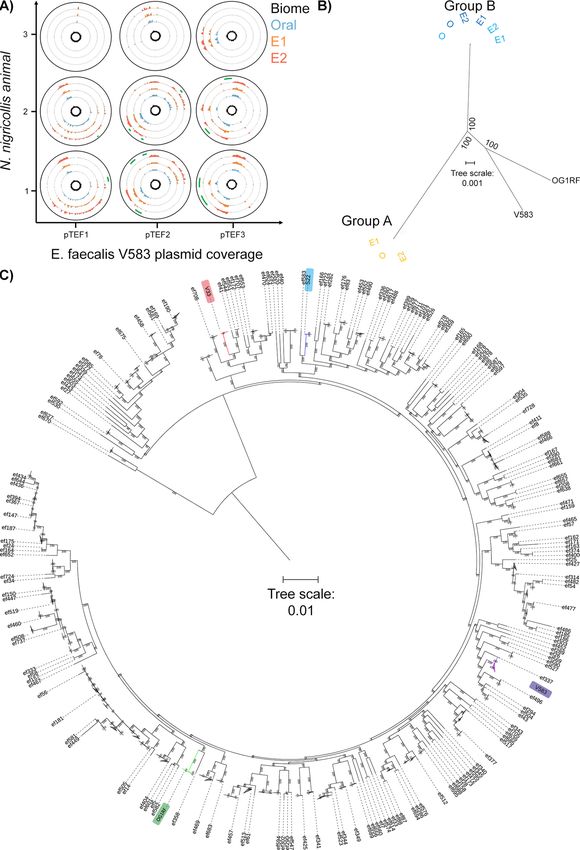
RESEARCH ARTICLE

Bacterial Adaptation to Venom in Snakes and Arachnida
Elham Esmaeilishirazifard,a,b* Louise Usher,a,b Carol Trim,c Hubert Denise,d§ Vartul Sangal,e Gregory H. Tyson,f Axel Barlow,g
Keith F. Redway,a John D. Taylor,a,b,h Myrto Kremyda-Vlachou,a Sam Davies,e Teresa D. Loftus,i Mikaella M. G. Lock,i Kstir Wright,a
Andrew Dalby,a Lori A. S. Snyder,j Wolfgang Wuster,k Steve Trim,i Sterghios A. Moschosa,b,e

a   Department of Biomedical Sciences, Faculty of Science and Technology, University of Westminster, London, United Kingdom
b   Westminster Genomic Services, Faculty of Science and Technology, University of Westminster, London, United Kingdom
c   School of Psychology and Life Sciences, Faculty of Science, Engineering and Social Sciences, Canterbury Christ Church University, Canterbury, Kent, United Kingdom
d   EMBL-EBI European Bioinformatics Institute, Wellcome Trust Genome Campus, Hinxton, Cambridge, United Kingdom
e   Department of Applied Sciences, Faculty of Health and Life Sciences, Northumbria University, Newcastle, Tyne and Wear, United Kingdom
f   Food and Drug Administration, Center for Veterinary Medicine, Office of Research, Laurel, Maryland, USA
g   Institute for Biochemistry and Biology, University of Potsdam, Potsdam, Germany
h   School of Environment and Life Sciences, University of Salford, Salford, Greater Manchester, United Kingdom
i   Venomtech, Ltd., Sandwich, Kent, United Kingdom
j   School of Life Sciences, Pharmacy, and Chemistry, Kingston University, London, United Kingdom
k   Molecular Ecology and Evolution at Bangor, School of Biological Sciences, College of Natural Sciences, Bangor University, Bangor, Wales, United Kingdom

Elham Esmaeilishirazifard and Louise Usher contributed equally to this article. Author order was determined on the basis of seniority.

ABSTRACT       Animal venoms are considered sterile sources of antimicrobial compounds
with strong membrane-disrupting activity against multidrug-resistant bacteria. However,
venomous bite wound infections are common in developing nations. Investigating the
envenomation organ and venom microbiota of five snake and two spider species, we
observed venom community structures that depend on the host venomous animal spe-
cies and evidenced recovery of viable microorganisms from black-necked spitting cobra

                                                                                                                                                                                         Downloaded from https://journals.asm.org/journal/spectrum on 11 August 2022 by 92.29.140.243.
(Naja nigricollis) and Indian ornamental tarantula (Poecilotheria regalis) venoms. Among
the bacterial isolates recovered from N. nigricollis, we identified two venom-resistant,
novel sequence types of Enterococcus faecalis whose genomes feature 16 virulence
genes, indicating infectious potential, and 45 additional genes, nearly half of which
improve bacterial membrane integrity. Our findings challenge the dogma of venom ste-
rility and indicate an increased primary infection risk in the clinical management of ven-
omous animal bite wounds.                                                                                                         Editor Olaya Rendueles Garcia, Institut Pasteur
                                                                                                                                  Ad Hoc Peer Reviewer        Bruno Amorim-
IMPORTANCE Notwithstanding their 3 to 5% mortality, the 2.7 million envenomation-                                                 Carmo, Federal University of Rio Grande do
related injuries occurring annually—predominantly across Africa, Asia, and Latin America—                                         Norte; Sylvie REBUFFAT, UMR 7245 CNRS -
                                                                                                                                  Muséum National d'Histoire Naturelle
are also major causes of morbidity. Venom toxin-damaged tissue will develop infec-
                                                                                                                                  Copyright © 2022 Esmaeilishirazifard et al. This
tions in some 75% of envenomation victims, with E. faecalis being a common culprit                                                is an open-access article distributed under the
of disease; however, such infections are generally considered to be independent of                                                terms of the Creative Commons Attribution 4.0
envenomation. Here, we provide evidence on venom microbiota across snakes and                                                     International license.
                                                                                                                                  Address correspondence to Sterghios A.
arachnida and report on the convergent evolution mechanisms that can facilitate ad-
                                                                                                                                  Moschos, sterghios.moschos@northumbria.ac.uk.
aptation to black-necked cobra venom in two independent E. faecalis strains, easily                                               *Present address: Elham Esmaeilishirazifard,
misidentified by biochemical diagnostics. Therefore, since inoculation with viable                                                 Cancer Research UK Cambridge Institute,
and virulence gene-harboring bacteria can occur during envenomation, acute infec-                                                 University of Cambridge, Cambridge, United
                                                                                                                                  Kingdom.
tion risk management following envenomation is warranted, particularly for immuno-                                                §
                                                                                                                                   Present address: Hubert Denise, UK Health
compromised and malnourished victims in resource-limited settings. These results                                                  Security Agency, London, United Kingdom.
shed light on how bacteria evolve for survival in one of the most extreme environ-                                                The authors declare no conflict of interest.
ments on Earth and how venomous bites must be also treated for infections.                                                        Received 1 December 2021
                                                                                                                                  Accepted 14 April 2022
KEYWORDS drug resistance evolution, extremophiles, genome analysis, microbiome,                                                   Published 23 May 2022
multidrug resistance, venom

May/June 2022 Volume 10 Issue 3                                                                                                                   10.1128/spectrum.02408-21          1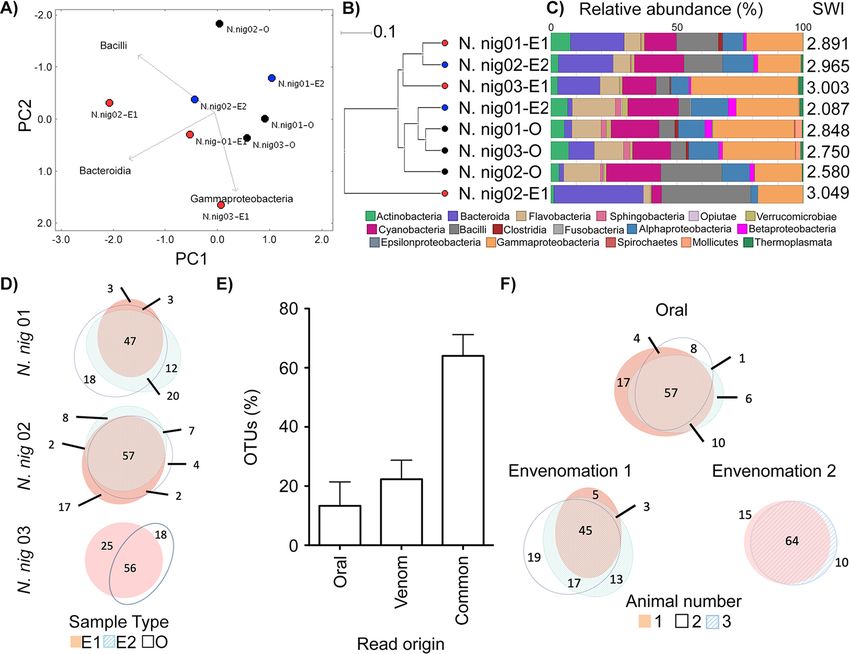
Bacterial Colonization and Adaptation to Animal Venoms                                                    Microbiology Spectrum

T    he rise of multidrug-resistant (MDR) bacterial infections suggests the end of the an-
     tibiotic golden era might be approaching fast. Discovery of novel antimicrobials is
therefore an urgent priority of exceptional socioeconomic value. Crude preparations of
animal venoms exhibit strong antibiotic potencies, including against clinical MDR bac-
terial isolates such as Mycobacterium tuberculosis (1). With antimicrobial properties
described for crotalid (pit viper) venom as early as 1948 (2), relevant compounds have
been isolated from most animal venoms, including those of spiders, scorpions, and
insects, as well as aquatic species. Examples include phospholipase A2 (PLA2) enzymes,
L-amino acid oxidases, cathelicidins, C-type lectins, and hydrophobic/cationic peptides,
as well as venom toxin domains (3), which may act by physically disrupting bacterial
cell membranes through pore formation (4, 5). Accordingly, venomous animal bite or
sting (envenomation) wound infections are considered rare (6) and are attributed to
secondary infection (7). Yet, over three-quarters of snake bite victims may develop
mono- or polymicrobial envenomation wound infections, characterized by Bacteroides,
Morganella, Proteus, and Enterococcus (8, 9)—bacterial taxa commonly found in the
gut. Indeed, Enterococcus faecalis and Morganella morganii have been independently
reported as the most common Gram-positive and Gram-negative envenomation
wound infections across several countries (8–10). Historically associated with the oral
snake microbiota (11), these bacteria are thought to originate from prey feces (12) per-
sisting in the snake oral cavity (10), with a diversity similar to that of the snake gut (13).
Yet, no “fixed” oral microbiota were observed in early systematic studies, beyond a sea-
sonal variation of diversity (14). Curiously, mouths of nonvenomous snakes were
reportedly more sterile than those of venomous snakes (14), a counterintuitive finding
independently reproduced elsewhere (10). More recently, the oral microbiota of the
nonvenomous Burmese python (Python bivittatus) has also been reported to be native
and not derived from prey guts (15).
    As venom glands are connected to the tip of envenomation apparatus via a persis-
tently open duct that is continuously exposed to the environment (16), the envenoma-
tion apparatus could be compared to clinical catheterization assemblies: a transcutane-
ous needle resting on a nonsterile environment, connected to a continually open duct,
leading to a liquid-containing vessel. Such devices develop biofilms within a few days,
making weekly catheter replacement necessary (17). Unlike the high flow rates of cath-

                                                                                                                                  Downloaded from https://journals.asm.org/journal/spectrum on 11 August 2022 by 92.29.140.243.
eters, however, the envenomation apparatus normally ejects venom only sporadically.
Captive snakes are often fed weekly and can fast for months, whereas large arachnids
typically are fed monthly. Wild animals may also undergo hibernation for several
months, when venom expulsion frequency can be assumed to be zero. Collectively,
these conditions offer opportunities for microbes to colonize venom across its concen-
tration gradient from the animal mouth to the venom gland, potentially driving the
evolution of resistance against venom antimicrobials.
    The aims of this study were to evaluate whether the microbiota of venom differs
from that of the envenomating apparatus, whether microbes can survive in venom,
and, if so, what genetic adaptations facilitate their survival in such extreme microenvir-
onments. Using culture and culture-free methods, we examined this hypothesis in
snakes and spiders and then employed antimicrobial susceptibility testing, whole-ge-
nome sequencing analysis, and comparative genomics to identify and characterize
venom tolerance and infectivity potential in representative novel E. faecalis isolates
from venom from captive Naja nigricollis (black-necked spitting cobra).

RESULTS
    The snake venom microbiota vary on account of host species and not on account
of the oral microbiota. Applying established culture-free methods (18) on commer-
cially available venom from Bothrops atrox (fer-de-lance; Viperidae) and a venom sam-
ple from a captive Bitis arietans (African puff adder), we first optimized microbial DNA
extraction for this unusual biological matrix (see Fig. S1 in the supplemental material).
Given animal availability, behavioral, and sampling limitations, such as the limited and
inconsistent venom yields of scorpions, we next focused on five snake and two spider

May/June 2022 Volume 10 Issue 3                                                                  10.1128/spectrum.02408-21   2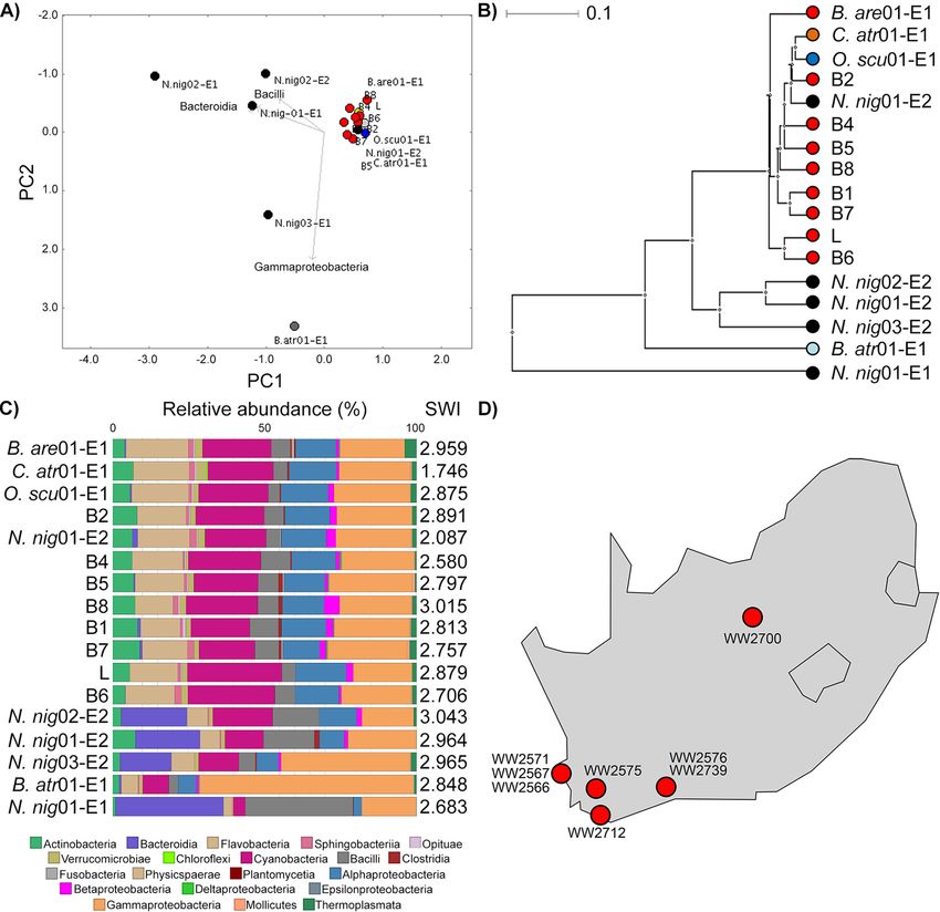
Bacterial Colonization and Adaptation to Animal Venoms                                                                  Microbiology Spectrum

TABLE 1 Animals sampled for the presence of microbiomes in venom
Common name                               Scientific name           Short name     Origin        Preservation method           No. of animals
Snakes
  Puff adder                              Bitis arietans           B. are         Captivity     Flash frozen                  1
                                                                   L              Commercial    Lyophilized                   1
                                                                   B1–B8          Wild          Air dried                     8
  Black-necked cobra                      Naja nigricollis         N. nig         Captivity     Flash frozen                  3
  Fer-de-lance                            Bothrops atroxa          B. atr         Captivity     Flash frozen                  3
  Western diamond rattlesnake             Crotalus atroxa          C. atr         Captivity     Flash frozen                  2
  Taipan                                  Oxyuranus scutellatusa   O. scu         Captivity     Flash frozen                  2

Spiders
  Indian ornamental                       Poecilotheria regalis    P. reg         Captivity     Flash frozen                  3
  Salmon pink                             Lasiodora parahybanab    L. par         Captivity     Flash frozen                  5
aVenom     produced by one animal only.
bYields   ranged from ,1 to 30 m L.

species (Table 1). We collected a swab (sample O) of the oral cavity (snakes) or fang sur-
face (spiders) and two consecutive envenomation samples (E1 and E2), expecting the
second venom sample to have fewer contaminants from bacterial plugs possibly forming
on the envenomation apparatus. In agreement with previous reports (11, 14), principal-
coordinate analysis (PCoA) and unsupervised clustering (Fig. S2) failed to discriminate the
swab microbiota by host species, suggesting the common diets and, most likely, water
sources in captivity had the biggest impact.
    This led us to hypothesize that captive animals would feature more closely related
microbiota in their venoms than commercial or wild samples. We therefore compared
the venom microbiota of all snakes using the same approach (Fig. 1). High Shannon-
Wiener indices indicated considerable diversity in snake venom microbiota; however,
surprisingly closer relationships were observed between B. arietans and other Viperidae,
despite samples spanning captive and wild animals; an exception was B. atrox venom,
which was characterized principally by Gammaproteobacteria. Focusing on B. arietans
also failed to cluster samples by origin (Fig. S3), despite the unknown providence of the
commercially available lyophilized sample and the disparate locations across South

                                                                                                                                                Downloaded from https://journals.asm.org/journal/spectrum on 11 August 2022 by 92.29.140.243.
Africa where wild B. arietans samples were collected (Fig. 1D); among the latter, the air-
drying method used for venom preservation could have been expected to substantially
compromise the microbiota signature. In contrast, N. nigricollis microbiota largely formed
a distinct cluster (Fig. 1) characterized by Bacteroidia (Bacteroidaceae), a taxon less com-
mon among Viperidae. This could reflect anatomical differences in elapid (cobra) fang
location at the front of the mouth compared to the sheathed nature of the longer,
hinged viperid fangs, whose tips rest at the back of the oral cavity. In contrast, spider
species did not seem to influence venom microbiota consistency and exhibited lower
biodiversity (Fig. S4). These results likely reflected vertebrate/invertebrate anatomical dif-
ferences and the limited venom yield from invertebrates (,1 to 30 m L) versus snakes
(100 to 1,000 m L).
    A fifth of the N. nigricollis venom microbiota is distinct from that of fangs.
Encouraged by the distinctive bacterial taxonomies in N. nigricollis venom, the avail-
ability of animals under controlled conditions, and the paired nature of the fang swab
and envenomation samples, we delved deeper into this data set. The fang microbiota
appeared to form a cluster distinct from that of venom microbiota (Fig. 2A and B), sug-
gesting the venom gland might be a distinct ecological niche (Fig. 2C). We therefore
asked if any bacterial taxa were unique to subsets of these samples. Operational taxo-
nomic unit (OTU) incidence analysis within each animal (Fig. 2D) suggested some 60%
of OTUs were shared between corresponding venoms and fangs. Yet, importantly, up
to 20% of these appeared to be unique to venom, and some 15% were unique to the
fang (Fig. 2E), indicating an OTU continuum between the two microenvironments, with
unique taxa in each site. Common sample types also featured a majority of common
taxa and OTUs unique to each site in each animal (Fig. 2F). However, taxa unique to

May/June 2022 Volume 10 Issue 3                                                                                10.1128/spectrum.02408-21   3
Bacterial Colonization and Adaptation to Animal Venoms                                                                                           Microbiology Spectrum

                                                                                                                                                                            Downloaded from https://journals.asm.org/journal/spectrum on 11 August 2022 by 92.29.140.243.
FIG 1 Snake venom microbiomes cluster on account of host species. Viperid venom microbiomes cluster separately from N. nigricollis, with the exception of
B. atrox, as determined by (A) PCoA, (B) an UPGMA tree (i.e., unweighted pair group method using average linkages), and (C) class-level taxonomic profiling
following 16S rRNA phylogenetic analysis. Dots in panels A and B are colored by species (red, B. arietans; black, N. nigricollis; light blue, B. atrox; orange, C. atrox;
dark blue, O. scutallatus), represent data from individuals in captivity, are labeled with the short species name, are enumerated for the individual, and are
identified for the envenomation number (E1 or E2) of the sample. The 8 wild B. arietans samples (red dots B1 to B8) and the commercially sourced, lyophilized B.
arietans sample (red L dot) are independently labeled. Sample B3 was removed from the analysis due to the yield of ;100 lower read depth from this sample
compared to all other B. arietans samples. Relative taxonomic diversity profiles in panel C are aligned to the UPGMA tree sample labels, with the Shannon-Wiener
index (SWI) of each sample indicated. The geographical origins of the wild B. arietans samples collected in South Africa are shown in panel D.

each sample type (O, E1, or E2) were rarely found across all animals. These results sug-
gested that although the microbiota between each snake fang and venom were
largely common, venom contained distinct organisms.
    The venom microbiota in snakes and spiders is viable. After identifying all culti-
vable and noncultivable bacterial species in swabs and venoms, we next proceeded to
examine if cultivable aerobes could be recovered from these samples, as an indication
of adaptation to venom. Testing for microbial viability (Table S1) yielded less growth

May/June 2022 Volume 10 Issue 3                                                                                                        10.1128/spectrum.02408-21       4
Bacterial Colonization and Adaptation to Animal Venoms Microbiology Spectrum
Downloaded from https://journals.asm.org/journal/spectrum on 11 August 2022 by 92.29.140.243.
FIG 2 The intra- and interindividual relationships of venom and oral microbiomes in N. nigricollis. Comparison of the oral and venom microbiomes in three
N. nigricollis individuals by (A) PCoA, (B) UPGMA tree, and (C) class-level taxonomic profiling following 16S rRNA phylogenetic analysis indicates separate
clustering of the microbiota in the two microenvironments. (D) Within-animal incidence comparisons of OTUs suggest (E) unique taxa exist within the oral
but also the venom microenvironments. (F) Between-animal comparisons per niche (E1, E2, and oral) indicate most OTUs are shared, but some OTUs are
unique to each animal for each site. Dots in panels A and B represent individual N. nigricollis (N. nig) animal data and are colored/labeled by sample type
(black, oral; red, envenomation 1 [E1]; blue, envenomation 2 [E2]). Relative taxonomic diversity profiles in panel C are aligned to the UPGMA tree sample
labels, with the Shannon-Wiener index (SWI) of each sample indicated. The “venom” histogram in panel E represents the sum OTU fraction found in the
two envenomation samples per individual (6 standard deviation).
with swab samples. Where this was significant, it was not usually matched by similar
growth from the corresponding venom samples, further suggesting that the venom
bacteria were probably not mouth contaminants. Strikingly, substantial and consistent
growth was encountered among N. nigricollis (Fig. 3A) and P. regalis (Table S1) samples
on blood agar. Unexpectedly, neither the wild (air-dried) nor the commercial (lyophi-
lized) venom samples yielded any growth, although colonies were obtained in blood
agar from the captive B. arietans fangs, underscoring the impact of venom handling on
microbial viability, at least for aerobic bacteria.
Clinical biochemistry tests identified the multiple, punctate white colonies from N.
nigricollis almost universally as Staphylococcus spp., albeit with assay confidence inter-
vals (CIs) below 50% (Table S2). In contrast, Stenotrophomonas maltophilia (80.4% CI) was
present in five out of six Poecilotheria regalis (all animals positive) and two Lasiodora par-
ahybana (salmon pink tarantula) venom samples, but not on any fang swabs. Perplexed
by the N. nigricollis results, we sequenced these isolates on the Ion Torrent Personal
Genome Machine (PGM).
May/June 2022 Volume 10 Issue 3 10.1128/spectrum.02408-21 5Bacterial Colonization and Adaptation to Animal Venoms Microbiology Spectrum
Downloaded from https://journals.asm.org/journal/spectrum on 11 August 2022 by 92.29.140.243.
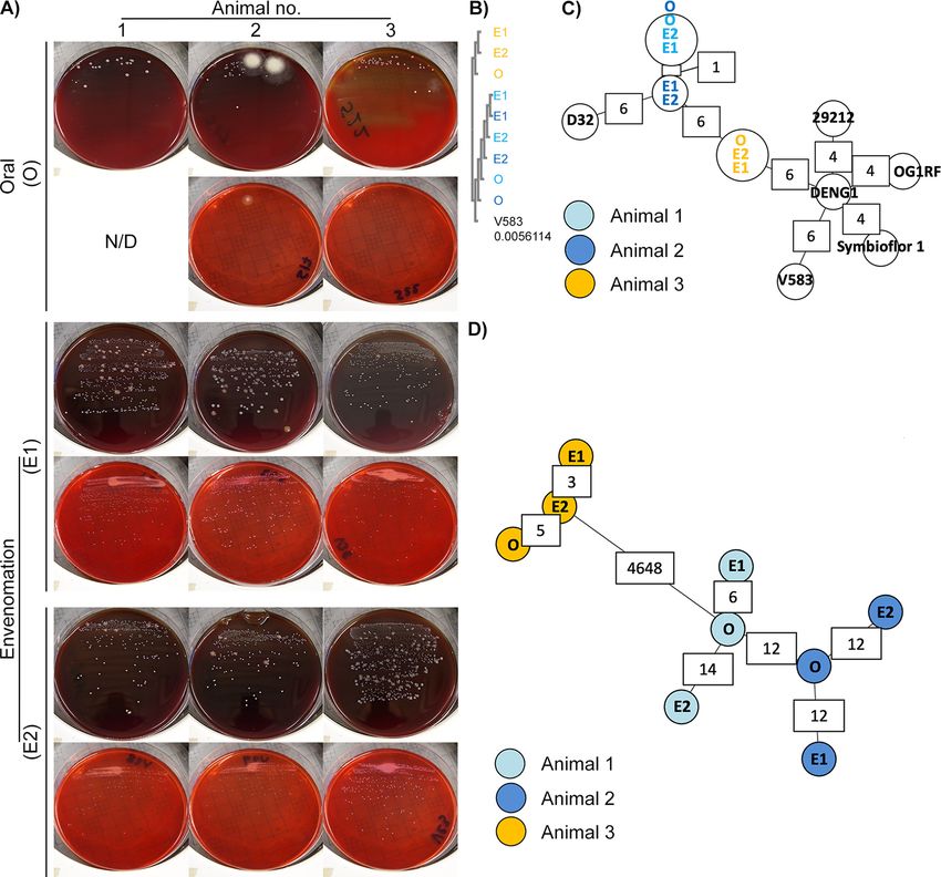
FIG 3 Whole-genome sequencing identifies viable bacteria in N. nigricollis venom as two animal-specific E. faecalis strains. (A) White punctate colonies were
recovered in blood agar (upper panels) and MacConkey agar (lower panels) blind cultures of individual oral swab (O) and two consecutive envenomation
samples (E1 and E2) obtained from three captivity N. nigricollis snakes. N/D, none detected. (B) Blind multiple-sequence alignment (ClustalO followed by
ClustalW phylogeny) of homologous sequences across the de novo assembled genomes against the E. faecalis V583 katA gene (distance to V583 katA
indicated in the V583 track) suggests two sequence groups reflecting the history and housing of the host animals. (C) Blind MST construction based on the
MLST of the N. nigricollis-derived isolates against nine E. faecalis reference genomes again separates samples into two distinct clusters that reflect the
history and housing of the host animals. Partially available allele data are included in this analysis, and instances of allelic differences between nearest
neighbors are annotated in white boxes. (D) Blind complete genome MLST against a custom schema generated using three closely related E. faecalis
reference genomes clusters these isolates by animal of origin (animal 1, light blue; animal 2, dark blue; animal 3, orange). The host animal color scheme
depicted in panel D is also used in panels B and C.
Viable bacteria in N. nigricollis venom are two new E. faecalis sequence types.
Resequencing against putative reference genomes (Table S2) demonstrated less than
6% base alignment across all isolates. Instead, BLASTn analysis of the largest de novo
assembled contigs identified E. faecalis V583 as the closest likely relative (.80% base
alignment, 51.2 coverage). This microbe is considered of mammalian origin and is
usually found in soils, waters, and foodstuffs probably arising from mammalian gastro-
intestinal tracts via feces. This was puzzling given the catalase-positive biochemistry of
May/June 2022 Volume 10 Issue 3 10.1128/spectrum.02408-21 6Bacterial Colonization and Adaptation to Animal Venoms Microbiology Spectrum
TABLE 2 Novel sequence types of E. faecalis recovered from fangs and venoms of N.
nigricollis
Sample Locus
Animal no. Isolate origina Blinding code gdh gyd pstS gki aroE xpt yqiL
1 O S22 22 6 31 13 11 35 8
E1 V36 22 6 31 13 11 35 8
E2 V29 22 6 31 13 11 35 8
2 O S17 22 6 31 13 11 35 8
E1 V31 22 6 31 13 11 35 8b
E2 V28 22 6 31 13 11 35 8c
3 O S3 18 1 New 24 83 47 New
E1 V33 18 1 New 24 83 47 New
E2 V23 18 1 New 24 83 47 New
aO,oral swab sample; E1, envenomation 1; E2, envenomation 2.
bSingle-base-pair
deletion in NGS data not validated by Sanger sequencing.
cHomopolymer single-base extension not validated by Sanger sequencing.
the isolates versus the generally accepted catalase-negative nature of E. faecalis (19),
but was explained by confirming the E. faecalis V583 katA gene, coding for a heme-de-
pendent cytoplasmic catalase (20), among all isolates at 99% identity. Blind multiple-
sequence alignment (MSA) further revealed two katA alleles: one among isolates from
animals 1 and 2 (allele 1) versus another found in animal 3 isolates (allele 2) (Fig. 3B)
differing from the V583 allele by less than 20 single nucleotide polymorphisms (Fig.
S5A). Interestingly, these alleles grouped isolates according to the origin and joint
housing histories of animal 3 (group A) versus animals 1 and 2 (group B).
To explore isolate relationships further, we generated minimum spanning trees
(MSTs) (Fig. 3C) by multilocus sequence typing (MLST) (Table 2), including at core ge-
nome level (cgMLST) (Fig. 3D; Fig. S5B to D) (21, 22). Comparisons to five complete
Enterococcus faecium genomes succeeded only for the gyd (alelles 16 and 19) and adk
(allele 18) loci. In contrast, MLST succeeded for all E. faecalis loci (Table 2), reinforcing
katA allele observations (Fig. 3B) and identifying two novel sequence types featuring
Downloaded from https://journals.asm.org/journal/spectrum on 11 August 2022 by 92.29.140.243.
two new alleles for pstS and yqiL (Fig. S6), as confirmed by Sanger sequencing. MLST
also indicated closer relationships to the E. faecalis strains OG1RF (87.5% 6 1.7% of
OG1RF cgMLST targets), D32 (78.8% 6 2.1%), and DENG1 (77.4% 6 1.5%). Pairwise com-
parisons of the resulting custom cgMLST schema, including 5,041 loci found across all
the N. nigricollis-derived isolates, further reinforced isolate grouping (Fig. 3C), suggesting
two independent E. faecalis strain acquisition events across these three animals.
Pangenomic and experimental evidence of E. faecalis isolate adaptation to
venom. Expanding cgMLST by an additional 3,060 loci found in some E. faecalis iso-
lates (Fig. S5D) identified 290 to 831 allelic differences within each animal. While 80.9%
of alleles varied between the two nearest-neighbor isolates from the two strains,
venom isolates from group B animals were divergent by 7.15 to 10.3% from their oral
isolate counterparts, indicating that genomic changes were occurring with increased
frequency, possibly in response to selective pressure applied by the venom. Given the
well-described plasticity of the E. faecalis genome (OG1RF versus V583, 2.74 versus
3.36 Mb), we next examined mobile element divergence as the potential source of this
adaptation. Detection of the pTEF2 gene repA-2 (plasmid initiator protein) (Table S3)
suggested only plasmid fragments were found in these genomes. As pTEF2 is one of
three E. faecalis V583 plasmids associated with vancomycin resistance (23), we next
confirmed the presence of fragments from the other pTEF plasmids in patterns consist-
ent with the isolate groupings (Table S4), and some sequences absent from group A
secondary envenomation isolates (Fig. 4A). As many of the genomic elements with
high (.95%) sequence identity to these plasmids were known, highly mobile sequen-
ces (e.g., the E. faecalis Bac41 bacteriocin locus) (24), these results suggested their par-
ticipation in the genomic divergence of E. faecalis within each animal. Moreover,
May/June 2022 Volume 10 Issue 3 10.1128/spectrum.02408-21 7Bacterial Colonization and Adaptation to Animal Venoms Microbiology Spectrum
Downloaded from https://journals.asm.org/journal/spectrum on 11 August 2022 by 92.29.140.243.
FIG 4 Comparative genomics of mobile and core genomic chromosomal elements of venom-tolerant E. faecalis. (A) Circos coverage
plots of the vancomycin resistance-associated V583 plasmids pTEF1, pTEF2, and pTEF3 in the E. faecalis isolates obtained from oral,
(Continued on next page)
May/June 2022 Volume 10 Issue 3 10.1128/spectrum.02408-21 8Bacterial Colonization and Adaptation to Animal Venoms Microbiology Spectrum
prompted by the detection of bacteriocin 41, a search for additional bacteriocins
revealed a distinct cadre of genes among the two isolate groups. Thus, group A fea-
tured a class II bacteriocin, MR10A, MR10B, and enterolysin A, while group B shared
carnocyclin A (bacteriocin IId), sonorensin, and a putative bacteriocin ABC transporter.
Such antimicrobial peptide findings indicated that these E. faecalis strains had addi-
tional competitive advantages against other oral/fang bacteria, which could contribute
to their positive selection among the microbiota attempting colonization of N. nigricol-
lis venom.
To ascertain if genes unique to these strains were responsible for adaptation to venom,
we undertook closer examinations of their draft genome assemblies. Group A isolates
shared ;2.9-Mb genomes (2,772 to 2,836 genes), with group B genomes ranging from
3.04 to 3.24 Mb in size (3,128 to 3,282 genes) (Table S3). Including the strains OG1RF and
V583, an E. faecalis pangenome consisting of 5,130 genes was derived, 1,977 of which
formed a core genome that retained venom isolate groupings distinct to OG1RF and V583
(Fig. 4B). After annotation, 235 genes were identified as specific to group A and 321 genes
as specific to group B. Most interestingly, among a set of 45 genes unique to both groups,
UniProt functional annotation indicated known functions for 46.7% of these (Table S5),
with a subset of 15 genes (35.7%) associated with cell wall/membrane integrity and an
additional 4-gene set (9.53%) associated with pathogenic foreign protein and toxin
defense. Significantly, pathway analysis via DAVID using Bacillus subtilis orthologues identi-
fied 8-fold enrichment (P = 0.018) in the two-component system pathway, specifically
genes responsive to cationic antimicrobial peptides, cell wall active antimicrobials, and
bacitracin efflux—mechanisms that are collectively compensatory to the well-established
antimicrobial activity of venoms.
To ascertain the origin of these isolates and expand our search for proteins with
functions opposing venom antimicrobial activity, we extended comparisons to 723
additional E. faecalis genome sequences in GenBank. This increased the pangenome
by 5-fold (26,412 genes), with only 342 genes highly conserved among all 734 strains.
A maximum likelihood tree from the core genome alignment separated the venom-
derived strains into different groups (Fig. 4C): placed within distinct subclades relative
to isolates of diverse origin globally, including animal, environmental, and human sour-
Downloaded from https://journals.asm.org/journal/spectrum on 11 August 2022 by 92.29.140.243.
ces, there was no obvious geographical or other link to the two venom-derived strains
(Fig. S7). Genome comparisons (70% identity threshold) in this wider context identified
42 genes unique to group A and 97 genes unique to group B (Table S6): among pro-
teins with annotated functions, genes with similar functions were also observed in
other E. faecalis strains (Tables S7 and S8) such as S3_02356 encoding a colanic acid
biosynthesis protein, functionally represented by gene ef95_02851 in strain 7330112-3.
The relevance of such genes in adaptation or tolerance to venom will require further
molecular characterization in the future.
We next looked for genes whose functions could counteract N. nigricollis venom
components, such as phospholipase A2 (PLA2), L-amino oxidase, and three-finger pep-
tides (PDB ID 3FTX) that facilitate disruption of membrane integrity in bacteria (25–29).
Putative candidates included homologs to acyltransferase-acyl carrier protein synthe-
tase (Aas), which was previously reported to protect bacterial cell envelope from
human PLA2 in Gram-negative bacteria (30), and homologs to cell wall (dltA) and cell
membrane (mprF) polyanions previously associated with sensitivity to human PLA2 in
FIG 4 Legend (Continued)
envenomation 1 (E1), and envenomation 2 (E2) samples from three N. nigricollis individuals reinforce the two sequence type
groupings and highlight within-animal variation (green arcs) indicative of sample-specific variation (lack of reads) across E2 samples
in animals 1 and 2. The central plot for each plasmid and animal reflects GC content. All data are represented in 50-nt blocks. (B)
Blind maximum likelihood tree of the core genomic alignments for the 6 N. nigricollis-derived E. faecalis isolates against the V583 and
OG1RF reference strains, with color coding referring to the origin of the isolates: light blue, animal 1; dark blue, animal 2; yellow,
animal 3. (C) Maximum likelihood tree from concatenated nucleotide sequence alignment of 865 core genes (381,319 bp) from 734
genomes after removing the sites with gaps. The best-fit GTR1I1G4 substitution model was used with 100,00 ultrafast bootstraps
and SH-aLRT tests. The tree was rerooted on the longest branch, and branch lengths of ,0.001 were collapsed. The scale bar shows
number of nucleotide substitutions per site. Branches in red, blue, purple, and green show group A, group B, and clades containing
strains V853 and OG1RF, respectively.
May/June 2022 Volume 10 Issue 3 10.1128/spectrum.02408-21 9Bacterial Colonization and Adaptation to Animal Venoms Microbiology Spectrum
Staphylococcus aureus (31). Interestingly, the mprF gene appeared disrupted into two
smaller proteins in one group B E. faecalis isolate (V31_01061 and V31_01062) (Table
S9). Three copies of sortase family proteins were also detected: sortase A has been
associated with the resistance to human PLA2 in Streptococcus pyogenes (32). A further
21 genes involved in oxidative stress response and encoding various antioxidative
enzymes, such as superoxide dismutase, catalase, and those associated with glutathi-
one metabolism, which would counteract reactive oxygen species produced by venom
L-amino acid oxidase activity (25), were also found: these proteins also contribute to
the virulence of E. faecalis strains (Table S9) (33–35).
Taken together, genome analyses of these E. faecalis strains indicated they were
well equipped to survive the stress imposed by N. nigricollis venom, a tenet supported
experimentally, since V583 growth was dose-dependently inhibited at a minimum con-
centration of 11.7 mg/mL (95% CI, 9.36 to 14.6 mg/mL) and noninhibitory concentra-
tion of 2.78 mg/mL (95% CI, 2.21 to 3.50 mg/mL) of filter-sterilized, freeze-dried N. nigri-
collis venom in brain heart infusion broth. In stark contrast, all the venom-derived strains
exhibited ,30% growth inhibition, even at concentrations of freeze-dried venom of 50
mg/mL (i.e., ;4 lower than the 208 mg/mL concentration of fresh N. nigricollis venom),
resulting in ambiguous noninhibitory concentration ranges of 25.2 to 44.0 mg/mL (no
CIs calculable), with the group A strain V33 exhibiting no susceptibility to the inhibitory
effects of venom (Fig. S8).
Projecting primary infection clinical risk from venom-tolerant E. faecalis isolates.
Since these viable E. faecalis strains could potentially infect envenomation wounds, we
next looked for known resistance determinants that might facilitate opportunistic pri-
mary infection. None of these strains had any acquired resistance genes to any antibiotic
classes (Table S10), although all isolates featured lsaA (Fig. S9), an intrinsic streptogramin
resistance gene (36): Since horizontally acquired genes are largely responsible for resist-
ance to vancomycin, aminoglycosides, macrolides, and tetracycline in Enterococcus (37),
these strains were considered susceptible to drugs in each of these drug classes. In addi-
tion, the absence of known resistance-associated mutations in gyrA (DNA gyrase), parC
(DNA topoisomerase), and the 23S rRNA genes also increased the likelihood of suscepti-
bility to oxazolidinones and fluoroquinolones. Thus, several available antimicrobials
Downloaded from https://journals.asm.org/journal/spectrum on 11 August 2022 by 92.29.140.243.
would likely be effective in treating infections caused by these strains of E. faecalis.
However, a gene related to macrolide export (macB5) was detected in the venom-toler-
ant strains (Table S5), and several virulence genes (38, 39) were also identified. Among
all isolates, these included conjugative plasmid transfer pheromone genes associated
with virulence (cCF10, cOB1, cpd, cad, camE), endocarditis and biofilm formation-associ-
ated pilus subunit genes (ebpA, ebpB, ebpC, srt, pil), a biofilm on plastic operon gene
(bop), quorum sensing (fsrA, fsrB, fsrC) and virulence-associated Fsr locus genes, such as
gelatinase (gelE) and serine protease E (sprE), and other important virulence genes, such
as those coding for hyaluronidases A and B (hylA, hylB), thiol peroxidase for oxidative
stress resistance (tpx), adhesin to collagen of E. faecalis (ace), endocarditis-specific anti-
gen/E. faecalis antigen A (efaA), and enterococcal leucine-rich protein A (elrA), which pre-
vents macrophage chemotaxis to E. faecalis and endocytosis. Furthermore, all six group
B isolates had the aggregation substance (agg) gene, which enhances macrophage
escape by suppressing respiratory burst. Therefore, despite the absence of cytolysin (e.g.,
cylA, cylB, cylM) and glycopeptide (e.g., vanA, vanC) resistance genes, both sequence types
appeared well equipped to establish infections in human patients following envenomation.
DISCUSSION
In contrast to the generally held view that venoms are both antimicrobial (1–4) and
sterile (6, 7, 40), despite contrasting reports since the 1940s (41), we show that microorgan-
isms can viably colonize venoms of both vertebrates and invertebrates. Moreover, signifi-
cant adaptation appears to be necessary in genes that counter the mechanisms of action
of known, venom-derived antimicrobial peptides and enzymes (4, 5) to attain resistance to
venom. Although we documented this in multiple isolates of two independently acquired
May/June 2022 Volume 10 Issue 3 10.1128/spectrum.02408-21 10Bacterial Colonization and Adaptation to Animal Venoms Microbiology Spectrum
E. faecalis strains, adaptation appeared to occur in parallel within each of the 3 black-
necked cobras from whence the isolates were obtained. It is unclear to what extent this
form of parallel convergent evolution extends beyond N. nigricollis and Enterococcus spp.
or other antimicrobial resistance mechanisms, such as antibiotic resistance genes found on
mobile genetic elements and working against last-resort antibiotics (42, 43). This work
therefore adds to the body of evidence (44) supporting further scrutiny of host-microbe
interactions in the venomous microenvironment in understanding microbial adaptation
mechanisms to extreme environmental challenges.
Identification of E. faecalis as the most prevalent culturable microbe across our European
N. nigricollis venom samples strikingly reflects three independent clinical reports across
Africa and Asia that this nonsporulating microbe is the most common Gram-positive infec-
tion cultured from infected envenomation wounds (8–10). Likewise, E. faecalis isolates were
the most common aerobic Gram-positive isolates in N. naja oral swabs (n = 6) (45).
Postenvenomation cellulitis and dermatitis, presumed bacterial in nature, were additionally
observed in 25% of a 16-case series of Steatoda nobilis (false widow spider) envenomations
in the United Kingdom and Ireland: one of these required intravenous penicillin and fluclox-
acillin treatment after hospital admission (46). S. nobilis chelicerae were previously found to
harbor 11 bacterial taxa and 22 separate bacterial species, including class 2 pathogens; 3 of
these 22 species showed multidrug resistance (47). Although explicit genomic evidence
connecting venom microbes to envenomation infection remains elusive, in an experimental
rabbit model of dermonecrosis (48) caused by Loxosceles intermedia (recluse spider) venom,
Clostridium perfringens recovered from the spider fang and venom enhanced disease symp-
toms. Stenotrophomonas-like bacteria were also found to dominate cone snail venom micro-
biomes (49), indicating that microbial venom adaptation may extend well beyond snakes,
spiders, scorpions, and snails. Furthermore, building upon the few instances of polymicro-
bial infection reported clinically (8–10), the reports on L. intermedia (48) and Conus (49) and
herein suggest that diverse microbes effectively cocolonize venom glands in a host-species-
specific manner, and thus envenomation wounds. Envenomation wound management (40)
should therefore extend beyond simply managing the severe tissue damage and necrosis
that might be caused by venomous bites to include clinical microbiology on envenomation
wounds upon presentation. This would be particularly relevant to individuals immunocom-
Downloaded from https://journals.asm.org/journal/spectrum on 11 August 2022 by 92.29.140.243.
promised through disease or malnutrition (e.g., in developing nations, where envenomation
incidence rates are high) or to children on the basis of venom/CFU dose by body weight.
Yet, common microbial diagnostic methods relevant to resource-limited settings
have mistaken E. faecalis for Staphylococcus, which could lead to unfavorable clinical
decision making. Although many of the same antibiotics, including vancomycin and
linezolid, would be considered for treatment of both staphylococci and enterococci,
there are some potential differences. For instance, oxacillin is often employed as a first-
line agent to treat Staphylococcus (50). This drug is not effective against enterococci, as
the use of penicillins for E. faecalis infections would typically involve ampicillin, usually
in combination with an aminoglycoside (51). In addition, cephalosporins such as cefo-
taxime are considered second-line therapies for coagulase-negative staphylococci such
as Staphylococcus epidermidis (52). However, enterococci are intrinsically resistant to
this class of drugs, and their prevalence in the gut tends to increase in response to
cephalosporin therapy (53). Thus, while the E. faecalis strains in this study did not have
any known acquired resistance determinants, if they were to cause infections, ensuring
their proper identification would be critical to issuing correct treatment and achieving
positive clinical outcomes.
It is unclear at present how frequent misidentification events might be. At least one
retrospective study reported higher incidence of Staphylococcus spp. in envenomation
wounds (12), and Blaylock’s seminal snake oral microbiota studies also reported
Proteus and Staphylococcus (14): both studies relied on the same methods we used in
this study and which misidentified the pathogen. Our results therefore further support
use of PCR/sequencing methods as they become more relevant to resource-limited set-
tings (54) and suited to the point of need (55), in line with World Health Organization
May/June 2022 Volume 10 Issue 3 10.1128/spectrum.02408-21 11Bacterial Colonization and Adaptation to Animal Venoms Microbiology Spectrum
ASSURED criteria. Understanding the sensitivity of these methods will be crucial in their
reliable implementation in envenomation care. It is therefore noteworthy that despite
the limited biomass levels in these samples, species-level OTU analysis on MG-RAST (56)
correctly identified E. faecalis as one of the principle aerobic isolates in N. nigricollis
venom. Thus, a simple phylogenetic or metagenomic approach, combined with local
herpetogeography knowledge, could quickly and accurately inform clinical action
regarding antivenom administration without relying on descriptions or capture of the
offending animal or unreliable antibody-based venom identification kits (57).
To conclude, we provide evidence that vertebrate and invertebrate animal venoms
host diverse, viable microbiota, with isolates genetically adapted to venom antimicro-
bials of medical interest against MDR. These results challenge perceptions on the sterility
of venom and absence of primary infection risk upon envenomation, pointing to modern
nucleic acid technologies to better inform envenomation care and antibiotic use.
MATERIALS AND METHODS
Animals and sampling. All samples analyzed in this study were provided by Venomtech, Ltd., with
the exception of freeze-dried B. arietans venom (Latoxan, Portes les Valence, France) and air-dried field-
collected samples of B. arietans venom collected in South Africa (Table 1). Briefly, captive animals were
housed in 2-m by 1-m wooden, glass-fronted vivaria with a large hide, thermal gradient, and water ad
libitum. All procedures for venom collection and swabbing were approved as unregulated under the
Animals (Scientific Procedures) Act 1976. Venom was collected by standard techniques; briefly snakes
were restrained behind the head and presented to a collection vessel. Snakes freely bit into the vessel
until envenomation was observed. Each snake was presented to two sterile collection vessels in succes-
sion: one for the first envenomation with potential fang plug and the other for the second flow (labeled
E1 and E2, respectively). While the snake was positioned over the second vessel, the oral cavity was
swabbed with a sterile swab with individual collection tubes (an invasive sterile swab with transport me-
dium) (DeltaLab; VWR, Lutterworth, United Kingdom). The venom collection vessels were clear, sterile,
125 mL polypropylene containers (Thermo Fisher Scientific, Ltd., Paisley, United Kingdom) covered by 2-
by 9 cm2 pieces of Parafilm stretched to fit (Thermo Fisher Scientific, Ltd.). The collection vessel was
secured to a bench during collection. After collection, aseptically dispensed aliquots were stored in indi-
vidual 1 mL sterile, DNA-free, polypropylene collection tubes (FluidX, Ltd., Nether Alderley, United
Kingdom), at 280°C. Samples collected in the field were from wild puff adders sampled as part of a pre-
vious phylogeographic study (58). Venom samples were collected using a method similar to that
described for captive animals, except that the entire venom sample was collected in a single collection
vessel. Samples were lyophilized by storing ,100 m L venom aliquots in a vacuum-sealed container that
was half-filled with silica gel for 1 to 2 days at room temperature. Following drying, venom samples
Downloaded from https://journals.asm.org/journal/spectrum on 11 August 2022 by 92.29.140.243.
were stored in a refrigerator at 5°C. Snake venom composition and its constituent proteins have been
shown to be remarkably resistant to alterations in storage conditions (59) and to degradation during
long-term storage (60). We have further observed that our air-dried, field-collected samples for rattle-
snake venoms show proteomic profiles identical to those obtained from other research groups using
freeze-dried venoms (61, 62). We thus reasoned that variation in storage conditions in this study would
be unlikely to have substantially altered the basic properties of the venom substrate available for micro-
bial growth.
Lasiodora parahybana and Poecilotheria regalis were housed in 5- and 8-L polypropylene boxes (Really
Useful Products, Ltd., Normanton, United Kingdom), respectively, with moist vermiculite (Peregrine Livefoods,
Ltd., Ongar, United Kingdom), a plastic hide, and a 5 cm water bowl. Arachnids were anaesthetized with a ris-
ing concentration of carbon dioxide, the fangs were swabbed with a sterile swab, which was then placed in
an individual 1 mL sterile, DNA-free, polypropylene collection tube (FluidX, Ltd.), and venom was subsequently
collected from arachnids by electrical stimulation. All samples were stored at 280°C. The same transport
swabs (VWR) as those used for snakes were also used for invertebrate oral/fang swabbing. Samples were
stored at 4°C and cultured within 24 h of collection.
Microbial culture. Aerobic microbial viability was determined by plating swabs or aliquoting 10 m L
volumes of venom samples onto plates containing oxalated whole-horse-blood agar, MacConkey agar,
or mannitol salt agar (Thermo Fisher Scientific) and incubating them at 30°C for 72 h. Biochemical isolate
identification was undertaken using API strips (20E, 20NE, and Staph), interpreted via the APIWEB inter-
face (bioMérieux, Basingstoke, United Kingdom). All isolates were stored on beads at 280°C at the
University of Westminster microbial isolate library. N. nigricollis subculture was performed by restoring
cryogenically stored bacteria on lysogeny broth agar (Thermo Fisher Scientific), which were grown for
48 h at 30°C, followed by single-colony overnight culture in lysogeny broth (Thermo Fisher Scientific)
using aerated culture (300 rpm). MICs and noninhibitory concentrations were determined by broth
microdilution assays (63) in brain heart infusion medium by measuring absorbance at the optical density
at 600 nm (OD600) on a Tecan Spark Cyto 96 plate reader (Tecan, Männedorf, Switzerland) and computed
in GraphPad Prism v.9.2.0 according to Lambert and Pearson (64). All bacterial agar and broth materials
were purchased from Formedium, Ltd. (Norfolk, United Kingdom).
DNA extraction. Neat venom samples or samples diluted in 18 MX water previously confirmed as
bacterial DNA free by 16S PCR were subjected to DNA extraction using TRIzol, PureLink genomic DNA
May/June 2022 Volume 10 Issue 3 10.1128/spectrum.02408-21 12Bacterial Colonization and Adaptation to Animal Venoms Microbiology Spectrum
kits, or MagMAX cell-free DNA kits (Thermo Fisher Scientific) according to the manufacturer’s instruc-
tions. For combined extraction of Gram-positive and Gram-negative bacteria from liquid samples,
diluted samples were split into equal volumes and processed according to the manufacturer’s Gram
wall-specific lysis protocols, with lysates combined prior to DNA binding onto columns by simple admix-
ture. DNA content was then analyzed by Nanodrop (Thermo Fisher Scientific) spectrophotometry, and
purified material was stored at 280°C until further analysis.
16S phylogenetic library preparation and sequencing. For short amplicon library preparation, the
hypervariable V3 region of the 16S rRNA gene was amplified from 20 ng of DNA using the primers
59-CCTACGGGAGGCAGCAG-39 and 59-ATTACCGCGGCTGCTGG-39 (Integrated DNA Technologies BVBA,
Leuven, Belgium) (18), 1 U Platinum PCR SuperMix, high fidelity (Thermo Fisher Scientific), and 10 m M
primer mix. The reaction mixtures were incubated at 94°C for 5 min, followed by 30 cycles of 30 s at
94°C, 30 s at 55°C, and 1 min at 72°C and then a final elongation at 72°C for 10 min using a Techne
Prime thermal cycler (ColePalmer, Staffordshire, United Kingdom). PCR products (193 bp) were con-
firmed by 2% (wt/vol) agarose gel electrophoresis in TAE (Tris-acetate-EDTA) buffer (Thermo Fisher
Scientific).
Next-generation sequencing (NGS) library preparation was carried out using the Ion Plus fragment
library kit according to the manufacturer’s instructions (Rev. 3; Thermo Fisher Scientific), except that
reaction mixtures were reduced to 1/5 volumes. Pooled libraries were diluted to ;26 pM for templating
on the Ion OneTouch 2 system (Thermo Fisher Scientific) using the Ion PGM Template OT2 200 v.2 kit
according to the manufacturer’s instructions (Rev. B; Thermo Fisher Scientific). Templated samples were
sequenced on the Ion Torrent Personal Genome Machine (PGM) (Thermo Fisher Scientific) system on a
single Ion 318 Chip (ThermoFisher Scientific) using the Ion PGM 200 sequencing kit according to the
manufacturer’s instructions (Rev G.; Thermo Fisher Scientific).
Whole-genome sequencing. DNA extracted from cultured isolates was mechanically sheared using
the Covaris S220 focused ultrasonicator (Covaris, Brighton, United Kingdom). NGS libraries were gener-
ated using the NEBNext fast DNA library prep set for Ion Torrent (New England Biolabs, Hitchin, United
Kingdom). Pooled samples were size selected with the LabChip XT (LabChip XT DNA 300 assay kit;
PerkinElmer, Seer Green, United Kingdom) and diluted to 26 pM for templating with the Ion OneTouch 2
system using the Ion PGM Template OT2 200 kit. Templated samples were sequenced on the Ion PGM
using the Ion PGM Sequencing 200 v.2 reagent kit (Thermo Fisher Scientific) and Ion 318 v.2 Ion Chip
(Thermo Fisher Scientific).
Bioinformatic analyses. Raw Ion Torrent sequencing data reads were quality controlled and demul-
tiplexed using the standard Ion Server v.4.0 pipeline (Thermo Fisher Scientific). Referenced and de novo
assemblies were carried out using TMAP v.4.0 and the SPAdes plugin in the Ion Server. Phylogenetic
data analyses were carried out after independent data deposition and curation on the MG-RAST v.3.0 pipe-
line (56) (project IDs MGP5177 and MGP5617), which uses a BLAST approach, and the EBI-METAGENOMICS
v.1 pipeline (65) (project ID ERP004004), which uses a hidden Markov model approach. Raw 16S sequenc-
ing reads were deposited in the European Nucleotide Archive (accession no. PRJEB4693). Quality control
for both resources included length and quality filtering followed by a dereplication step in which sequen-
ces with identical 50 nucleotides (nt) in 59 positions were clustered together. MG-RAST taxonomy annota-
Downloaded from https://journals.asm.org/journal/spectrum on 11 August 2022 by 92.29.140.243.
tion involved RNA identification using VSearch, and assignments using a custom database generated by
90% identity clustering of the SILVA, GreenGenes, and RDP prokaryotic databases. EBI-METAGENOMICS
identified rRNA using hidden Markov models present in the RDP databases and assigned taxonomy using
QIIME and the GreenGenes prokaryotic database.
For postprocessing analyses, the EBI-curated data set was analyzed using MEGAN v.5.5.3 (66). Classical
multilocus sequence typing (http://efaecalis.mlst.net/) and cgMLST (21, 22) were carried out using Ridom
SeqSphere1 v.4.0 running on a 2-core, 10-GB RAM, 500-GB hard disk Biolinux v.8.0 installation on a
VirtualBox virtual machine instance on a 16-GB RAM, 1-TB hard disk Apple iMac. Extended cgMLST analysis
to include partially detected loci excluded loci annotated as “failed” due to sequencing error, suggesting
genuine E. faecalis genomic divergence occurring within each animal. Plasmid detection was carried out
using the PlasmidFinder v.1.3 server (67), followed by NCBI BLASTn analysis to detect shorter fragments
(e.g., the same 398-nt fragment of repA-2 in animal 3 isolates (,40% of the full-length gene) at 90.1% iden-
tity to the plasmid-borne reference sequence). Single-gene comparisons and multiple-sequence analyses
were carried out using TCoffe and MView on the EMBL-EBI server, with base conservation visualized by
BoxShade v.3.3.1 on mobyle.pasteur.fr. Genome-level plasmid coverage analyses were carried out by NCBI
BLASTn, and comparisons were visualized using Circos v.0.69-4.
The sequencing reads were assembled using SPAdes v.3.9.0 (68), and the draft assemblies were
annotated using Prokka (69) before NCBI deposition (BioProject accession no. PRJNA415175). The ge-
nome sequences of E. faecalis strains V583 and OG1RF (accession no. NC_004668.1 and NC_017316.1,
respectively) as well as 723 other E. faecalis strains were obtained from GenBank and were reannotated
using Prokka to have an equivalence of annotation for comparative analyses. The genomes were com-
pared using the program Roary, with a protein similarity threshold of 70% (70, 71). A maximum likeli-
hood tree was constructed from the core genomic alignment using IQ-Tree (72), with 100,000 ultrafast
bootstraps and 100,000 SH-aLRT tests. The tree was visualized using Interactive Tree Of Life (iTOL) (73).
To identify acquired resistance genes, nucleotide BLAST analysis was performed on the ResFinder
(74) and NCBI (https://www.ncbi.nlm.nih.gov/pathogens/) resistance gene databases, using cutoffs of
50% length and 85% identity to known resistance determinants. Additional BLAST analysis was per-
formed to identify single nucleotide polymorphisms in the quinolone resistance-determining region
(QRDR) of gyrA and parC (75). Additional mutational analysis was performed on region V of the 23S
rRNA-encoding genes (76). Virulence genes were identified using a combination of VirulenceFinder 2.0
May/June 2022 Volume 10 Issue 3 10.1128/spectrum.02408-21 13Bacterial Colonization and Adaptation to Animal Venoms Microbiology Spectrum
(https://cge.cbs.dtu.dk/services/VirulenceFinder/), with default parameters of 60% length and 90% iden-
tity to known Enterococcus virulence genes (77–79), and manual BLAST, with an E value cutoff of 1025.
Bacteriocins and ribosomally synthesized and posttranslationally modified peptides were mined using
BAGEL4 (http://bagel4.molgenrug.nl) (80).
BLASTp was performed in Ensembl Bacteria (release 38), against E. faecalis V583 and E. faecalis
(GCA_000763645) to obtain further gene IDs from significant matches. Bacillus subtilis orthologue gene
IDs were collated as this species is the closest relative to E. faecalis (VetBact.org), with the most compre-
hensive genome annotation required for Gene Ontology and KEGG pathway analysis. From the 45 genes
unique to venom isolates, useable B. subtilis gene IDs were obtained for 20, of which 18 of these were
successfully converted to ENTREZ gene IDs using the functional annotation tool (DAVID Bioinformatics
resource 6.8) (81, 82), selecting B. subtilis as the background species.
Data availability. Phylogenetic data were deposited in the MG-RAST (project IDs MGP5177 and
MGP5617) and EBI-METAGENOMICS servers (project ID ERP004004). Raw 16S sequencing reads were de-
posited in the European Nucleotide Archive (accession no. PRJEB4693). Annotated draft E. faecalis ge-
nome assemblies were deposited in NCBI (BioProject accession no. PRJNA415175).
SUPPLEMENTAL MATERIAL
Supplemental material is available online only.
SUPPLEMENTAL FILE 1, PDF file, 2.4 MB.
SUPPLEMENTAL FILE 2, XLSX file, 1 MB.
ACKNOWLEDGMENTS
We thank Pamela Greenwell and Caroline Smith for invaluable input on nonstandard
DNA extraction methodology options suited to unusual samples, Patrick Kimmit for
input on microbial characterization, and Peter Gibbens for housing and venom
collection from captive N. nigricollis and B. arietans.
This work was funded by the University of Westminster, University of Northumbria,
and Venomtech, Ltd. The views expressed in this article are those of the authors and do
not necessarily reflect the official policy of the Department of Health and Human
Services, the U.S. Food and Drug Administration, or the U.S. Government.
M.M.G.L. and T.D.L. sampled and C.T. and S.T. prepared the library of captive animal
venoms. W.W. and A.B. collected and prepared the wild snake samples. E.E., J.D.T., and
S.A.M. optimized and performed the DNA extractions and 16S PCR. J.D.T. and E.E.
performed the preliminary and main study library preps and next-generation
sequencing experiments, respectively. A.D., H.D., L.A.S.S., and S.A.M. performed the
Downloaded from https://journals.asm.org/journal/spectrum on 11 August 2022 by 92.29.140.243.
phylogenetic data quality control, curation, and analysis. K.F.R. and S.A.M. performed
the microbial cultures and biochemical characterization. M.K.-V. and L.U. grew the E.
faecalis isolates and performed the whole-genome sequencing. M.K.-V., K.W., and S.A.M.
performed the E. faecalis isolate genomic characterization and MLST1 analysis. G.H.T.
performed E. faecalis resistome analysis. V.S. performed the E. faecalis isolate
pangenome data reduction, and S.T. identified the venom resistance Gene Ontology
subset. G.H.T., V.S., and S.A.M. identified the virulence genes, and S.A.M. identified the
bacteriocin content in E. faecalis isolates. S.A.M. conceived the study and designed
experiments with S.T. S.D. performed the MIC/NIC curve assays. All authors contributed
equally to the overall interpretation of the data set and manuscript preparation.
We declare no conflict of interest.
REFERENCES
1. Xie JP, Yue J, Xiong YL, Wang WY, Yu SQ, Wang HH. 2003. In vitro activities 5. Perumal Samy R, Stiles BG, Franco OL, Sethi G, Lim LHK. 2017. Animal ven-
of small peptides from snake venom against clinical isolates of drug-re- oms as antimicrobial agents. Biochem Pharmacol 134:127–138. https://
sistant Mycobacterium tuberculosis. Int J Antimicrob Agents 22:172–174. doi.org/10.1016/j.bcp.2017.03.005.
https://doi.org/10.1016/s0924-8579(03)00110-9. 6. Talan DA, Citron DM, Overturf GD, Singer B, Froman P, Goldstein EJ. 1991.
2. Glaser HSR. 1948. Bactericidal activity of Crotalus venom in vitro. Copeia Antibacterial activity of crotalid venoms against oral snake flora and other
1948:245. https://doi.org/10.2307/1438710. clinical bacteria. J Infect Dis 164:195–198. https://doi.org/10.1093/infdis/
3. Samy R, Gopalakrishnakone P, Satyanarayanajois S, Stiles B, Chow V. 2013. Snake 164.1.195.
venom proteins and peptides as novel antibiotics against microbial infections. 7. Powers DW. 2005. Stings and bites: what to do about envenomation inju-
Curr Proteomics 10:10–28. https://doi.org/10.2174/1570164611310010003. ries. Emerg Med Serv 34:67, 69–75.
4. Stocker JF, Traynor JR. 1986. The action of various venoms on Escherichia 8. Wagener M, Naidoo M, Aldous C. 2017. Wound infection secondary to
coli. J Appl Bacteriol 61:383–388. https://doi.org/10.1111/j.1365-2672.1986 snakebite. S Afr Med J 107:315–319. https://doi.org/10.7196/SAMJ.2017
.tb04300.x. .v107i4.12084.
May/June 2022 Volume 10 Issue 3 10.1128/spectrum.02408-21 14You can also read


