Berkeley Optometry UNIVERSITY OF CALIFORNIA - VOL. 1, NO. 1 | FALL 2008 - University of ...
←
→
Page content transcription
If your browser does not render page correctly, please read the page content below
UNIVERSITY OF CALIFORNIA                                                VOL. 1, NO. 1   | FALL 2008

Berkeley Optometry                                     magazine

                      ADVANCING EDUCATION AND RESEARCH AT BERKELEY OPTOMETRY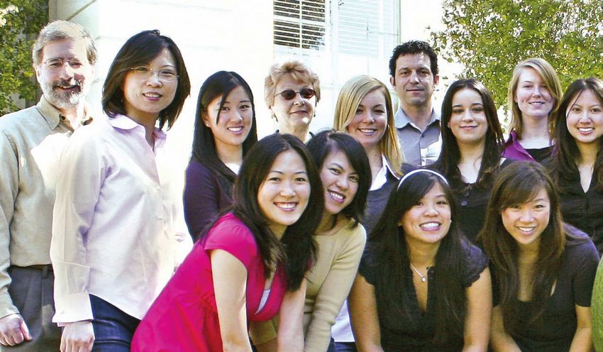
EXECUTIVE EDITOR
                 Lawrence Thal

                MANAGING EDITOR
                                                                                                dean’s note
               Jennifer C. Martin

                    EDITOR
                Barbara Gordon

                ASSISTANT EDITOR
                 John Fiorillo

            EXECUTIVE ART DIRECTOR
                 Molly Sharp

                  ART DIRECTOR
                  John Fiorillo

                PHOTOGRAPHY
          John Fiorillo and Ken Huie
                                                        T           HE FUNDAMENTAL MISSIONS OF THE UNIVERSITY
                                                        of California are teaching, research, and public service. Berkeley Optometry
                                                        excels in all of these.
             PRODUCTION MANAGER                            Our greatest responsibility as educators in a public university is to foster
                 Molly Sharp                            graduates who are capable of providing for the public good and have the com-
                                                        mitment to do so. The message we strive to impart is that our graduates have a
           WRITERS/CONTRIBUTORS
   John Fiorillo, Meng Lin, Austin Roorda,
                                                        responsibility toward humankind as well as to their profession. That responsi-
             Christine Wildsoet                         bility is embodied within the Optometric Oath:

                   DESIGN
                                                              “I will provide professional care for those who seek my services, with
              ContentWorks, Inc.
                                                               concern, with compassion, and with due regard for their human
                                                               rights and dignity. I will place the treatment of those who seek my
Published by University of California, Berkeley,               care above personal gain and strive to see that none shall lack for
   School of Optometry Office of External                      proper care. I will do my utmost to serve my community, my coun-
       Relations and Professional Affairs
                                                               try, and humankind as a citizen as well as an optometrist.”
  Phone: 510-642-2622, Fax: 510-643-6583
         Send comments and letters to:
                                                           These are not just words that are recited at meetings or graduations; they
            optodev@berkeley.edu
                                                        represent a philosophy that should be embodied within every public insti-
          Submit your class notes to:
                                                                                            tution. Our students come here not only
            optodev@berkeley.edu
                                                                                            because they are smart; they come because
     Send change of address or e-mail to:                                                   they are passionate about helping others.
           jmichael@berkeley.edu
                                                                                            Whether they are native or foreign born, priv-
 Submit School of Optometry gifts on-line at:                                               ileged or disadvantaged, have suffered per-
http://givetocal.berkeley.edu/makeagift/optometry/                                          sonal hardship or not, our charge is to inspire
                   or mail to:                                                              that sense of responsibility toward serving
           Berkeley Optometry Fund
                                                                                            mankind. I believe we do a good job of that.
            302 Minor Hall # 2020                                                           As I travel throughout the country and have
           Berkeley, CA 94720-2020                                                          the opportunity to talk to our graduates, I
                 510-642-2643                                                               am proud of what they do as professionals,
                  Fall 2008                                                                 as members of their communities, and as
           Volume 1, Number 1                                                               parents and grandparents helping to prepare
© 2008 Regents of the University of California
                                                        DENNIS LEVI, OD, PHD, FAAO
                                                                                            future generations.
  Berkeley Optometry is a trademark of the              Dean, School of Optometry              At Berkeley Optometry, we continue
      University of California, Berkeley,                                                   to make an impact on the world through
            School of Optometry.                        ground-breaking optometric research and by reaching out to provide quality
         Not printed at state expense.                  eye care. I hope that will be evident as you read this and future issues of Berkeley
                                                        Optometry Magazine. I look forward to hearing from you and always welcome
 Front cover photos courtesy of John Fiorillo,
                                                        your thoughts at dlevi@berkeley.edu.
    Ken Huie, John Quick, David Schmitz,
               Peg Skorpinski.

  This magazine is printed on recycled-content paper.
UNIVERSITY OF CALIFORNIA                                                                                      VOL. 1, NO. 1     | FALL 2008

Berkeley Optometry                                                             magazine
                         in this issue
                                               fea t u res
                                               3   THANKS TO BERKELEY . . .
                                                   Facts and statistics that illustrate the outstanding institution your alma mater has become.

                                               4   THE EARLY YEARS OF BERKELEY OPTOMETRY By John Fiorillo
                                                   Over a century ago, optometrists inspired by the profession’s potential launched the effort
                                                   that resulted in the birth of Berkeley Optometry. Here’s a look at the people and events
                                                   that shaped our school.

                                              12   NEW VIEWS OF THE HUMAN RETINA By Austin Roorda
                                         4         How the adaptive-optics scanning laser ophthalmoscope (AOSLO) sheds new light on
                                                   optical disorders and potential treatments.

                                              14   RESEARCH ON MYOPIA By Christine Wildsoet
                                                   Studying the causes and progression of myopia is opening up new directions for treatment.

                                              16   BERKELEY OPTOMETRY’S CLINICAL RESEARCH CENTER By Meng Lin
                                                   We’re proud to present the stunning accomplishments of Berkeley Optometry’s CRC.

                                              23   BUILDING FOR EXCELLENCE AND LEADERSHIP
                                                   Berkeley Optometry’s vision for the future.

                                         12   departments
                                               2   LETTER FROM THE EDITOR
                                               9   STUDENT FOCUS
                                                   A glimpse of our professional future.

                                              10   NEWS & NOTES
                                              19   BERKELEY OPTOMETRY—A FISCAL SNAPSHOT
                                                   Ever wonder where our school’s funds come from and go to?

                                              20   FACULTY SPOTLIGHT
                                         16        A look at the newest faculty educating our profession.

                                              22   BERKELEY OPTOMETRY ALUMNI BENEFITS
                                                   A trip to China, free legal service consultations, and more.

                                              24   CLASS NOTES
                                                   We know you’ve been wondering what your classmates and colleagues have been up to…

                                              27   SCHOOL SUPPORT
                                                   A complete listing and recognition of our many generous supporters.
 Noll & Tam Architects

                                              39   MEET TAMMY, DIRECTOR OF PHILANTHROPY
                                         23        Getting to know Tammy, an important pillar of our support.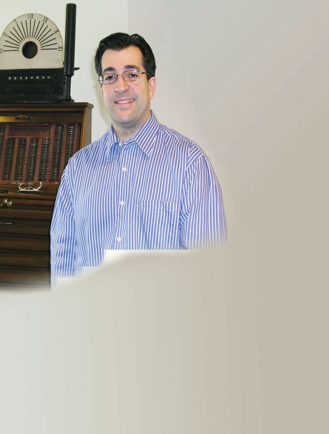
letter from the editor

                  W                ELCOME TO OUR NEW MAGAZINE, Berkeley Optometry.
                  Inside you will discover how your school is touching countless lives—
                  students, patients, educators, and colleagues. We are all extremely grate-
                  ful for the support from alumni and friends of the School of Optometry.
                  We thank you and encourage your continued support of the work we do
                  in preparing future generations of practitioners and educators.                    LAWRENCE THAL, OD, MBA, FAAO
                                                                                                     Assistant Dean,
                        Early last year we surveyed our graduates to determine what it is you        External Relations and Development
                  want from our school in order to better serve your needs. (For lack of bet- School of Optometry
                                                                                                     LarryThal@Berkeley.edu
                  ter contact information, we used the California State Board of Optometry           510-643-0786
                  mailing list for our graduates who maintain a California license. We realize
                  this misses many of you, so please e-mail us or use the insert in this magazine to update your contact informa-
                  tion.) Fortunately, three members of the Class of 2009, Eileen Ng, Matt Wilkening, and Ben Cheung, were also
                  interested in this subject and were willing to take this on as an OD Project. While they have not finished writing
                  up their results, what is clear at this point is that our continuing education programs, alumni directory, and social
                  and networking events were highest on your list of expectations. It was also clear from your comments that we
                  have not done a very good job communicating with you, and we are doing the following to fix that:

                       ■    We are sending broadcast e-mails to those for whom we have good addresses. If we do not have yours,
                            please let us know by e-mailing jmichael@berkeley.edu.
                       ■    We have completed an updated University of California, Berkeley, School of Optometry directory—a
                            first step in improving our mutual abilities to communicate with each other. It will be available in
                            December.
                       ■    We have published the first issue of Berkeley Optometry Magazine and have plans to do an annual issue
                            for all alumni and friends of the school.
                       ■    We have many social events planned—a trip to China, football tailgates, opportunities to meet with the
                            dean, and more. We will communicate those by e-mail, so be on the lookout.

                        While we know you want better and more frequent communication from us, be assured that we also want
                  to hear from you. Throughout the magazine you will see phone numbers and e-mail addresses to make that
                  easier. Please, stay in touch and let us know how we are doing to meet your needs as alums! We know that our
                  responsibilities to you do not end upon your graduation.
                        If you have a private practice, please consider putting Berkeley Optometry Magazine in your waiting room
                  after you are finished reading it. Perhaps the magazine will encourage some of your younger patients to consider
                  a career in optometry. And please send us your comments—in future issues we would like to replace this page,
                  Letter from the Editor, with Letters to the Editor.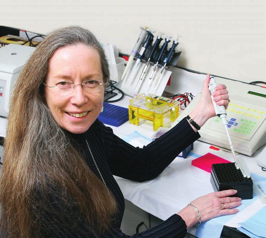
thanks to berkeley...

ACADEMIC EXCELLENCE (from U.S. News &
World Report doctorate program rankings;                                               Optometry National Boards NBEO Examination Pass Rates
highest possible score is 5; mean rankings listed):                                                       BERKELEY 2007
      UC Berkeley . . . . . . . . . . . . . . . . . . . .            4.8
      Stanford . . . . . . . . . . . . . . . . . . . . . . . .       4.8
      MIT . . . . . . . . . . . . . . . . . . . . . . . . . . . .    4.8                                          Berkeley: 87.3%
      Princeton . . . . . . . . . . . . . . . . . . . . . . .        4.6           PART I: BASIC SCIENCE
      Harvard . . . . . . . . . . . . . . . . . . . . . . . .        4.6                                          National: 74.8%
      Yale . . . . . . . . . . . . . . . . . . . . . . . . . . . .   4.4
      Michigan . . . . . . . . . . . . . . . . . . . . . . .         4.4
      Wisconsin-Madison . . . . . . . . . . . . . .                  4.3
                                                                                                                  Berkeley: 100%
      University of Chicago . . . . . . . . . . . .                  4.3         PART II: CLINICAL SCIENCE
      Cornell . . . . . . . . . . . . . . . . . . . . . . . . .      4.3                                          National: 91.5%
      Columbia . . . . . . . . . . . . . . . . . . . . . . .         4.3
      UCLA . . . . . . . . . . . . . . . . . . . . . . . . . .       4.2                                          Berkeley: 100%
      Texas-Austin . . . . . . . . . . . . . . . . . . . .           4.0           PART III: PATIENT CARE
97% of Berkeley’s graduate programs rank                                                                          National: 96%
among the top 10 in their fields.

TOP UNIVERSITIES (from “World University                                   BERKELEY UNDERGRADUATE STUDENTS IN GENERAL
Rankings” by The Times of London, based on a
                                                                           Berkeley is the opportunity of a lifetime for many of our students: 36% of our students come
three-year average, 2004–06):
                                                                           from families with annual incomes under $45,000. In fact, Berkeley educates more students
     1. Harvard
                                                                           from these income brackets than all the Ivy League schools put together.
     2. MIT
     3. Cambridge                                                            • 87% come from public schools.
     4. Oxford                                                               • 30% are the first in their family to attend a four-year college.
     5. UC Berkeley                                                          • 75% of all undergraduates receive financial aid.
      6. Stanford
                                                                             • 98% of entering freshmen are among the top 10 of their high-school class.
                                                                             • Berkeley has more Peace Corps volunteers than any other university since the start of the
BERKELEY’S ENDOWMENT HAS MORE THAN                                             program.
DOUBLED in the past decade from $1.1 billion                                 • There are 450,000 UC Berkeley alumni worldwide.
to $2.9 billion (data as of June 30, 2008). (Note
                                                                             • In 2007–08 over 90,000 donations accounted for more than $409 million in private
that it still lags far behind those of peer private
institutions.)                                                                 support to UC Berkeley.
     Harvard . . . . . . . . . . . . . . . . $34.9 billion
     Yale . . . . . . . . . . . . . . . . . . . . $22.5 billion
     Stanford . . . . . . . . . . . . . . . $17.2 billion                  BERKELEY OPTOMETRY STUDENTS
     Princeton . . . . . . . . . . . . . . $15.8 billion                   Applicants for the class that started in August 2008: 246                 1    Pacific Islander
     MIT . . . . . . . . . . . . . . . . . . . . $9.9 billion                 Enrolled: 68
     Berkeley . . . . . . . . . . . . . . . . $2.9 billion                                                                                           4    Hispanic
                                                                              Mean age at admission: 23
                                                                              Number of women: 50                                                    1    East Indian / Pakistani

UC BERKELEY HAS PRODUCED 41 NOBEL                                             Number of men: 18                                                      13   Not Stated
LAUREATES among its faculty and alumni. The                                   Number of states represented: 9
first of these was Ernest Lawrence, who, at the                                                                                                      12   Non-Hispanic White
                                                                              Average GPA: 3.53
age of 29, unlocked the gates to the world of the                             Average OAT score: 360                                                 37   Asian
atom with his invention of the cyclotron.

SUCCESS IN FACULTY RECRUITMENT AND                                         PATIENT CARE
RETENTION: Despite fierce competition from                                 At the Meredith Morgan and Tang Eye Centers on the Berkeley Campus alone, there are
elite private universities, UC Berkeley has had                            more than 75,000 patient visits each year. The total number of patients cared for by Berkeley
a 72% success rate in recruiting new faculty in                            Optometry students is significantly increased as a result of their experiences during the three
the past decade, and in the past five years has                            or four 10-week external rotations they complete during the fourth year. The numbers of
retained almost 70% of faculty whom other                                  patient encounters for the average Berkeley Optometry student during training are 560 for
institutions tried to recruit.                                             third-year students and 2,000 for fourth-year students.

                                                                                                                                                  optometry.berkeley.edu      |     3
the early years of

                           B E R K E L E Y
                           OPTOMETRY
                  “All the material gain
            I have in this world I must credit                               By John Fiorillo
       entirely to optometry. The little knowledge
   I have, by which I am able to render fair service
. . . has come to me by virtue of those who sacrificed
before me. Therefore, if success in life is to leave the
world a little better for having been in it, then I feel
honor bound to give to that which has given to me.”
                —GEORGE L. SCHNEIDER, JANUARY 18, 1923

                              A MEETING WITH UC PRESIDENT BENJAMIN WHEELER
                              In 1907, a committee of three optometrists from the California State Association
                              of Optometrists met with Benjamin Ide Wheeler, president of the University of
                              California, about establishing a “Course in the Science of Optometry.” The com-
                              mittee consisted of George Schneider of Berkeley, Charles Wood of Oakland, and
                              Frederick Chinn of Sacramento. This would be the first of many meetings between
                              optometrists and the University, comprising a 16-year undertaking leading to the
                              inaugural optometry curriculum in August 1923.
                                 Although President Wheeler responded favorably, he wanted to consult his “ocu-
                              list” friends. Schneider, Wood, and Chinn immediately understood that there would
                              be troublesome obstacles to overcome, not least of which was determining the
                              role, if any, of the often antagonistic medical profession. They also had to persuade
                              administrators and faculty to support a professional curriculum at the University,
                              as well as finance the course and recruit students.
                                 Schneider and his allies would succeed only after tireless effort and resilience
                              in the face of repeated setbacks and opposition from both outside and within the
                              profession of optometry. The early advocates of a “Course in the Science of Optom-
                              etry” believed passionately in the righteousness of their cause—for them it was
                              a mission of historical proportions. They wanted to advance optometry through

44 || BERKELEY
      BERKELEY OPTOMETRY
               OPTOMETRY                                                                                       FALL
                                                                                                               FALL 2008
                                                                                                                    2008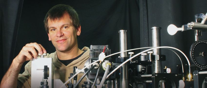
professional and scientific instruction, to put                                                   any requirements to demonstrate proficiency in
it on an equal footing with other health-care                                                     the practice of vision care. It was not until 1901
professions. These were remarkable men, dedi-                                                     that states began passing statutes defining and
cated to the proposition that both the profession                                                 regulating the profession (California did so in
and the public would benefit from an affiliation                                                  1903), and two decades would pass before all the
with the University—an institution of interna-                                                    states had optometry laws on their books.

                                                                                                                                                       Photograph, Berkeley Optometry Archives
tional repute with exacting standards of education                                                   These developments took place in a rapidly
and growing achievements in research.                                                             changing world. In the San Francisco Bay Area,
   George Schneider was foremost among these                                                      a land boom and resulting population explo-
pioneers—a visionary optometrist who would                                                        sion created a need for more expert vision
guide the process from start to finish, and who                                                   care. The expansion of the University’s student
would become the first lecturer in optometry at                                                   body, faculty, research, and infrastructure also
                                                         George L. Schneider (1874–1928)
the University of California. The early history of                                                opened possibilities for a professional program
                                                         First Lecturer at Berkeley Optometry     in optometry.
Berkeley Optometry serves, in part, as a biography
of Schneider’s professional life. Through it all, he                                                 The mobilization for a curriculum was thus
stands out as a person of ideals, patience, persistence, cleverness,             intimately associated with urban development, educational prog-
kindness, and vision. Without him there might never have been a                  ress, legal protections, and professional advancement. Advocates
School of Optometry on the Berkeley campus.                                      of the course understood these larger issues as they labored to
                                                                                 elevate the profession, which was, in their view, a prerequisite to
THE PROFESSION BEFORE BERKELEY OPTOMETRY                                         establishing a curriculum at a world-class university.
When Schneider and his colleagues began their campaign, optom-
etry was struggling to survive and define itself as a profession.
                                                                                                                                         THE ROLE OF ORGANIZED OPTOMETRY
Special interests threatened to foil attempts to organize into                                                                           Optometric practice was changed in California largely through
optometric associations or gain legitimacy through statutory                                                                             the efforts of the more forward-thinking members of the profes-
regulations. The medical profession in particular was determined                                                                         sion. The lobbying by organized optometry for a curriculum was
to maintain its hegemony in health care and the marketplace by                                                                           critical to winning over University officials and faculty. Yet these
excluding optometrists from all but the manufacture of eyeglasses.                                                                       activists did more than voice their support—they also agreed to
Standing alongside these medical activists were many opticians                                                                           pay for the costs of the inaugural courses. In 1922–23, Schneider
and optometrists. They were among the most vocal of obstruc-                                                                             organized a campaign to collect pledges totaling more than $9,000
tionists, not wanting to jeopardize their lucrative businesses by                                                                        (a considerable sum in those days) in support of the first year’s
changing the rules or increasing the number of better-trained                                                                            instruction. In the second year, all operating costs were met by
practitioners. Advocates of a university curriculum also faced                                                                           the assessment of higher annual license renewal fees (raised from
widespread prejudice from a public that viewed optometry with                                                                            $2 to $10, with $8 going to support the curriculum and research).
suspicion. After all, until the start of the 20th century, anyone could                                                                  This represented an extraordinary instance of private practitioners
practice optometry—there were no educational prerequisites or                                                                            entirely financing a professional program at a public university.
                                                                                       Copy of photograph, Berkeley Optometry Archives

                                                                                                                                                                                                                               Photograph by McCurry, Bancroft Library, UARC PIC 11n:19

This portrait was taken circa 1926–27 at the Athens Athletic Club in Oakland,
California. The men, all practicing optometrists and members of local and state
optometric associations, had been active in lobbying for the curriculum in optom-                                                        Le Conte Hall, the home of the Department of Physics, circa 1925–26, with the
etry at the University. (Left to right, front row, seated): Charles Mueller, William                                                     Campanile and a corner of Bacon Hall (no longer standing) in the background.
Fraser, Herbert Schultz, Fred Laufer, George Schneider, and Edward Hardy; (Back                                                          Beginning in 1923, the curriculum in Optometry was assigned temporary offices and
row, standing): Morris Kittredge, Fred Watson, Charles Wood, Wallace Doig, William                                                       clinic space on the attic floor, plus an optics lab with spectacle-making equipment
Burns, and Paul Eckley.                                                                                                                  in the basement.

                                                                                                                                                                                                 optometry.berkeley.edu   |                                  5
Curriculum in Optometry brochure, Berkeley Optometry Archives
    Clinical rooms on the third floor of Le Conte Hall in the 1930s. Nine refracting rooms were equipped through gifts of apparatus and machinery made by individuals and organizations.
    (L) Clinic room with complete American Optical Company unit and, at the left, the Ferree-Rand Perimeter; (R) Clinic room equipped with Bausch and Lomb Ophthalmological Unit
    and Instruments.

    PHYSICS-OPTOMETRY                                                              board examinations, and find gainful employment. Anything less
                                                                                   would be seen as a failure of the curriculum to prepare its gradu-
    In the fall of 1923, Berkeley Optometry offered its first classes.
                                                                                   ates for a professional life. Minor also believed (correctly) that
    The fledgling program was, however, a poor relation to well-
                                                                                   gaining the endorsement of University administrators and faculty
    established University departments, and it had much to prove.
                                                                                   for an independent school would require long years of effort.
    Optometry was not an independent department or school, nor
                                                                                      Support from California optometrists remained critical for the
    even a division within a home department—it was merely a two-
                                                                                   survival and expansion of the program. Organized optometry was
    year curriculum for junior and senior undergraduates. Initially,
                                                                                   the curriculum’s financial lifeline, for in the early years the sole
    students graduated with a BS degree in Physics and a Certificate in
                                                                                   support of the program came from the redirected $8 portion of
    Optometry; in 1929, the major was revised to Physics-Optometry.
                                                                                   state license renewal fees. Should the program fail in the view of
    The program was thus a curious hybrid of physics courses and
                                                                                   optometrists, they might call in their fees legislatively and force
    practical/theoretical optometry, making it vulnerable to criticism
                                                                                   the University’s hand—to either support the program on its own
    from skeptics among faculty and administrators across the cam-
                                                                                   or shut it down.
    pus. To put it bluntly, people were watching, and some were all too
                                                                                      Minor’s concerns went beyond monetary considerations. He
    eager to see the experiment fail.
                                                                                   wanted optometrists to believe that the program would advance
       The Department of Physics was an ambivalent host who con-
                                                                                   the profession for the benefit of both the practitioner and the
    sidered optometry’s occupation of rooms in Le Conte Hall to be,
                                                                                   public. With the approval and occasional direct involvement of
    at best, a temporary expedient. It would tolerate the intrusion
                                                                                   optometrists at large, he could build the professional program
    only as long as space was available. In the meantime, it expected
                                                                                   and guarantee its long-term survival. This period in the history of
    optometry students to hold up their end of the bargain by taking
                                                                                   Berkeley Optometry was characterized partly by Minor’s effort to
    all the required physics courses and performing, on average, at
                                                                                   foster an ongoing cooperative relationship with optometrists and
    the same level as physics majors. None of these
                                                                                                    their professional organizations.
    conditions were met, and despite parallel expan-
                                                                                                       For 23 years, Minor directed and expanded
    sion of the physics and optometry programs
                                                                                                    the program, working toward independence from
    and the growing need for more space, another
                                                                                                    physics while improving the range and depth of a
    quarter century would pass before the interlop-
                                                                                                    curriculum suitable for the practice of optometry.
    ers found lodgings in a separate building.
                                                                                                    He did this despite a rather unsteady start. At first,
                                                                                                    student recruitment faltered—a cause for great
                                                                                                                 Photograph, Berkeley Optometry Archives

    RALPH MINOR’S TENURE AND LEGACY                                                                 concern in an unproven curriculum, but then it
    Berkeley Optometry began when the University                                                    took off. Enrollment for all optometry registrants
    appointed Ralph Minor in 1923 as “Professor of                                                  increased from 13 students in 1929 to 64 in 1933.
    Physics in charge of Optometry.” Minor’s chal-                                                  In 1934, Minor graduated the so-called “Wonder
    lenge was to transform what amounted to an                                                      Class.” Eighty percent (11 of 14 graduates) were
    experiment in professional instruction into an                                                  honor students, an astonishing percentage within
    independent school of optometry. Undergradu-           Ralph S. Minor (1876–1961)               a single program at the University. This group rep-
    ates had to complete the curriculum, pass the state    First Dean of Berkeley Optometry         resented the highest scholastic achievement of any

6     |   BERKELEY OPTOMETRY                                                                                                                                                   FALL 2008
Photograph from the Emily Boettler scrapbook, Berkeley Optometry Archives
The “Wonder Class” of 1934. (L to R, front row): Prof. Ralph J. Minor, Head of Curriculum; Edward I. Goodlaw; King C. Louie; Audra Mae Booth; Chauncey M. Smith; and Halley E.
Stephenson; (middle row): Walter A. Robinson; U. Sinclair Ives; Meredith W. Morgan Jr.; Harold A. Jacobsen; C. Roger Brissman; and Raymond L. Ng; (top row): Frederick L. Mason (Lecturer
in Optometry); Herbert S. Player; George H. Voorhies; and Robert D. Newton [not pictured: Harold W. Tower).

class during the first 10 years of the program. Minor and others                                 department) while introducing more instruction and training
frequently cited the “Wonder Class” as evidence of bona fide schol-                              in the physiological and pathological aspects of vision. He also
arship among its graduates, which boosted the effort to expand the                               oversaw the re-equipping of refracting rooms and the optics
program and gain independence. Included among these graduates                                    laboratory, as the number of patients receiving eye-care services
was Meredith W. Morgan Jr., destined to become the third dean of                                 increased.
Berkeley Optometry.                                                                                 The research component, too, needed expansion and guid-
                                                                                                 ance in order to produce vision science investigations that Uni-
DEVELOPING THE FACULTY AND CURRICULUM                                                            versity faculty and administrators, optometrists, and the public
                                                                                                 could appreciate and support. Physiological optics became a
From the beginning, Ralph Minor displayed a talent for recruit-
                                                                                                 primary focus. As he was nearing retirement, Minor would rely
ing excellent faculty. Frederick Mason (1886–1954) was the first,
                                                                                                 increasingly on Kenneth Stoddard (PhD Stanford University,
accepting a position as full-time instructor in 1924. (Schneider
                                                                                                 OD Berkeley Optometry ’36), who by June 1938 had left private
continued his private practice in downtown Berkeley and thus lec-
                                                                                                 clinical practice to join the optometry faculty. Minor admired the
tured part-time.) The irascible Mason became an outstanding fig-
                                                                                                 energetic direction provided by Stoddard, who succeeded Minor
ure in the history of Berkeley Optometry, notable for introducing
                                                                                                 as dean in 1946, the inaugural year for the Graduate Program in
high standards of scholarship and advancing professional devel-
                                                                                                 Physiological Optics (now called Vision Science).
opment. A taskmaster who was as demanding of himself as of
others, Mason had a significant and enduring impact on an entire
generation of optometry students. Minor also established Berke-
                                                                                      ALUMNI AND STUDENTS
ley Optometry’s first instructional alliance with medicine when he                    Optometry alumni and student associations were also launched during
appointed Milton Shutes, MD, as a lecturer in ocular pathology.                       Minor’s tenure. The Optometry Alumni Association of the University
Others would follow, particularly                                                                                     of California began in 1926 as a small,
Marshall Atkinson (’40, MD UCSF                                                                                       loosely knit group holding educa-
                                                                                                                                    Photograph from the Emily Boettler scrapbook, Berkeley Optometry Archives

’46), who resisted sustained criti-                                                                                   tional meetings in Le Conte Hall.
cism and threats of sanctions from                                                                                    Angus McLeod ’25, from the first
organized medicine. (Atkinson had                                                                                     graduating class, and Albert Reinke
committed the unpardonable sin                                                                                        ’26 were instrumental in establish-
of teaching ocular pathology in an                                                                                    ing the association and making it a
optometry school.) He remained                                                                                        more formal organization by writ-
undeterred, believing that medicine                                                                                   ing its first by-laws in 1931.
should cooperate with optometry                                                                                          The student association was
for the benefit of public health.                                                                                     closely allied with the Omega Delta
   Minor worked continually on                                                                                        National Fraternity of Optome-
curriculum development, gradu-                                                                                        trists until 1941, when they drafted
ally toning down the emphasis on           Frederick Mason (left) speaking with Ralph Minor at the Berkeley Optometry the first constitution for the Asso-
mathematics and physics (much              Commencement in May 1936, the same year in which Mason published his       ciated Optometry Students of the
to the displeasure of the physics          influential textbook Principles of Optometry.                              University of California.

                                                                                                                                                                                                                optometry.berkeley.edu   |                        7
INDEPENDENCE                                                                        new home for optometry (he seemed to prefer UCLA), but
                                                                                        he would not decide without hearing from the California
    On July 1, 1941, the Regents finally approved an independent                        State Association of Optometrists (once again, a remarkable
    “School of Optometry” (although it was still housed in the physics                  acknowledgment by the University of the role played by orga-
    building). Minor was named its first Director (he would become                      nized optometry). Berkeley alumni and other sympathetic
    the first Dean in 1945, one year before his retirement).                            optometrists quickly rallied to protect the Berkeley site. In
      Now that the program had evolved into a professional school,                      September 1943, the state association voted unanimously in
    Minor continued to work for the funding of a new building. On                       favor of Berkeley.
    November 16, 1939, he and Thomas Peters (1886–1956; president                          In early 1947, the state legislature drafted assembly bill
    of the California State Association of Optometrists, 1938–40)                       AB-2118 to appropriate $300,000 (to be added to the funds
    wrote a report providing the association membership with a                          already raised by organized optometry) for a new optometry
    cogent summary of events leading up to an independent school                        building. On July 8, 1947, Governor Earl Warren (1891–1974,
    of optometry and the possibility of a new optometry build-                          future Chief Justice of the U.S. Supreme Court) signed the
    ing. California optometrists, as they                                                                                 bill. By this time, however, post-
    had in 1923, responded enthusiasti-                                                                                   war inflation had made it unfea-
    cally, and by May 1941 they had                                                                                       sible to construct a building of
    pledged seed money totaling more                                                                                      sufficient size and quality for a
    than $80,000. Despite the tragedies                                                                                   mere $380,000. President Sproul
    of World War II and the ensuing                                                                                       and the Regents then provided
    economic disruptions, leaders in                                                                                      an alternative by approving the

                                                                                                                                                    Photograph, Berkeley Optometry Archives
    the state association kept their eyes                                                                                 conversion of Durant Hall into
    on the prize, collecting the money                                                                                    the optometry building.
    behind the pledges during the years                                                                                      More than a half century of
    1942–1946 as they waited for the                                                                                      program change and develop-
    opportunity to promote the new                                                                                        ment would follow the early
    building once again.                                                                                                  period of Berkeley Optometry—
      As if the war were not enough          Students conducting visual function studies in the physiological optics      a story that will be told in detail
    of a diversion, the school narrowly      laboratory in Le Conte Hall, circa late 1940s–early 1950s, using haploscopes in a forthcoming book.* After
    escaped relocation when, in 1942,        mounted on laboratory tables.                                                more than 80 years of clinical
    some UCLA alumni, aided by fac-                                                                                       and research program develop-
    ulty at the private Los Angeles School of Optometry (later the                      ment and facilities enhancement, Berkeley Optometry—once
    Southern California College of Optometry), attempted to per-                        only the dream of pioneering optometrists—stands as the
    suade UC president Robert G. Sproul that construction of the                        premier institution of its kind, graduating the best trained
    proposed building should be moved to the UCLA campus.                               optometrists in the profession and hosting a world-renowned
    Sproul was willing to accept either Berkeley or UCLA as the                         vision science research program. ■

                                  *This text has been excerpted and adapted from John Fiorillo’s forthcoming Berkeley Optometry—A History.
                                                                                             Reproduced from the Berkeley Gazette, June 23, 1948,
                                                                                             from negatives in the Berkeley Optometry Archives

                                                                                                                                                                                                    Photograph, Berkeley Optometry Archives

    (L) View of the “Optometry Building” during the 25th Anniversary Celebration and Dedication on June 22, 1948. The audience (partly visible on the left) was composed of faculty,
    staff, students, and members of the American Academy of Optometry (whose annual congress was held in San Francisco that year). The building, formerly called the Emergency
    Classroom Building and then Durant Hall, would undergo five years of interior remodeling before Berkeley Optometry enjoyed full occupancy. It was rededicated as Ralph S. Minor
    Hall in 1970.

8   |   BERKELEY OPTOMETRY                                                                                                                                                                    FALL 2008
student focus
DORA SZTIPANOVITS working at a small office to be a great way to learn about optometry, and
Born in Budapest, Hungary in 1980, Dora he decided to become an optometrist. With an undergraduate scholarship
Sztipanovits moved to the U.S. at the age in hand, Alvaro returned to Florida for college before being accepted into
of three, after her father received a visiting Berkeley Optometry, where he could be close to family again.
professorship at Vanderbilt University. Not Now in his final year at Berkeley Optometry, Alvaro has discovered a pas-
able to visit their former home until 1989, sion for community service. At Berkeley Optometry, Alvaro learned about
the family has returned every other sum- Volunteer Optometric Services to Humanity (VOSH), and he saw that VOSH
mer since then. Dora grew up bilingual; her went to countries like Nicaragua to provide vision services to the indigent.
close ties to Hungary, her language, and her Alvaro found a genuine desire to help the people of Nicaragua, and in VOSH
frequent trips there created a lasting bond he found the perfect vehicle. His participation in VOSH, with two trips to
with her culture. Nicaragua and one to the Dominican Republic, led him to become even more
In 2002 Dora graduated from Washington University in St. Louis with a BS in active in school and the community. He has served as president of VOSH
biomedical engineering. She then obtained a master’s degree in the same field at Berkeley Optometry, and he has been honored to receive the Thomas
at Vanderbilt University. Her interest in optometry stemmed from her back- B. Hewitt Volunteer Public Service grant. Alvaro believes that his volunteer
ground in biomedical engineering, specifically in the area of orbital naviga- experiences at Berkeley Optometry have given him skills that can benefit
tion systems. During graduate school, she discovered an interest in vision and his community both locally and worldwide. His current plans are to pursue
more importantly, a desire to work in direct patient care. She is honored to private practice, but Alvaro always intends to remain active with VOSH.
be one of the recipients of The Dr. John R. and Norma M. Austin Optometry
Student Support Fund, which is given to students who demonstrate a high
PATTY OH
level of academic distinction.
Patty grew up in Seattle in the pre-Starbucks/
Introduced to a number of specialties at Berkeley Optometry, Dora decided Microsoft/Nirvana era. More hippy than
to delve into ocular disease. During her internship at the Omni Eye Center in trendy, she went to Reed College in Portland,
Atlanta, she was exposed to pre and post-operative care, witnessed multiple Oregon to get her bachelor’s degree in biol-
cataract extractions, and assisted in the management of ocular diseases. In ogy. She then continued southward to the Bay
2008–2009 she will be doing a residency in ocular and refractive surgery in Area, where she worked as a research assis-
Nashville. Her goal is to obtain more experience in a variety of refractive tant at UCSF and UC Berkeley. At Stanford
and ocular surgery procedures so that she can offer proper pre and post- University Patty earned her MS in biology, but
operative care independently in the future. Optometry has vast potential then she decided to make a career switch.
for growth in surgical co-management, and she looks forward to bringing her Unsure where her future lay, she worked a few
knowledge and skills to this area. years in the high-tech industry and then lived in Brazil for two years. She then
Dora’s research focused on the development of an orbital endoscopic system decided to move back to the Bay Area to pursue optometry.
to deliver neuro-protective agents to the optic nerve. Optic neuropathies Believing it is important to take time from studies or work to participate
are hard to treat due to the difficulty reaching the optic nerve in the tight in the community, Patty continues to be active in public service. In her first
neurovascular environment of the orbit. By combining an orbital endoscope year at Berkeley Optometry she served as the Class of 2010 Philanthropy
with a flexible electromagnetic tracking system, it is possible to quickly Chair, helping to organize and recruit students for numerous philanthropic
and accurately locate the optic nerve. Her project focused on assessing the activities, including nine vision screenings. As a second-year student,
accuracy of such a tracked endoscopic system as well as implementing it in Patty was the school’s Bay Area Optometric Council Representative, serv-
phantom and animal models. ing to bridge the local optometric professional societies with Berkeley
Optometry. Since the professional societies have a large philanthropic
component, many of her responsibilities extended to community vision
ALVARO CASTILLO screenings. During her first two years of optometry school, Patty volun-
Alvaro was born in Nicaragua, but his family teered at diabetes health fairs, children’s health fairs, senior citizen vision
moved to the United States when he was screenings, Latino health screenings, Special Olympics exams, and vision
one year old to escape the political turmoil services for the indigent. In January 2007 she traveled to Palau as part of
in Nicaragua. After Alvaro completed high the Volunteers of Optometric Services to Humanity (VOSH) program. There
school in Florida, he moved with his family she and other students examined nearly a thousand Palauans and distrib-
to Fresno, where his father had gotten a job. uted hundreds of pairs of glasses.
Interested in healthcare but not yet decided Patty feels fortunate to have found a career that values public service. Her
on a specific career, Alvaro spent a couple of public service activity and academic achievement have resulted in several
months looking for a job. During this time awards: the 2006–2007 UCBSO Student Leadership and Service Grant, the
he found he needed to see an optometrist, 2007 Summer NEI T35 Research Grant, the 2008 Great Western Council of
where he overheard a conversation among the office staff about hiring Optometry Scholarship, and the 2008 Raymond L. Eng Family Professional
someone for data entry. He applied for the position and was hired. He found Student Support Award.
optometry.berkeley.edu | 9news & notes
Photograph, Bob Knight Photography
10 | BERKELEY OPTOMETRY FALL 2008optometry.berkeley.edu | 11
NEW VIEWS OF THE HUMAN RETINA
                                        BY AUSTIN ROORDA, PHD
                                        Associate Professor Austin Roorda joined the Berkeley Optometry and Vision Science faculty in
                                        January 2005. His innovative applications of modern optics to the study of the human visual
                                        system have the potential of improving the diagnosis and understanding of ocular disease and
                                        dysfunction. Shown here, Dr. Roorda is at work on the adaptive-optics scanning laser opthal-
                                        moscope (AOSLO), which can record video with cellular-level resolution in living human eyes.

                                                                               astigmatism, but this term is rapidly being replaced by high-order
THE DEVELOPMENT OF RETINAL IMAGING
                                                                               aberrations, which can be quickly and accurately measured using a host
After one year of training, optometry students will learn that the optical     of commercially available devices called wavefront sensors. No sooner
principles behind ophthalmoscopy are quite simple. In fact, the optics of      had wavefront sensors become available to measure these high-order
the eye were well described by Kepler in the 1600s. A series of technical      aberrations than efforts were underway to correct for them. Wavefront-
insights involving a partially silvered mirror and a properly directed light   guided refractive surgery, for example, uses aberration measurements
source led to the invention of the direct ophthalmoscope by Helmholtz          to generate a custom correction that will not only relieve your need for
in 1850. Since then, ophthalmoscopy has improved by employing better           glasses but also give you sharper vision. You can even purchase spectacle
ways to get illumination into the eye and to properly capture the light        lenses that will correct your high-order aberrations.
exiting the eye. The development of photography and the electronic flash          In the Ophthalmic and Visual Optics Lab at Berkeley Optometry, we
lamp, for example, represented major advances.                                 employ a technique called adaptive optics, or AO, to correct for high-
    One of the most significant developments in ophthalmoscopy was the         order aberrations. Our correction is done on a bench-top, so it is not
invention of the scanning laser ophthalmoscope (SLO). This is a special-       portable, but it does enable us to test vision with images that are sharper
ized version of a scanning laser microscope, which builds an image by          than the patient has ever experienced in their lifetime. Moreover, it
recording the scattered light from a focused spot as it is scanned across      allows us to see inside the eye with a resolution that is unprecedented.
the region of interest. The only difference between a microscope and an
ophthalmoscope is that in a microscope, one uses a high-quality objec-
tive and can look at whatever sample is placed on the microscope stage,        ADAPTIVE OPTICS: FROM THE TELESCOPE TO THE OPHTHALMOSCOPE
whereas in an SLO, the optics of the eye serve as the objective lens and       AO was developed by astronomers, who were faced with blurry images
the retina is always the sample.                                               of distant objects caused by turbulence in the earth’s atmosphere (the
    Having the optics of the eye serve as the objective lens in an SLO         twinkling-star phenomenon). To overcome the problem, they developed
imposes a major limit on performance. High-quality microscope objec-           ways to measure the effects of the turbulence and invented deformable
tive lenses are well designed for one specific task—that of focusing light     mirrors to correct for them. Today, most major telescopes employ some
to a small spot. The optics of the eye, however, are designed for a multi-     type of AO system and, as a result, large ground-based telescopes can take
tude of tasks, including wide-angle viewing, dynamic focusing (accom-          pictures with resolutions rivaling that of the Hubble Space Telescope.
modation), operating in a variety of lighting conditions, and lasting the         These technical developments by astronomers set the stage for vision
lifetime of the person using it. As such, the eye rivals any manufactured      scientists to attempt the same. In 1996, David Williams, Donald Miller,
imaging system in functionality; but when it comes to pure image qual-         and Junzhong Liang completed the world’s first adaptive-optics ophthal-
ity, it leaves a lot of room for improvement.                                  moscope. They showed the first extensive mosaics of cone photorecep-
    The primary limits to image quality in the eye imposed by defocus and      tors ever taken from living eyes.
astigmatism are relatively easy to overcome. Spectacles have been used            After completing a PhD at the University of Waterloo (where I had
for centuries, contact lenses in the last 50 years, and more recently, cor-    been frustrated in my attempts to take high-resolution images with a
neal reshaping has been accomplished using lasers. But there are imper-        modified SLO), I had the opportunity to work with the AO ophthalmo-
fections in the eye that are not correctable with conventional methods.        scope at the University of Rochester. By 1998, we managed to map the
In optometric lingo, such eyes have been classified as having irregular        trichromatic cone mosaic, representing the first major scientific outcome

12   |   BERKELEY OPTOMETRY                                                                                                                    FALL 2008
faculty research
of this new technology. However, that ophthalmoscope had limitations
in image frame rate as well as image contrast that scanning laser methods
could potentially overcome. So I spent three years at the University of
Houston developing the adaptive-optics scanning laser ophthalmoscope,
or AOSLO. Completed in 2001, the AOSLO could record real-time
videos of the living retina on a microscopic scale. The real-time aspect
allowed us to see single leukocytes coursing through the smallest retinal
capillaries and track the motion of the eye better than any other device.
Moreover, the AOSLO was confocal, making optical sectioning of the
retina possible.
A NEW WAY TO SEE EYE DISEASE
By offering microscopic views of the inside of the eye, AO imaging is
driving a paradigm shift in how ophthalmoscopy is used for clinical and
basic science. In collaboration with Jacque Duncan, an ophthalmologist
from UC San Francisco, we have been measuring the structural proper-
ties of patients with a variety of retinal degenerations. Little is known
about how many of these diseases progress on a cellular scale. In fact, the Optical Researcher Yuhua Zhang and Associate Professor Austin Roorda standing beside
tissue structure of some conditions, such as NARP (neuropathy, ataxia, AOSLO generation II. The AO system employs a compact micro-electro-mechanical
retinitis pigmentosa) has never been studied. Seeing these cells in living (MEMS) deformable mirror.
eyes, therefore, can shed new light on how these diseases progress.
In an era where our understanding of the genetic basis of eye disease is
blossoming, AO imaging will be useful in determining the complex ways
in which these mutations affect the retina. Furthermore, knowledge of
the molecular underpinnings of these diseases is beginning to generate
novel therapies, and AO imaging represents possibly the most sensitive
technology to measure their outcomes. Without sensitive outcome mea-
sures, the development of therapies is hampered. Dry AMD, for example,
progresses so slowly that the amount of time and money required to
determine whether a drug is effective is prohibitive for drug companies.
Sensitive, cellular-level imaging biomarkers for the disease have the
potential to reduce the time and cost significantly.
The image spans 1.2 degrees (~0.36 microns) in a region about 300 microns from the
foveal center (located up and to the left of the image). The left image is without adap-
THE FUTURE
tive optics. The right image is taken after AO is turned on. The image is brighter and has
The potential applications of AO are far-reaching, as any ophthalmic imag- higher contrast and improved resolution, revealing the array of cone photoreceptors
ing modality can benefit. Some labs are already working on incorporating in the eye.
AO into OCT systems. As the technology matures, costs will fall and AO
may become part of routine clinical practice. Your standard retinal camera,
for example, might use a basic AO system to quickly correct for defocus
and astigmatism. Your phoropter may eventually employ an AO system
with a built-in autorefractor to replace the myriad of lenses they current
use. Not only could the AO phoropter converge quickly to the desired
refractive correction, but the AO system could demonstrate what vision
would be like if high-order aberration were corrected. You could also have
patients preview multifocal or progressive-addition lens designs.
At the other end of the spectrum, new developments in AO will A B
also facilitate the most advanced ophthalmic imaging systems. Already,
researchers are using AO with autofluorescent imaging as well as extrin-
sic fluorescent dyes to help visualize otherwise invisible cells in the ret-
Three optical sections of a human
ina, like ganglion cells. By offering microscopic access to the retina, AO retina. (A) shows the photoreceptor
systems will be used not only to image the anatomy but also to measure mosaic. Focusing up (B) reveals the
functional properties of the cells that we see. blood vessels and (C) the nerve fibers
In the Ophthalmic and Visual Optics Lab we continue to work in that run across the retinal surface.
both directions, from the most applied to the most advanced appli-
cations, and Cal is the best place to develop and use this technology.
Berkeley Optometry leads the way in basic and clinical vision research
C
and offers access to the best researchers in other disciplines, both on
campus and in the Bay Area. ■
optometry.berkeley.edu | 13faculty research
Educated in Australia, Professor
Wildsoet joined Berkeley Optometry
in 2000. Professor Wildsoet teaches
systemic and ocular pharmacology as
well as clinical optometry. She is also
Research on
heavily involved in mentoring student
researchers, at both the undergraduate
Myopia and graduate levels. The overriding
goal of research in the Wildsoet lab
is the development of preventative
treatments for myopia.
BY CHRISTINE WILDSOET, PhD, FAAO
M
yopia, or short-sightedness, while not a modern disease, is students and UC Berkeley’s Undergraduate Research Apprenticeship
now present in epidemic levels in Asia—over 90% in some Program, providing mentors for future clinicians and scientists.
university student populations. In the U.S. in 1999, spec- Animal studies have taught us a great deal about how refractive errors
tacles, contact lenses, and doctor visits accounted for $8.4 billion/year, are regulated during development in the process known as emmetropiza-
and LASIK surgery another $2.1 billion/year in healthcare costs. Spec- tion. From chicks, we learned that young eyes grow in response to their
tacles alone cost $15 billion in 2002, with over 150 million Americans visual environment. When spectacles lenses are fitted to chicks to impose
using corrective lenses to compensate for refractive errors. refractive errors, their eyes adjust their growth to compensate. Amazingly,
Myopia is a leading cause of blindness. Although there is general their eyes can distinguish hyperopia (where the eye is too short for its
agreement that human myopia is mainly due to excessive elonga- optical power) from myopia (where the eye is too long) and increase or
tion of the eye, the causes of that elongation are poorly understood. decrease eye growth appropriately. We also learned that eye growth is regu-
Genetic factors likely play a role, but myopia’s increasing prevalence lated within the eye—the brain is not needed, although the nature of the
suggests environmental factors may also contribute. This notion is visual information is critically important. These observations have rein-
not new. In Juler’s 1904 Handbook of Ophthalmic Science and Practice, vigorated myopia research, raising many questions about how eyes decode
there is a section devoted to the treatment of progressive myopia: “All defocus and the signal pathways underlying eye-growth regulation.
work must be stopped,” he advised, “No reading or writing is to be Our lab is currently using chick and guinea pig models, in vitro cell
permitted.” Other equally impractical recommendations followed. But and tissue models, and human subjects to study fundamental questions
the urgency of Juler’s concern is understandable, because progressive such as these:
myopia can lead to sight-threatening staphylomas, or scleral bulges. • What aspects of visual experience influence the development of
The Wildsoet Lab studies refractive development, particularly myo- myopia?
pia. We are a multidisciplinary team—clinician researchers, molecu- • How are growth signals relayed from the retina (where image defo-
lar biologists, bioengineers, and optical engineers—working toward cus is detected) to the sclera (which grows to correct defocus)?
new understandings and therapies for this old problem. We perform
• Can myopic growth be inhibited pharmacologically or with engi-
human studies to learn more about the problem, and we use animal
neered tissue scaffolds?
models, cell cultures, and molecular biological techniques to investi-
gate the mechanisms underlying myopia and test potential therapies. • What are the roles of optical aberrations, accommodation anom-
To supplement our approaches and anchor us to real-world problems, alies, and peripheral refractive errors in myopia development, and
we collaborate with other research laboratories as well as with clini- can they be manipulated optically to control myopia?
cal and industrial researchers. Our collaborators include researchers • Does myopia affect retinal structure and gene expression in ways
from the Moscow Helmholtz Research Institute of Eye Diseases, where that predict myopia-related pathologies?
refractive surgery (radial keratotomy) originated. Our lab also partici- We have three on-going current projects of particular interest, described
pates in the NEI-funded Summer Research Training program for OD on the next page.
14 | BERKELEY OPTOMETRY FALL 2008Myopic defocus
Animal studies use form deprivation, as shown, or negative lenses Jv
(hyperopic defocus) to induce myopia, and positive lenses (myopic Choroidal thickening,
defocus) to induce hyperopia. The retinal pigment epithelium is decreased scleral growth RPE
presumed to play a key role in relaying to the choroid and sclera growth
modulatory signals generated by the retina, which is responsible for Decreased vitreous chamber depth: Choroid
decoding the visual image. induced hyperopia
Sclera
Form deprivation
and
hyperopic defocus
CAN CONTACT LENSES CONTROL MYOPIA PROGRESSION? Choroidal thinning,
An unexpected but potentially important result from our animal increased scleral growth
studies was that bifocal spectacle lenses are able to slow eye growth.
It was therefore fascinating to learn that Berkeley Optometry Vitreous chamber enlargement:
alumnus Thomas Aller (OD ’83) had noted in his clinical practice induced myopia
a slowing of myopia progression with bifocal soft contact lenses.
Similar to chicks reared with our special “multizone” spectacle
lenses, myopic children wearing concentric bifocal contact lenses
showed little or no increase in their eye length, whereas children
wearing standard soft contact lenses showed the familiar pattern
of progression. We are currently testing optimized lens designs known for their role in regulating blood glucose. Our result fits
in chicks and guinea pigs. Accommodation (near focusing abil- with their additional growth modulatory roles, and leaves open the
ity) and peripheral (as opposed to central or foveal) refractive question of whether myopia is a product of insulin resistance, like
errors are thought to be involved in myopia progression, so in Type 2 diabetes, as others have speculated. We plan to target IGF-
separate studies, we are testing the effects of bifocal contact lenses 1 in research into drugs for controlling myopia progression. This
on these factors. Disentangling such multiple mechanisms is a work involves a team of researchers led by Professor Wildsoet, and
major research challenge. Clinician scientists Janice Tarrant, MS, including scientist Zhan Zhang, PhD, Vision Science graduate stu-
OD, and Maria Liu, MD, OD, are directing these studies. They are dent Yan Zhang, MD, and optometry student Thien Nguyen, BA.
also studying orthokeratology (ortho-K), which uses rigid contact
lenses worn overnight to flatten the cornea. There are intriguing USING TISSUE ENGINEERING TO STRENGTHEN THE MYOPIC SCLERA
parallels between ortho-K effects and those of bifocal soft contact
We are also engineering new materials to reinforce eyes damaged
lenses, not least of which is that ortho-K slows eye growth.
by high myopia. The weakened scleras of highly myopic eyes can
become unstable and develop bulges called staphylomas, with seri-
ous visual consequences. Surgical reinforcement with cadaveric
tissue is the only available treatment. We are making and testing
injectable hydrogel polymers that stiffen after implantation. These
Spectacle lens fitted can then be used as tissue scaffolds to facilitate cell attachment,
to a young chick, used migration, and proliferation, and also as vehicles for drug deliv-
in myopia research to ery directly to the sclera. Similar to the materials used in contact
create rather than correct lenses, they are well tolerated by the body. Bioengineer James Su is
refractive errors. leading this project, and bioengineer Prema O’Brien is developing
a bioreactor to screen drugs for their anti-myopia potential.
MULTIDISCIPLINARY COLLABORATIONS ARE THE KEY TO OUR SUCCESS
Our multidisciplinary, translational research relies on the expertise
of many collaborators: Professors Kevin Healy and Song Li from the
Bioengineering Department at the University of California, Berke-
GENES AND RETINAL PATHOLOGY IN MYOPES ley; Brian Ward, MD, a Los Altos–based retinal surgeon involved
In our animal studies, we discovered that the gene for vascular in scleral support surgery; Thomas Aller, OD, a San Bruno–based
endothelial growth factor (VEGF), a target of therapy for age- optometrist; Jie Song, PhD, from the University of Massachusetts
related macular degeneration, shows increased expression in Medical School (formerly of the Molecular Foundry); Brad Boyce,
myopic eyes. This result provides a possible explanation for the PhD, and Reese Jones, PhD, of Sandia Laboratories; Vicki Nguyen,
connection between myopia and retinal pathology. VEGF gained PhD, from the Mechanical Engineering Department at Johns Hop-
notoriety as a mediator of retinal neovascularization and is the kins University; Sally McFadden, PhD, of the School of Psychol-
target of two drugs (Macugen and Lucentis) now widely used to ogy of the University of Newcastle, Australia; Elena Tarruta, MD,
treat age-related macular degeneration. We also found the expres- and Elena Iomdina, PhD, from the Moscow Helmholtz Research
sion of the receptor for insulin-like growth factor 1 (IGF-1) to be Institute of Eye Diseases, Russia; and Likun Ai, MD, of the Beijing
increased in myopia. Insulin and related growth factors are better Tongren Hospital, Capital Medical University, China. ■
optometry.berkeley.edu | 15Berkeley
Optometry’s Clinical
MENG C. LIN, OD, PHD, FAAO Research
DIRECTOR OF THE CRC
Center DR. LIN AND HER RESEARCH TEA
M
THE BIRTH OF THE CRC intrigued by the challenge of establishing a clinical research center
that could accommodate a broad spectrum of research with direct
T
he private sector has long had a keen interest in opportuni-
ties to collaborate with Berkeley Optometry’s world-class applications to improved patient care.
research faculty. Indeed, industry has funded a variety of Given very modest startup funds (helped in part by the Sarver
studies in our faculty’s research laboratories. Some of these inves- Foundation and CIBA Vision), Dr. Lin recruited an expert research
tigations have produced significant results, leading to important staff and simultaneously searched for funds to pay their salaries
discoveries in both fundamental and applied science. Until the for the long term. She developed project proposals whose study
spring of 2004, however, Berkeley Optometry did not have an designs were guided by her knowledge of industry needs as well
independent research center capable of accommodating a wide as her desire to answer questions about ocular surface response
array of industry sponsored, patient-based clinical trials and prod-
to contact lenses. As Dr. Lin’s expertise was focused on the effects
uct development studies. Consequently, the school had to decline
of contact lenses on tear film and corneal physiology, she sent
most industry proposals for patient-based investigations because
her first research proposals to contact lens companies eager to
of insufficient space and personnel within its individual basic sci-
ence laboratories. find ways to improve contact lens products and lens care. Within
All this changed on April 15, 2004, when Berkeley Optom- months, industry responded enthusiastically to her proposals, and
etry established the University of California, Berkeley, Clinical the CRC bloomed from one person with a dream to a research
Research Center (CRC). The CRC’s initial objective was to bridge team that today numbers 20. The acquisition of fully equipped,
the gap between clinical and basic science research through indus- state-of-the-art imaging systems with automated data-manage-
try driven, patient-based research known as translational research. ment software followed a similar trajectory. With these, the CRC is
Dean Dennis Levi and former Associate Dean Suzanne Fleiszig able to complete 45–60 patient visits per day and promptly com-
selected Meng C. Lin as director of the CRC. As both a clinician plete data management and analysis, efficiency that is attractive to
and research scientist, Dr. Lin was extremely well qualified to lead R&D divisions in biomedical device companies.
the CRC. She had earned her OD degree from The Ohio State Uni- Despite the eager response from the private sector, changes in
versity School of Optometry in 1996 and a PhD in Vision Science
University procedure were necessary for the CRC to respond with
from Berkeley Optometry in 2002. She first conducted patient-
the procedural agility and speed required by industry. Dr. Lin
based research during a residency in advanced contact lens and
ocular diseases at the University of Alabama School of Optometry negotiated with the assistant vice chancellor of research, as well
(1996–97). Dr. Lin accepted a position as a post-doctoral research as with representatives of Extramural Funds Accounting and the
fellow at Berkeley Optometry (1997–99) and entered the Vision Industry Alliance Office, to establish practical and efficient meth-
Science graduate program in 1999. She was appointed Assistant ods for collaborating with the private sector. The new procedures
Clinical Professor in 2001 and Senior Research Optometrist in represent a critical and sustaining achievement for the CRC and
2002 before accepting the directorship of the CRC. Dr. Lin was Berkeley Optometry.
16 | BERKELEY OPTOMETRY FALL 2008You can also read


