BICEPS TENDON INJURY DALTON CARTER 10/15/2019 RAD 4001 REVIEWED BY: DR. PRITISH BAWA - MCGOVERN MEDICAL SCHOOL
←
→
Page content transcription
If your browser does not render page correctly, please read the page content below
Biceps Tendon Injury

           Dalton Carter
            10/15/2019
             Rad 4001
    Reviewed By: Dr. Pritish Bawa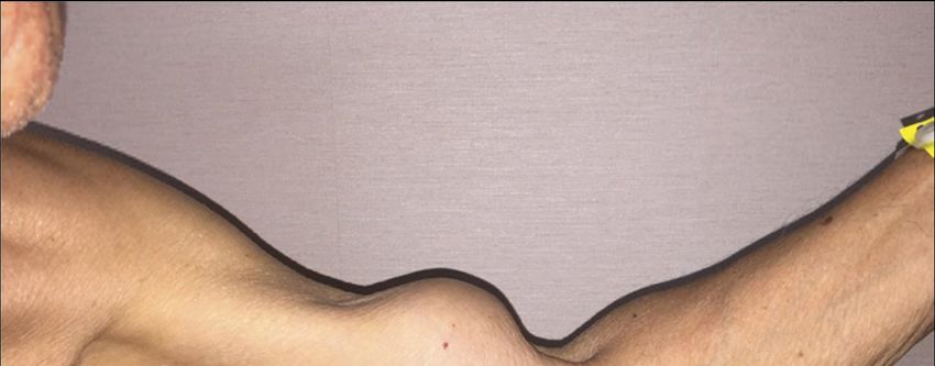
Clinical History
• Patient is a 40yo male with a noncontributory medical history,
  presenting with CC of pain and difficult flexion of the right upper
  extremity.
   • Patient stated that he was at work lifting a pallet of marine batteries when he
     felt a pop.
   • Patient began to experience severe pain and swelling. At which time he
     sought medical care.
   • Initial exam was significant for edema and mild bulging of the anterior
     portion of the upper arm.
   • Initially x-ray was performed at an outside facility as was found to be
     inconclusive. An MRI of the right elbow was performed for greater specificity.

                                McGovern Medical School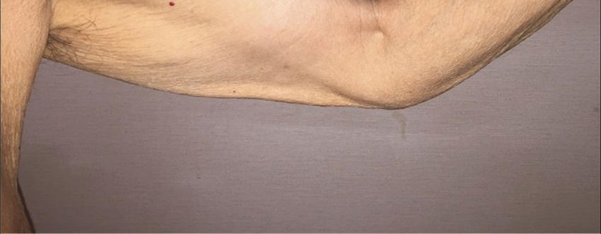
Differential Diagnosis
• Partial/Complete Biceps tendon injury
• Impingement syndrome
• Distal humeral/Radial head fracture
• Rotator cuff injury

                           McGovern Medical School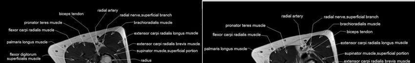
Normal Imaging Axial
• Include relevant imaging with description of the modality, date
  acquired, labeled arrows of normal and abnormal anatomy
• Optional: Side by side comparison of multiple images
   • Showing progression or imaging changes over time
   • Comparison with normal anatomy
      • (please include copy URL for each image obtain through online sources)

                                   McGovern Medical School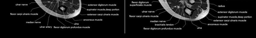
MRI Axial

            McGovern Medical School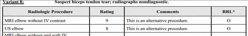
Normal Imaging Sagittal
           Biceps Muscle and
           Tendon                            Triceps

                           Brachialis

                       McGovern Medical School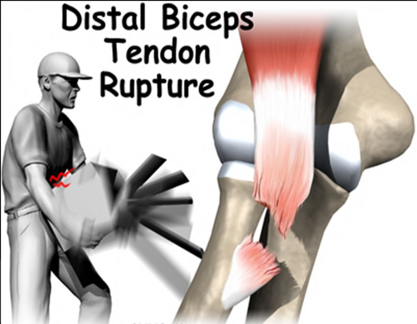
MRI Sagittal

               McGovern Medical School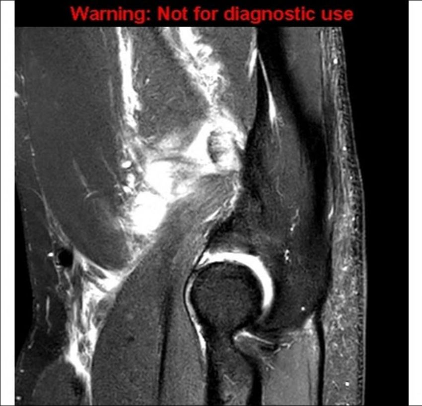
Key imaging findings and diagnosis
• Complete rupture of the distal right biceps tendon from its insertion
  at the radial tuberosity. Mild proximal retraction.

                            McGovern Medical School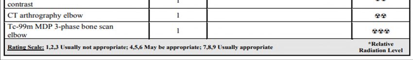
Discussion
• Age, overuse, smoking and corticosteroid use contribute to tendon degeneration
and tendinopathy. Sudden excessive load may break tendon structures, primarily
at the bony attachment. This is thought to be due to a watershed zone of vascular
supply at this area. Proximal injuries are more common in older patients while
distal injuries are seen more frequently in younger patients.
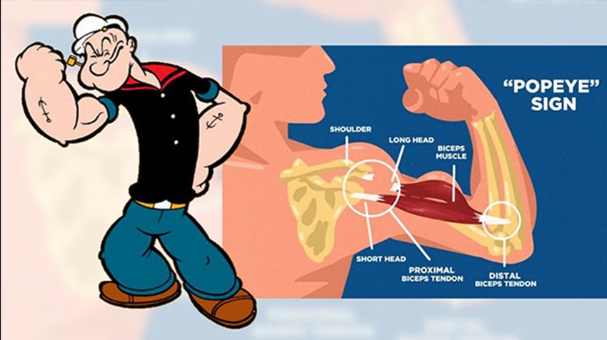
• Tendinous rupture is most often diagnosed clinically on physical exam by the
observation of muscle retraction (Popeye sign). In addition to this the examiner
can attempt to hook their finger under the tendon while the arm is in 90 degrees
of flexion. If the examiner is unable to do so this is further confirmation of a tear.
• Partial ruptures may present with similar, but subtle, symptoms and physical
presentation is usually less a significant weakness or no palpable defect,
sometimes leading to delayed diagnosis.
• Further work up can include: ultrasound, radiographs and much more commonly
MRI for showing the degree of tear.
McGovern Medical School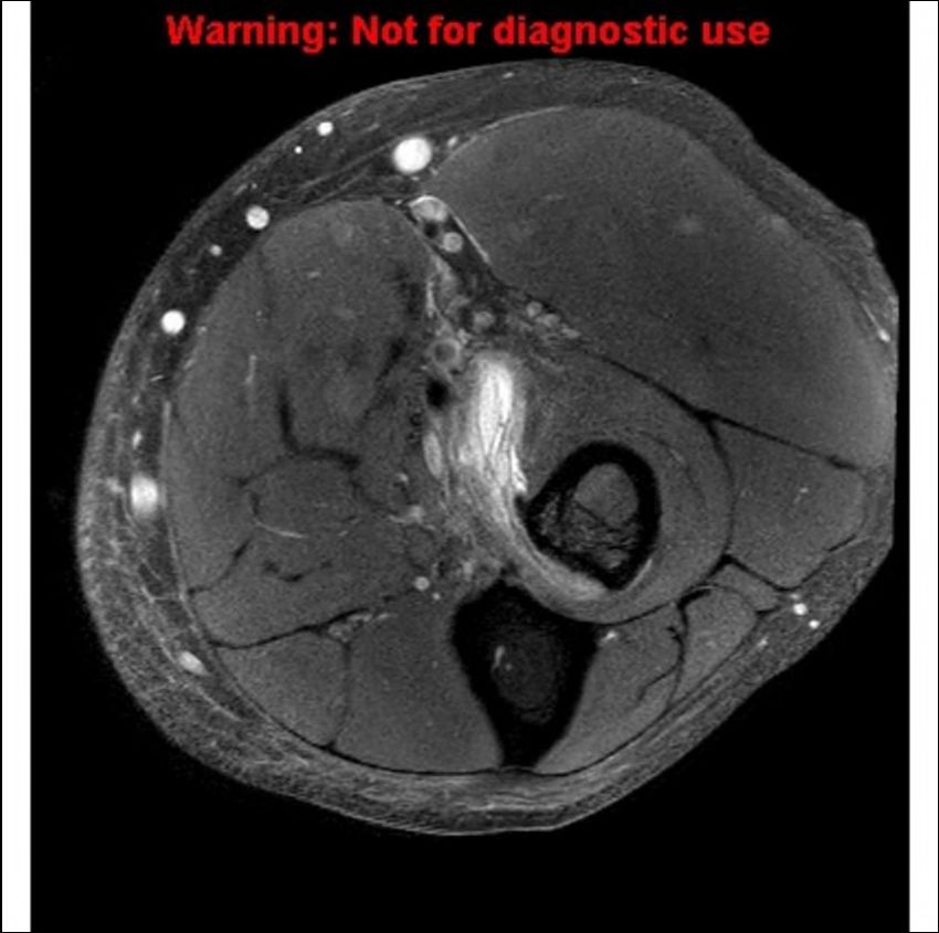
McGovern Medical School
Continued discussion
• Rupture of the biceps tendon affects the strength of flexion and supination
  at the elbow. Despite this there is no absolute indication for surgery.
• Surgical intervention is most commonly performed in younger patients that
  require strenuous use of the upper extremity such as athletes.
• Prognosis is dependent on prompt evaluation, diagnosis, rehabilitation and
  successful operation in severe cases.
• Chronic biceps tendon rupture is defined as tendon tear for more than 4
  weeks. Chronic rupture may be due to missed diagnosis or failure of
  conservative treatment. Partial tear or other coexisting pathology may
  complicate the diagnosis.

                              McGovern Medical School
Final Diagnosis
• Complete rupture of the right biceps tendon at the distal insertion.
McGovern Medical SchoolTreatment and Outcome
• Repair of the tendon was repaired roughly one month post injury,
with removal of the protective cast four weeks later. The patient then
underwent a total of 4 weeks of physical therapy, which was
successful in reconditioning the extremity for normal daily activities.
Precautions were given for heavy lifting and strenuous us in the short
term.
• Patients with complete rupture often return to near full function of
the extremity post repair as long as there is no underlying nerve
damage. This can be seen in the relatively quick improvement in our
patient post-op.
• No post-op imaging was available.
McGovern Medical SchoolACR appropriateness Criteria
• Discuss whether case was in accordance to the ACR appropriateness
guidelines
• Can include a screenshot of the table with the modality completed
highlighted
• Discuss the cost of the imaging throughout the entirety of the case
• Must include url or reference to the cost calculations
McGovern Medical SchoolTake Home Points
• Biceps tendon tears can be striking on initial
physical exam.
• Despite this imaging is sometimes required,
as some cases have only mild retraction
despite complete rupture.
• MRI is the image of choice when grading soft
tissue/tendinous injuries such as this.
• The key to prevention of this injury is to
educate the patient on modifying the risk
factors. After the injury is diagnosed, work
specific or sports specific training is often
recommended before returning to the
original activity. For most patients full
recovery is possible within 8-12 weeks.
McGovern Medical SchoolReferences
• De Maeseneer M, Boulet C, Pouliart N, et al. Assessment of the long head of the biceps tendon of
the shoulder with 3T magnetic resonance arthrography and CT arthrography. Eur J Radiol
2012;81:934-9.
• Chan TW, Dalinka MK, Kneeland JB, Chervrot A. Biceps tendon dislocation: evaluation with MR
imaging. Radiology 1991;179:649-52.
• Dubrow SA, Streit JJ, Shishani Y, Robbin MR, Gobezie R. Diagnostic accuracy in detecting tears in the
proximal biceps tendon using standard nonenhancing shoulder MRI. Open Access J Sports Med
2014;5:81-7.
• https://www.ncbi.nlm.nih.gov/books/NBK513235/#_article-18256_s9_
• https://radiopaedia.org/articles/biceps-brachii-tendon-rupture?lang=us#nav_radiographic-features
• https://www.seattleshoulderdoc.com/biceps-tendon-tear-at-the-elbow-distal-biceps-rupture.html
• http://radsource.us/distal-biceps-tendon-rupture-elbow/
• https://mrimaster.com/anatomy%20elbow%20axial%20%20.html
McGovern Medical SchoolQuestions?
You can also read