Case Report Lymphocutaneous Sporotrichosis Refractory to First-Line Treatment
←
→
Page content transcription
If your browser does not render page correctly, please read the page content below
Hindawi
Case Reports in Dermatological Medicine
Volume 2021, Article ID 9453701, 5 pages
https://doi.org/10.1155/2021/9453701

Case Report
Lymphocutaneous Sporotrichosis Refractory to
First-Line Treatment

          Walter Belda Jr. ,1,2 Luiz Felipe Domingues Passero ,3,4
          and Ana Thereza Stradioto Casolato1
          1
            Dermatology Department, University of São Paulo, Medical School, Clinics Hospital, São Paulo, Brazil
          2
            Laboratory of Pathology of Infectious Diseases, Medical School, University of São Paulo, São Paulo, Brazil
          3
            São Paulo State University (UNESP), Institute of Biosciences, São Vicente, Presidente Prudente, Brazil
          4
            São Paulo State University (UNESP), Institute for Advanced Studies of Ocean, São Vicente, Presidente Prudente, Brazil

          Correspondence should be addressed to Walter Belda Jr.; walterbelda@uol.com.br and
          Luiz Felipe Domingues Passero; felipepassero@yahoo.com.br

          Received 23 June 2021; Revised 21 September 2021; Accepted 23 September 2021; Published 6 October 2021

          Academic Editor: Hristo Dobrev

          Copyright © 2021 Walter Belda et al. This is an open access article distributed under the Creative Commons Attribution License,
          which permits unrestricted use, distribution, and reproduction in any medium, provided the original work is properly cited.
          Sporotrichosis is a fungal infection endemic in Latin America and has been attributed to the thermodimorphic fungus of the genus
          Sporothrix. Transmission to humans occurs during a traumatic injury with soil or organic material; additionally, lesions caused by
          infected cats play an important role in the epidemiology of the disease. The classic treatment of sporotrichosis is performed with
          itraconazole or potassium iodide; second-line medications, such as amphotericin B and terbinafine, can alternatively be used in cases of
          first-line drug failure. In the present study, a patient with lymphocutaneous sporotrichosis in the right upper limb exhibited intolerance
          to itraconazole and potassium iodide, additionally during the period of use; these drugs did not control skin lesions. In this patient,
          amphotericin B deoxycholate and its liposomal version were used in this patient; and complete recovery of the lesions was observed.

1. Introduction                                                            and starts as a paponodular lesion at the site of inoculation
                                                                           and develops into ulcerated nodules that drain the sero-
Sporotrichosis is an infectious disease caused by the ther-                purulent material. Frequently, this lesion spreads, and more
modimorphic fungus that belongs to the genus Sporothrix,                   nodules can be formed following the lymphatic vessels.
and although cosmopolite, it is endemic in Latin America                       The gold standard for diagnosing sporotrichosis is to
[1]. The main forms of transmission to humans are asso-                    identify and isolate the etiologic agent, Sporothrix sp., from
ciated with traumatic injuries that occur with contaminated                cutaneous lesions. Histopathological analysis can be sug-
soil or organic material; infected cats are considered im-                 gestive, but it has low sensitivity due to a reduced number of
portant vectors of this disease and are responsible for epi-               fungal entities; however, it can increase after staining the
demic outbreaks in Latin America, such as Brazil [1, 2].                   tissue with PAS or Gomori; immunolabeling the etiologic
    The clinical manifestations of sporotrichosis are classi-              agent in histological sections can also enhance the sensitivity
fied as cutaneous and extracutaneous. Among the cutaneous                   of diagnosis [2].
forms, sporotrichosis can be further classified as lympho-                      Oral itraconazole, potassium iodide, and terbinafine as
cutaneous, localized cutaneous, and cutaneous caused by                    well as intravenous amphotericin B have been used in the
multiple inoculations. In extracutaneous clinical form, dif-               treatment of sporotrichosis. In cutaneous and lymphocu-
ferent organs or systems can be affected. Furthermore,                      taneous forms itraconazole is recommended as the first-
disseminated sporotrichosis can affect the skin, lungs, si-                 choice drug for all patients [3, 4] at 200 mg per day until
nuses, liver, kidney, eyes, heart, and genitalia [3]. Classical            clinical cure. Patients receiving itraconazole therapy exhibit
sporotrichosis manifests between 2 and 4 weeks after injury                some tolerable side effects, such as headaches, nausea,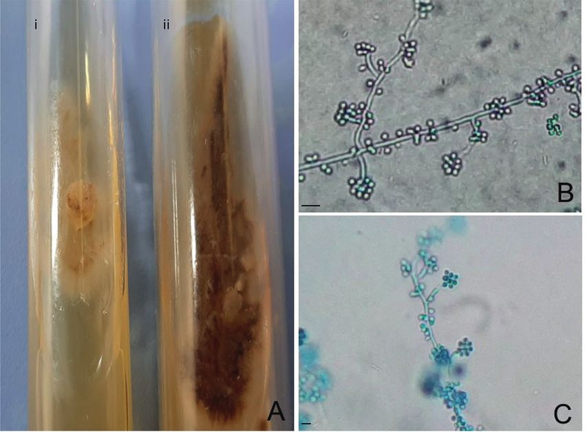
2 Case Reports in Dermatological Medicine
vomiting, and epigastric pain. In cases of failure of treat- showed chronic granulomatous dermatitis with focal necrosis
ment, 400 mg of itraconazole is used per day, divided into and neutrophil exudation of neutrophils (Figures 1(b) and
two doses. Additionally, itraconazole can be replaced by 1(c)).
potassium iodide, initially with 3–5 drops three times a day, Fite-Faraco and Grocott staining did not evidence the
until 40–50 drops (4–6 g/day). This drug was chosen as the presence of alcohol-acid-resistant bacilli or fungi, respec-
first-choice treatment for lymphocutaneous and localized tively. The immunohistochemistry reaction was not positive
clinical forms of sporotrichosis, but was replaced by itra- for BCG. Bacterioscopy and direct fungi research as well as
conazole because the dose, counted in drops, is not as ac- culture of aerobic and anaerobic bacteria were negative. Skin
curate as itraconazole. In addition, frequent side effects of samples cultured in agar-dextrose Sabouraud medium sup-
potassium iodide include metallic taste in the mouth and plemented with chloramphenicol at 25°C allowed the growth
gastrointestinal intolerance [3, 4]. Fluconazole has been of a whitish membranous culture, with a blackened peripheral
considered the second-line treatment, but the efficacy of this halo (Figure 2(a)). Microscopically septate hyaline hyphae
drug is low compared to itraconazole and potassium iodide present in the apex conidiophores and bouquet-like struc-
[5]. In severe forms of sporotrichosis, amphotericin B tures were observed, suggesting a fungus belonging to the
deoxycholate or the liposomal version has been used as the genus Sporothrix (Figures 2(b) and 2(c)). At 37°C, a white/
first-line drug in treatment, but after improvement of the yellowish colony with a smooth aspect was observed; addi-
lesions, it is replaced with itraconazole [6]; furthermore, tionally, yeasts were visualized at this temperature. Tech-
patients with a compromised immune system also may be niques of molecular biology were not used to identify species.
treated with amphotericin B when presenting with fixed Based on these findings, lymphocutaneous sporotri-
cutaneous sporotrichosis [7]. In patients, amphotericin B chosis was diagnosed, and potassium iodide was prescribed
can induce mild and severe side effects, such as fever, chills, at 1 g/mL; it was started with three drops per day and
headache, malaise, hypokalemia, hypomagnesemia, car- gradually increased as the patient tolerated the metallic taste.
diotoxicity, and nephrotoxicity [8], which in fact is a huge After one month of treatment, and using 25 drops of po-
drawback associated with the use of such a drug. tassium iodide per day, the patient reported lack of appetite,
In the present case report, a patient with lymphocutaneous nausea, and vomiting. Potassium iodide was replaced by
sporotrichosis, intolerant to first-line drugs, potassium iodide itraconazole, which was administered orally for 12 days at
and itraconazole, was successfully treated with amphotericin B 100 mg/day; as the patient became tolerant, the dose of this
and presented a significant improvement of lesions. antifungal drug was increased to 200 mg/day. The patient
reported constant nausea and emetic events after seven days
2. Case Report of treatment; however, levels of alanine and aspartate
transaminases, alkaline phosphatase, and gamma-glutamyl
The case was a female patient, 61 years old, with lesions in the transferase were normal. Fifty days after the beginning of
right upper limb that began 8 months before her appointment itraconazole therapy, the patient reported severe gastroin-
in the medical service of Hospital das Clı́nicas, Faculdade de testinal alterations, and the intravenous antiemetic drug
Medicina da Universidade de São Paulo, Brazil. She was born metoclopramide was administered to the patient; constant
in Barros Mendes (Bahia, Brazil), but residing in São Paulo hydration was recommended. Ten days after this event, no
city (São Paulo State, Brazil) for the last 45 years, working as a improvement of skin lesions was observed. Due to the side
teacher. She has three healthy cats that regularly visit vets; effects of both first-line drugs and the resistance of the
according to her report, the cats did not scratch her before the patient to take oral medicines, such as terbinafine or flu-
onset of cutaneous lesions. One year before the appearance of conazole, she was admitted to the Hospital das Clı́nicas to
skin lesions, she traveled to the countryside of São Paulo state initiate a treatment with systemic amphotericin
(Registro town), and although she had contact with the At- B. Additionally, it is important to observe that all these side
lantic forest, skin injury was not observed. Additionally, effects were associated with the therapy with potassium
between travel and appointment at the Hospital das Clı́nicas, iodide or itraconazole since the patient had no underlying
she had no skin injuries or contact with plants and soil. One diseases. Electrolytes, renal, and hepatic biochemical tests as
month after the appearance of the lesion, she was admitted to well as electrocardiogram were performed, before beginning
the Dermatology Service of the Hospital das Clinicas. The treatment. Amphotericin B deoxycholate at 50 mg/day
lesions were ulcerated, and the borders showed erythema and (diluted in 500 mL of sodium chloride 0.9% and glucose 5%
were infiltrated; the lesions were also covered with yellowish solution for 6 hours) was given during three days due to the
and hematic crusts. Furthermore, some ascendant subcuta- inaccessibility of the liposomal formulation. At the begin-
neous nodular lesions were detected that suggest lymphangitis ning of treatment, episodes of nausea and vomiting were
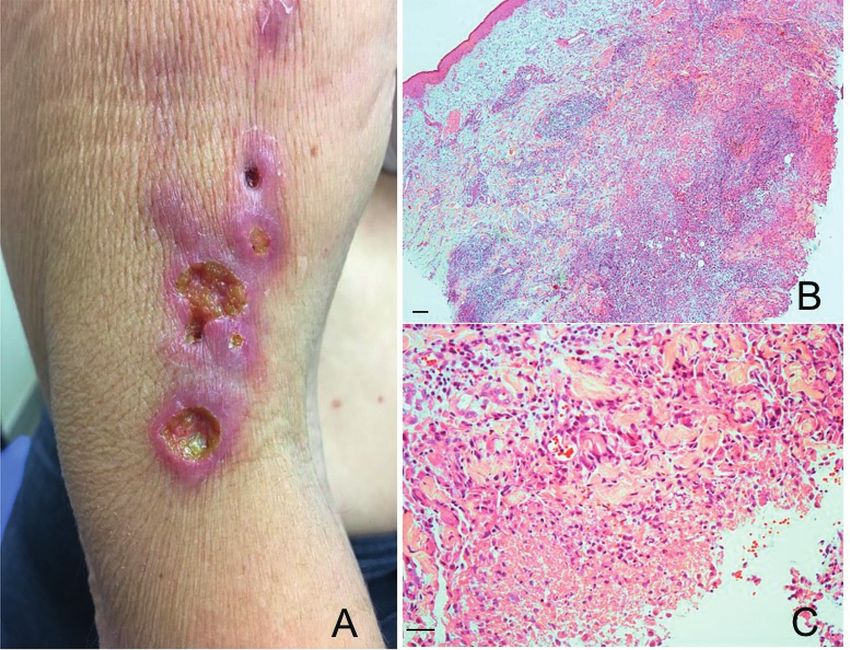
in the right upper limb (Figure 1(a)). Mobile and painless reported, but antiemetic medicines controlled these side
lymphadenopathy was detected in the right axillary area. The effects. Biochemical tests were performed and revealed an
patient complained of pain in the lesions, and discrete hyaline increase of serum potassium and creatinine. Correction of
exudation was verified, and no other local and systemic electrolyte disturbance was carried out; and to compensate
symptoms were observed. Skin fragments were collected to the biochemical alterations, the treatment was suspended for
identify the etiologic agent of the lesions, as the morphology three days. A second set of treatment was performed with
of the lesions suggested lymphocutaneous sporotrichosis, skin liposomal amphotericin B seven days after the infusion with
tuberculosis, or leishmaniasis. The histopathological study amphotericin B deoxycholate, and it was administered at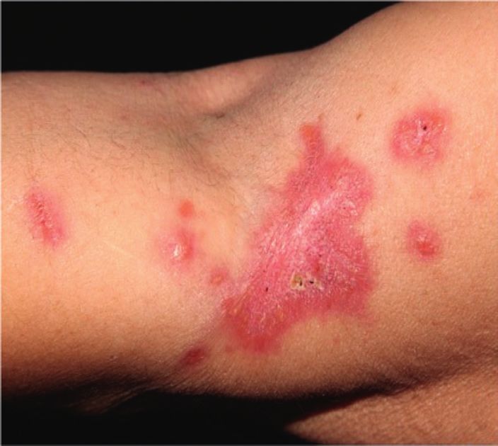
Case Reports in Dermatological Medicine                                                                                                   3

Figure 1: (a) Ulcerated lesions with borders showing erythema and infiltration. (b) Histological section showing the dense inflammatory
infiltrate with the presence of granulomas; magnification 100x. (c) Focal necrosis and neutrophil exudation; magnification 200 ×. His-
tological sections were stained with hematoxylin and eosin. Bars � 5 μm.

Figure 2: Skin samples were cultured in an agar-dextrose Sabouraud medium. (a) (i) In the sample cultured at 37°C, the fungi presented
whitish/yellowish color with leveduriform aspect, and (ii) at 25°C, the fungi presented a brownish color that corresponded to the filamentous
form of (b) fungi with conidiophore bouquet-like structures (c), magnification of 400x and 200x, respectively. Bars � 5 μm.

150 mg/day (diluted in 500 mL of sodium chloride 0.9% and               (Figures 3(a) and 3(b)). A timeline containing the lesion
glucose 5% solution for 6 hours) during 17 days. Clinical               appearance, diagnosis (Figure 4(a)), and treatment with
improvement of the lesions was observed during treatment                iodide potassium, itraconazole (Figure 4(b)), or
4                                                                                                                       Case Reports in Dermatological Medicine

                                            (a)                                                                          (b)

Figure 3: (a) Morphological aspect of the lesions after infusion of 150 mg of amphotericin B deoxycholate and 2600 mg of liposomal
amphotericin B presenting re-epithelization and absence of exudate. (b) Morphology of the lesion after infusion of 2600 mg of liposomal
amphotericin B and 150 mg of amphotericin B deoxycholate, showing complete re-epithelization of ulcerated lesions and absence of exudation.
                                                                        development
                                              Countryside

                                                                                            appoitment
                                                                                            Medical
                                                                        Lesion
                                              travel

                                                                                                             Time (months)
                                                  0                        12                 13
                                                                                           (a)                      Treatment
                                                                                                                    failure

                            Potassiumiodide                 Itraconazole                            Itraconazole
                              3 – 25 drops                   100 mg/kg                               200 mg/kg
                                                                                                                                Time (days)
                           0                30                                    42                                  90
                                                                                           (b)
                                        Amphotericin B                                       Liposomal
                                                                    Treatment
                                         deoxycholate                                      amphotericin B
                                                                    suspension
                                           50 mg/kg                                          150 mg/kg
                                                                                                                      Time (days)
                                    0                           3                     10                      27
                                                                                           (c)

Figure 4: Timeline describing the appearance of the lesion (a); treatment with the first-line drugs potassium iodide and intraconazole (b);
and treatment with amphotericin B (c).

amphotericin B (Figure 4(c)) summarizes the whole treat-                                         Similarly, potassium iodide, although an old drug, has been
ment performed in this patient. The treatment with lipo-                                         efficient at treating human cases [10]. Side effects of such
somal amphotericin B did not change the levels of blood                                          drugs have been associated with gastrointestinal toxicity, as
biochemical parameters.                                                                          reported by the patient who was intolerant and presented
    The patient was infused with a total dose of 2600 mg of                                      lack of appetite, nausea, and vomiting without alteration of
liposomal amphotericin B and 150 mg of amphotericin B                                            biochemical parameters [11]. Although highly toxic,
deoxycholate until complete clinical cure, with re-epitheli-                                     amphotericin B is prescribed for patients with severe clinical
zation of the ulcerated lesion and involution of lymph-                                          forms, such as disseminated sporotrichosis [12–14]. Despite
adenopathy. The follow-up has been performed monthly,                                            the toxicity of amphotericin B, this drug was used in lym-
and relapses were not recorded.                                                                  phocutaneous clinical forms exclusively because the patient
                                                                                                 was intolerant to both first-line drug options. Although not
3. Discussion                                                                                    the standard for the lymphocutaneous clinical form, in-
                                                                                                 travenous treatment with amphotericin B was maintained,
Itraconazole has been used as first-line treatment in cuta-                                       while improvement in clinical signals was observed.
neous and lymphocutaneous forms of sporotrichosis [9].                                           Amphotericin B topically applied was effective at healing
Case Reports in Dermatological Medicine 5
fixed cutaneous sporotrichosis in an immunosuppressed [11] S. L. Xue and L. Li, “Oral potassium iodide for the treatment of
patient that did not tolerate the first-line treatment [7]. In sporotrichosis,” Mycopathologia, vol. 167, no. 6, pp. 355-356,
the present report, although the patient has no underlying 2009.
diseases, the only tolerated treatment was amphotericin [12] C. A. Kauffman, B. Bustamante, S. W. Chapman, and
B. However, further studies need to be performed to un- P. G. Pappas, “Clinical practice guidelines for the manage-
ment of sporotrichosis: 2007 update by the infectious diseases
derstand the reasons of the refractoriness of the patient to
society of America,” Clinical Infectious Diseases, vol. 45,
the first-line drugs. Based on these findings, amphotericin B no. 10, pp. 1255–1265, 2007.
can be considered an interesting example of a successful [13] P. E. Bunce, L. Yang, S. Chun, S. X. Zhang, M. A. Trinkaus,
nonconventional way to treat lymphocutaneous sporotri- and L. M. Matukas, “Disseminated sporotrichosis in a patient
chosis in patients intolerant to first-line drugs. with hairy cell leukemia treated with amphotericin B and
posaconazole,” Medical Mycology, vol. 50, no. 2, pp. 197–201,
Data Availability 2012.
[14] K. Ishida, R. A. Castro, J. J. Torrado et al., “Efficacy of a poly-
The data used to support the findings of this study are aggregated formulation of amphotericin B in treating systemic
available from the corresponding author upon request. sporotrichosis caused by Sporothrix brasiliensis,” Medical
Mycology, vol. 56, no. 3, pp. 288–296, 2018.
Conflicts of Interest
The authors declare that there are no conflicts of interest
regarding the publication of this article.
Acknowledgments
The authors would like to thank HCFMUSP-LIM50.
References
[1] R. Orofino-Costa, P. M. de Macedo, A. M. Rodrigues, and
A. R. Bernardes-Engemann, “Sporotrichosis: an update on
epidemiology, etiopathogenesis, laboratory and clinical
therapeutics,” Anais Brasileiros de Dermatologia, vol. 92, no. 5,
pp. 606–620, 2017.
[2] R. Arenas, C. Sánchez-Cardenas, L. Ramirez-Hobak, L. Ruı́z
Arriaga, and M. Vega Memije, “Sporotrichosis: from KOH to
molecular biology,” Journal of Fungi, vol. 4, no. 2, p. 62, 2018.
[3] V. K. Mahajan, “Sporotrichosis: an overview and therapeutic
options,” Dermatology Research and Practice, vol. 2014, Ar-
ticle ID 272376, 13 pages, 2014.
[4] R. O. Costa, P. M. de Macedo, A. Carvalhal, and
A. R. Bernardes-Engemann, “Use of potassium iodide in
dermatology: updates on an old drug,” Anais Brasileiros de
Dermatologia, vol. 88, no. 3, pp. 396–402, 2013.
[5] L. Garcı́a Carnero, N. Lozoya Pérez, S. González Hernández,
and J. Martı́nez Álvarez, “Immunity and treatment of spo-
rotrichosis,” Journal of Fungi, vol. 4, no. 3, p. 100, 2018.
[6] G. Verma, S. Verma, and R. Rattan, “Lymphocutaneous
sporotrichosis of face with verrucous lesions: a case report,”
Indian Dermatology Online Journal, vol. 10, no. 3, p. 303, 2019.
[7] V. K. Mahajan, K. S. Mehta, P. S. Chauhan, M. Gupta,
R. Sharma, and R. Rawat, “Fixed cutaneous sporotrichosis
treated with topical amphotericin B in an immune suppressed
patient,” Medical Mycology Case Reports, vol. 7, pp. 23–25,
2015.
[8] R. Laniado-Laborı́n and M. N. Cabrales-Vargas, “Ampho-
tericin B: side effects and toxicity,” Revista Iberoamericana de
Micologı́a, vol. 26, no. 4, pp. 223–227, 2009.
[9] M. B. de Lima Barros, R. de Almeida Paes, and
A. O. Schubach, “Sporothrix schenckii and sporotrichosis,”
Clinical Microbiology Reviews, vol. 24, no. 4, pp. 633–654,
2011.
[10] S. Xue, R. Gu, T. Wu, M. Zhang, and X. Wang, Oral potassium
iodide for the treatment of sporotrichosis, vol. 2009, no. 4,
Article ID CD006136, 2009.You can also read