Cerebral Visual Impairment Characterized by Abnormal Visual Orienting Behavior With Preserved Visual Cortical Activation - IOVS
←
→
Page content transcription
If your browser does not render page correctly, please read the page content below

Eye Movements, Strabismus, Amblyopia and Neuro-Ophthalmology

              Cerebral Visual Impairment Characterized by Abnormal
              Visual Orienting Behavior With Preserved Visual Cortical
              Activation
              John P. Kelly,1,2 James O. Phillips,1,3 Russell P. Saneto,4 Hedieh Khalatbari,5
              Andrew Poliakov,5 Kristina Tarczy-Hornoch,1,2 and Avery H. Weiss2
              1
                Roger H. Johnson Vision Clinic, Seattle Children’s Hospital, Division of Ophthalmology, Seattle, Washington, United States
              2
                University of Washington, Department of Ophthalmology, Seattle, Washington, United States
              3
                University of Washington School of Medicine, Department of Otolaryngology, Seattle, Washington, United States
              4
                Seattle Children’s Hospital, Department of Neurology/Division of Pediatric Neurology, Neuroscience Institute, Seattle,
              Washington, United States
              5
                Seattle Children’s Hospital, Department of Radiology, Seattle, Washington, United States

              Correspondence: John P. Kelly,            PURPOSE. Children with cerebral visual impairment (CVI) often have abnormal visual
              Roger H. Johnson Vision Clinic,           orienting behaviors due to impaired or damaged visual cortex. Alternatively, visual-
              Seattle Children’s Hospital,              cortical function is intact but visual information is not transformed downstream into an
              OA.5.342, Seattle, 98105 WA, USA;         appropriate oculomotor output (visuomotor dysfunction). We examined visual, anatomic,
              john.kelly@seattlechildrens.org.          and oculomotor assessments to distinguish visuomotor dysfunction from CVI associated
              Received: January 15, 2021                with severely reduced visual-cortical response.
              Accepted: April 21, 2021
              Published: May 13, 2021                   METHODS. We reviewed the medical records from children with CVI having abnormal
                                                        visual orienting behaviors, normal ocular examinations, and born near term. Relevant
              Citation: Kelly JP, Phillips JO, Saneto   data were visual evoked potentials (VEPs), Teller card acuity, eye movements recorded
              RP, et al. Cerebral visual impairment     by video-oculography (VOG), and neuroimaging (magnetic resonance imaging [MRI])
              characterized by abnormal visual
              orienting behavior with preserved
                                                        including diffusion tensor imaging (DTI) tractography.
              visual cortical activation. Invest        RESULTS. Thirty subjects had visuomotor dysfunction based on a normal VEP; of these 33%
              Ophthalmol Vis Sci. 2021;62(6):15.        had a normal MRI and 67% had white matter abnormalities associated with metabolic
              https://doi.org/10.1167/iovs.62.6.15      disease and/or decreased volume of brain parenchyma. VOG recordings showed smooth
                                                        pursuit gains were uniformly reduced and saccades were dysmetric but followed the
                                                        main sequence. Ten subjects had severe CVI based on VEPs at noise levels; visual acuities
                                                        and MRI findings overlapped those of the visuomotor dysfunction group. Developmental
                                                        delay, seizures, microcephaly, and hypotonia were common across all groups. All subjects
                                                        with an abnormal conventional MRI had abnormal metrics on DTI tractography from the
                                                        occipital lobe.
                                                        CONCLUSIONS. A subset of patients with CVI have abnormal visual orienting behaviors
                                                        despite a normal VEP (visuomotor dysfunction). A majority have abnormal white matter
                                                        metrics on tractography suggesting a downstream defect in sensorimotor transformation.
                                                        Clinically, visuomotor dysfunction is indistinguishable from severe CVI.
                                                        Keywords: visual evoked potentials (VEPs), cerebral visual impairment (CVI), neuroimag-
                                                        ing, eye movements

              C    erebral visual impairment (CVI) is a frequent cause of
                   vision loss in children born in developed countries.1
              CVI is a complex disorder that can be broadly defined as
                                                                                       with CVI cannot communicate verbally or are developmen-
                                                                                       tally delayed, visual acuity can be assessed by preferential-
                                                                                       looking tests (e.g. Teller acuity cards) or by visual evoked
              impaired visual orienting behaviors with evidence of a corti-            potentials (VEPs). The VEPs provide objective assessment
              cal deficit that cannot be accounted for by ocular pathol-               of visual-cortical function in children with CVI.2,3,7 The
              ogy or an oculomotor disorder.1,2 Common causes of CVI                   VEP reflects population activity in both visual striate and
              include hypoxic/ischemic injury or cerebral infarct (often               visual association cortices8–10 predominately from central
              in the setting of prematurity), hydrocephalus, meningitis,               15 degrees of the visual field.11,12
              and cortical malformations.1–6 CVI is frequently associated                  Abnormal visual orienting behaviors in CVI are often
              with other neurological deficits, such as cognitive delay,               attributed to reduced vision from damage or dysfunction
              cerebral palsy, and seizures. Magnetic resonance imaging                 of striate and extra-striate cortex. In this case, spatial and
              (MRI) helps to localize and define the underlying brain                  temporal information from visual cortex is too degraded
              pathology in CVI. Because most children who present                      for planning and execution of precise visually guided eye

              Copyright 2021 The Authors
              iovs.arvojournals.org | ISSN: 1552-5783                                                                                              1

                                   This work is licensed under a Creative Commons Attribution 4.0 International License.

Downloaded from iovs.arvojournals.org on 07/10/2021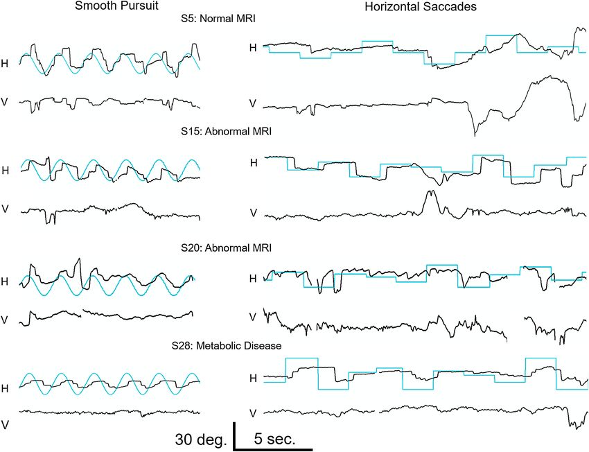
Visuomotor Dysfunction                                                           IOVS | May 2021 | Vol. 62 | No. 6 | Article 15 | 2

              movements. Alternatively, striate and extra-striate cortex        cal specialists for concerns of reduced vision, lack of visual
              could be functionally intact but transmission of visual           orienting, or poor tracking to high contrast toys, with or
              sensory information to downstream visual cortical and             without associated neurological deficits. Inclusion criteria
              oculomotor areas is diminished or completely interrupted.         for the visuomotor dysfunction group were (1) clinically
              Specifically, there can be an anatomic or functional discon-      abnormal visual tracking/orienting behaviors to targets at
              nection to parietal cortex, frontal cortex, the cerebellum,       near or distance (see the eye movement section below)
              vermis, midbrain, and brainstem. Given an inability to gener-     that could not be explained by ocular motor apraxia or
              ate appropriate visually guided behaviors, visual acuity          other motility disorders; (2) a VEP in the normal range
              using preferential-looking acuity cards may be difficult to       confirmed by objective analysis of electroencephalogram
              interpret because the test requires reliable gaze shifts.1 For    (EEG) epochs; (3) normal dilated fundus examination; (4)
              example, some children with CVI, who were initially diag-         gestational age ≥ 34 weeks; (5) neuroimaging by MRI
              nosed as being visually impaired on preferential-looking          without evidence of intraventricular hemorrhage, hypoxia-
              acuity cards, were eventually found to have normal visual         ischemia, cerebral infarct, traumatic brain injury, hydro-
              acuity after repetitive testing.13,14 Therefore, the VEP can be   cephalus, or significant cortical migration defect (e.g. pachy-
              critical for assessment of vision in these children if there is   gyria, polymicrogyria, and schizencephaly); and (6) devel-
              a defect in sensorimotor transformation into visually guided      opmental assessment by a pediatrician. Results of genetic
              behaviors.                                                        and metabolic testing were recorded for subjects subse-
                  Evidence that some children with CVI have a defect            quently found to have white matter changes in the presence
              in sensorimotor transformation to downstream oculomo-             of metabolic disease or deficiencies of mitochondrial elec-
              tor areas have been published in several case studies.15–17       tron transport (defined as 20% of control values).22 Inclu-
              Bodis-Wollner et al.15 reported a “blind” child that had a        sion criteria for the severe CVI group was a VEP wave-
              normal VEP response but damage to extra-striate areas in          form and signal at background noise levels (see the VEP
              the occipital lobe. The abnormal behaviors were attributed        section below) but required DTI studies, and did not exclude
              to disconnection between the striate visual cortex and the        hypoxic-ischemic injury.
              downstream cortical areas. Wygnanski-Jaffe et al.16 reported          All eye examinations were performed at Seattle Chil-
              a subset of children with CVI having robust responses on          dren’s Hospital that included external examination, motility,
              VEPs despite appearing “clinically blind.” These authors          and dilated fundus examination. Preferential-looking assess-
              noted several of their subjects had delayed myelinization or      ments by Teller Acuity Cards were performed by trained
              volume loss of deep white matter. Previously, we described a      observers in the Ophthalmology Clinic. To standardize visual
              group of infants with extensive neuronal migration defects        acuity measured at different ages, best corrected visual
              beyond visual cortex (polymicrogyria) having robust VEPs          acuity was converted to age-corrected log minimum angle
              despite subnormal visual tracking behaviors.17 In further         of resolution (logMAR).23 The logMAR value represents the
              support of defects in sensorimotor transformation, there are      logarithmic deficit relative to the average normal acuity for
              case reports of premature children with CVI showing abnor-        the equivalent age. For example, if the normal average was
              mal white matter tractography from visual cortex to dorsal        3.1 cycles/degree, a subject with 1.6 cycles/degree acuity of
              and ventral processing streams on diffusion-tensor imaging        the same age would have an age corrected logMAR value of
              (DTI).18–20                                                       0.3. Another child of the same age with 0.31 cycles/degree
                  The purpose of this study was to characterize chil-           acuity would have an age corrected logMAR value of 1.0,
              dren with CVI in which visual-cortical function is intact         and so forth. Reporting values in age-corrected logMAR is
              but visual information is not transformed into an appro-          more accurate than Snellen optotype acuity (the latter is not
              priate oculomotor output (“visuomotor-dysfunction”). We           always equivalent) and indicates the amount of visual-acuity
              hypothesized there would be consistent disorders across           deficit compared to normative values for age.
              eye movement studies (saccadic, smooth pursuit, optokinetic           All subjects had clinical assessment of eye movements.
              nystagmus [OKN], and gaze holding) in those with visuo-           The spectrum of abnormal visual tracking/orienting behav-
              motor dysfunction. We also searched for abnormal white            iors was comparable to that described by Jan et al.14 In
              matter tracts connected with striate cortex. To address these     addition to assessing versions, the vestibulo-ocular reflex
              hypotheses, we reviewed cortical function assessed by VEPs,       could be examined qualitatively to confirm the oculomotor
              preferential-looking visual acuity, ophthalmological clinical     system was intact. Our specific criteria were one or more
              findings, oculomotor recordings, and white matter metrics         of following: (1) gaze holding was defined as abnormal if
              using DTI tractography. Because abnormal visual orienting         nystagmus was present; (2) smooth pursuit was abnormal
              behaviors in CVI should be distinguished from that caused         if there was an absence (saccadic tracking or no response)
              by visual cortical dysfunction, we compared the visuomotor-       or a reduction (reduced gain) in smooth eye movement
              dysfunction group to children with CVI having DTI imag-           in response to a sinusoidally moving target; (3) saccades
              ing and severely reduced VEPs (“severe CVI”). Confounding         were abnormal if they were absent, dysmetric (hypo- or
              factors were reduced by excluding subjects with complica-         hyper-metric), slow, or had abnormal latency in response
              tions of prematurity, cortical migration defects, or delayed      to sequentially presented targets; and (4) OKN was abnor-
              visual maturation.3,21                                            mal if the response was absent, nystagmus was poorly
                                                                                formed, or the gain was abnormal. When possible, eye
              METHODS                                                           movements were recorded using binocular infrared video-
                                                                                oculography (Sensorimotoric Instruments, Berlin) as previ-
              The research was conducted in compliance with accredited          ously described.24 The subject viewed a 60 degrees projec-
              institutional review board approval. Patient records from         tion screen at 80 cm. Saccades were elicited by random
              Seattle Children’s Hospital were retrospectively reviewed.        5 degrees to 20 degrees horizontal target steps, and smooth
              All subjects were referred to Seattle Children’s Hospital         pursuit by sinusoidal target drift ±10 degrees along the
              Ophthalmology Clinic from a wide range of outside medi-           horizontal meridian at peak velocities of 10, 20, and

Downloaded from iovs.arvojournals.org on 07/10/2021
Visuomotor Dysfunction IOVS | May 2021 | Vol. 62 | No. 6 | Article 15 | 3
30 degrees/sec. OKN was elicited by square wave gratings a stationary and deterministic VEP signal (having constant
(0.1 cycles/degree; 80% contrast) drifting horizontally or amplitude and latency throughout the averaging process). In
vertically at constant velocities of 15, 30, and 45 degrees/sec. contrast, eye movement artifacts and abnormal background
Eye movements were analyzed using custom software (http: EEG generate noise variations in both amplitude and phase
//faculty.washington.edu/jokelly/voganalysis). in the Fourier transform.25,27 Epochs were converted into
VEP recordings followed published procedures25 using magnitude and phase plots at temporal frequencies from 5.5
International Society for Clinical Electrophysiology of Vision to 21.9 Hz. Then the signal at each frequency was taken as
(ISCEV) recommendations.12 The reference was at Cz, the ratio of its magnitude to the 95% confidence circles in
ground at Pz, and an active electrode at Oz. Additional loca- the phase plot. A signal-to-noise ratio (SNR) was defined as
tions at either O1, O2, T5, T6, Inion, and POz were used the sum of all ratios across all temporal frequencies. The
to analyze hemispheric symmetry or EEG artifacts and were algorithm then reiteratively selects EEG epochs with consis-
not reported. Subjects viewed a raster monitor (Eizo TX-C7, tent amplitudes and phases (lower 95% confidence circles)
22 × 17 degrees, 100 Hz frame rate) in darkness. Patterns until there is no improvement in SNR. A large SNR indicates
were reversing-checks (163, 84, 18, and 9 arc minutes, EEG epochs have large amplitude components with accurate
80% contrast, 2.74 reversals per second) and onset-offset of phase-locking to the stimulus presentation. After the reiter-
sinewave grating onset (0.5 cycles/degree; 80% contrast; on ation, the accepted epochs are averaged in the time domain
for 150 ms then off for 500 ms at constant mean luminance). to generate the FT-selective VEP. If the SNR was ≤ 1.3 after
However, inadequate fixation limited VEP data analysis to FT-selective averaging, then the signal was not statistically
163 arc minute checks and 0.5 cycles/degree grating across different from background noise,25 in which case, the algo-
all subjects. Additional check sizes for extraction of grating rithm lacks a phase-locking signal and outputs the standard
acuity26 were available in a subset of subjects but only used average. After each averaging method, VEPs were digitally
to confirm detectable responses to smaller check sizes. Fixa- filtered 1.5 to 41.3 Hz, then scored for latency (time to the
tion was aided by a small toy at the center of the display prominent positive peak after 80 ms) and amplitude (volt-
and VEPs were recorded during an alert state. The record- age difference between this peak and the preceding negative
ing was paused when fixation (by corneal light reflex) was deflection if present, or baseline). Control data were taken
not directed to the stimulus. Each time the recording was from published data using an identical analysis.25
resumed, an additional trial was added for averaging. There The MRI protocol included high-resolution sagittal T1
was a minimum of 40 stimulus presentations per stimulus, magnetization-prepared rapid acquisition of gradient echo
which could be doubled if the child was inattentive. (MPRAGE; repetition time [TR] ms = 1450–1950; echo time
Given the abnormal visual behaviors and seizures in [TE] ms = 2; field of view (FOV) mm, 190–256), axial and
these subjects, specialized averaging methods were used. coronal T2-weighted spin-echo (TR = 3200–5720 ms; TE =
For each stimulus, EEG epochs were overlaid on top of 75–386 ms; FOV 146–250), axial fluid-attenuated inversion
each other. Epochs with spikes or large background noise recovery (FLAIR). All MRIs were reviewed by a pediatric
were individually removed by visual inspection. The Oz neuroradiologist.
location then underwent “standard” averaging. The same DTI scans were acquired on Trio or Prisma scanners
epochs then underwent discrete Fourier transforms for “FT- at 3 Tesla (Siemens, Erlangen, Germany) using a single-
selective” averaging.25 FT-selective averaging also assumes shot echo planar imaging sequence (TR = 4300–6813 ms;
TABLE. Summary Data
VMD With Normal MRI VMD With Abnormal MRI VMD with Metabolic Disorder Severe CVI All Controls
N 10 14 6 10 26
Mean age 2.2 (1.6) 1.5 (1.1) 2.5 (3.7) 1.1 (0.6) 2.1 (1.5)
logMAR 1.9 1.9 1.4 2.6
Range 3.1 to < 0.23 cy/deg 6.5 to < 0.23 cy/deg 9.8 to < 0.23 cy/deg 1.3 to < 0.23 cy/deg
Stable gaze 100% 86% 50% 50%
Strabismus 40% 57% 67% 50%
Microcephaly 20% 36% 50% 50%
DD 90% 93% 100% 100%
Hypotonia 50% 93% 100% 70%
Seizures 50% 64% 33% 80%
Check VEP† Mean (SD) Mean (SD) Mean (SD) Mean (SD) Mean (SD)
Amplitude 38.0 (20.4) 38.2 (14.9) 22.5 (7.7) 5.0 (2.9)*** 36.6 (11.2)
SNR 72.4 (26.1) 73.6 (20.3) 68.0 (6.2) 1.2 (0.6)*** 112.1 (61.6)
Latency 98.0 (8.3) 114.4 (27.4) 109.3 (16.6) 126.4 (31.2) 102.9 (7.2)
Onset VEP†
Amplitude 43.9 (26.4) 34.8 (24.8) 29.3 (11.3) 10.9 (13.4)** 33.8 (11.0)
SNR 67.8 (19.0) 54.6 (23.6) 63.3 (21.0) 12.3 (24.0)*** 70.1 (30.6)
Latency 115.0 (30.9) 130.5 (61.7) 103.8 (11.8) 130.3 (35.3) 100.4 (11.6)
Age in years; logMAR visual acuity corrected for age; cy/deg is grating acuity in cycles/degree; microcephaly is < 5% of normative data
for occipital frontal circumference.
VMD = visuomotor dysfunction; CVI = cortical visual impairment; DD = developmental delay.
† Averaging using the FT-selective method; P values statistically significant from controls on t-test after Bonferroni correction.
** = P < 0.001.
*** = P < 0.0001.
Downloaded from iovs.arvojournals.org on 07/10/2021Visuomotor Dysfunction IOVS | May 2021 | Vol. 62 | No. 6 | Article 15 | 4
FIGURE 1. Sample eye movement recordings in four subjects (#5 with a normal MRI, #15 with delayed myelination, #20 with delayed
myelination and partial agenesis of the corpus callosum, #28 with delayed myelination and respiratory complex I, IV deficiency; top to
bottom). The left side shows smooth pursuit task of a target sinusoidally moving at 30 degrees/s. Horizontal (H) and vertical (V) eye position
traces are shown with the target position (blue traces). The right side shows targeted saccade. Both the horizontal (H) and vertical (V) eye
position traces are shown with the target position (blue traces).
TE = 65–96 ms; and FOV = 190–230 mm). The b-values ciated fiber tract from the HCP842 atlas using Hausdorff
for DTI were 0 and 1000 s/mm2 . Two DTI acquisition distances. Streamlines were retained only if they matched
schemes occurred during the study period: (1) a sequence the atlas target track. If the automated tracking method
of 30 gradient-encoding directions with in-plane resolution found less than 30 fiber tracts, the region was manually re-
of 2 mm and slice thickness of 2.2 mm (matrix size 112 seeded, and the anisotropy threshold set at 0.17 based on
× 112) with 55 to 65 sections covering the entire brain; pediatric subjects with abnormal white matter.30 Fiber track-
(2) a sequence of 10 gradient-encoding sampling direc- ing was chosen for projections to and from the occipital
tions with in-plane resolution of 1.8 mm and slice thick- lobe that included the optic radiations (ORs), inferior fronto-
ness of 3.3 to 3.5 mm (matrix size 128 × 128) with 34 to occipital fasciculus (IFOF), superior longitudinal fasciculus
50 sections covering the entire brain. DTI data and deter- (SLF), vertical occipital fasciculus (VOF), and corpus callo-
ministic fiber-tractography were analyzed using DSI Studio sum (CC). Additional tracks included the cortico-pontine
(http://dsi-studio.labsolver.org), which showed good valida- tracts; fronto-pontine (FP), occipito-pontine (OP), parieto-
tion with weak MR signals.28,29 Scans had to meet qual- pontine (PP), temporo-pontine (TP), and white matter of the
ity criteria of DSI Studio. Tracking parameters were default cerebellum (Cbl) and vermis. Fiber tracts or U-shaped fibers
anisotropy threshold, 60 degrees angular threshold, 0.1 mm were removed if clearly aberrant. For isolation of the OR,
step size, track length between 30 and 400 mm, and a maxi- we removed fibers anterior to the lateral geniculate body
mum of 10,000 seeds. DSI Studio performed automatic seed- and fiber crossing regions near Meyer’s loop. Fibers of the
ing of white matter tracts based on the HCP842 tractography splenium were only included in the reconstruction of the CC.
atlas with an internal fractional anisotropy threshold. The The CC, OP, TP, and PP tracts were reviewed to exclude fibers
software used nonlinear registration of subject data to MNI from the corticospinal tract, which appear as long tracts
space, then placed seeds within the HCP842 tract volume. vertically extending from the corona radiata and past the
The generated streamlines were compared to each asso- pons. We report the average fractional anisotropy (FA), radial
Downloaded from iovs.arvojournals.org on 07/10/2021Visuomotor Dysfunction IOVS | May 2021 | Vol. 62 | No. 6 | Article 15 | 5
amplitude, latency, and SNR in the range of controls (after
FT-selective averaging). The remaining 10 subjects classified
as having severe CVI based on a VEP waveform and SNR
at background noise levels. The severe CVI group served as
a comparison group only (Table). The visuomotor dysfunc-
tion group was further separated into three groups. The
first group (n = 10) had a normal MRI (“normal MRI”). The
second group (n = 14) had an abnormal conventional MRI
(“abnormal MRI”) predominated by bi-hemispheric abnor-
mality of the white matter (delayed myelination, abnormal
signal, and thinning of the corpus callosum). The third group
(n = 6) had low-normal VEP amplitudes with an inborn
error of metabolism also with predominately white matter
signal abnormalities on MRI (“metabolic disorder”). The
spectrum of MRI abnormalities in the severe CVI group over-
lapped that of the visuomotor dysfunction groups. Almost
all subjects in each group had developmental delay in
gross motor and cognitive skills. A majority of subjects had
hypotonia; seven had spasticity or hypertonia, and three
had dystonia. Genetic testing or biochemical assay in 25
subjects revealed a heterogenous range of chromosomal,
genetic, or metabolic disorders (Supplementary Table S1).
Seven subjects had genetic mutations associated with infan-
tile epilepsy.
The Table summarizes clinical findings, visual acuity, and
VEP data for the four groups. All logMAR acuities represent
age-corrected values. At presentation, 22 of the 40 subjects
had no response to the low vision Teller card (worse than
0.23 cycles/degree) and were assigned a logMAR value of
3.0. Differences in logMAR were not statistically significant
between groups (P > 0.25, Mann Whitney test; P = 0.42,
Kruskal Wallis test). Follow-up visual acuity was available
in 19 of the visuomotor dysfunction subjects. For these
subjects, logMAR averaged 2.0 at presentation and averaged
1.3 at their last visit after a mean follow-up of 2.7 years.
Follow-up visual acuity was available in 11 subjects who
FIGURE 2. The relationship between saccade peak velocity and had measurable acuity at presentation. Of these subjects,
saccade amplitude (main sequence) for horizontal saccades. (A) A logMAR averaged 0.84 at presentation and averaged 0.76 at
subject with a normal MRI, (B) 9 subjects with an abnormal MRI, their last visit after a mean follow-up of 2.5 years. Despite
and (C) 12 control subjects. Open circles and solid line fits are for abnormal eye movements (lack of visual orienting / track-
saccades generated in the correct direction. Crosses and dotted line
ing, random conjugate saccades, hypometric saccades, and
fits are for nontargeted saccades generated in the incorrect direc-
tion. The lines are third-order polynomial fits. absence of smooth pursuits), four subjects were found to
have mild acuity deficits for age (logMAR 6 months (ranges Gaze-holding was mostly stable across subjects. For others,
when VEPs are stable with age).25,31 gaze-holding was either nontargeted roving eye movements,
DTI imaging controls were matched for age, DTI proto- conjugate jerk nystagmus, or rotary nystagmus of < 10
col, and acquisition date within 6 months, from a database of degrees amplitude (Table). None had characteristics of
children without visual disorders, with normal neuroimag- congenital oculomotor apraxia. Strabismus was frequent in
ing (T1-MPRAGE, T2, T2/FLAIR, and ADC) done for isolated all groups. Due to technical considerations and coopera-
seizures, developmental delay, Rolandic epilepsy, unex- tion, assessments of eye movements by video-oculography
plained isolated events, or migraine. were limited to 11 subjects (n = 11 for OKN, n = 10 for
horizontal saccades, and n = 8 for smooth pursuit), all of
RESULTS whom were in the visuomotor dysfunction group. Figure 1
shows eye movement data from 4 subjects. For horizon-
Out of 550 records, 40 subjects met the inclusion criteria tal smooth pursuit, subjects had saccadic tracking that was
(16 boys; mean age at first visit = 1.4 years, median = primarily hypometric. One subject (#20 with delayed myeli-
0.9 years of age). All subjects were nonverbal at the time nation) responded with a brief period of a targeted hypomet-
of VEP testing. Visual acuity, MRI, and laboratory findings ric saccade and low gain smooth pursuit. Average horizontal
are listed in Supplementary Table S1. Thirty subjects were smooth pursuit gain across subjects was 0.28, 0.22, and 0.17
classified as having visuomotor dysfunction based on VEP for velocities of 10, 20, and 30 degrees/s, respectively (Visuomotor Dysfunction IOVS | May 2021 | Vol. 62 | No. 6 | Article 15 | 6
FIGURE 3. Sample visual evoked potential (VEP) waveforms compared to age-appropriate controls. Stimuli were reversing checkerboards.
For each panel, the upper trace shows the VEP after FT-selective averaging while the lower trace is the raw average. The same subjects
shown in Figure 1 are shown in this figure. (A) Waveforms from the subject with a normal MRI. (B, C) Waveforms from two subjects with
abnormal MRI. (D) Waveforms from the subject with complex I, IV deficiency. (E) Sample waveforms from 7 controls of 0.3 to 0.9 years age.
(F) Waveforms from 7 controls of 1 to 3 years age. (G) Range of waveforms from subjects with severe CVI.
of our controls24 ). Subjects also had large vertical compo- Visual Evoked Potentials
nents not seen in our control children on horizontal tasks.
There were no significant differences in smooth pursuit gain Thirty-six controls were selected to match the age range of
between subjects with or without MRI abnormalities. each group (t-test for age differences; P > 0.83). Figure 3
Saccades elicited to horizontally stepped targets were plots representative VEP waveforms, including the same
highly variable (see Fig. 1). Subject 5 (top row) intermittently subjects shown in Figure 1 (VEPs recorded 3–6 months
generated hypometric saccades to leftward target steps with before eye movements). Figure 3 demonstrates the VEPs
prolonged saccade latency. Subject 15 generated normo- in the visuomotor dysfunction were similar to controls
metric, horizontal saccades with prolonged latency. Subject despite abnormal visual acuity and oculomotor behaviors.
20 showed only one hypometric saccade in response to a FT-selective averaging generated larger responses without
stepped target. Subject 28 generated multiple hypometric altering peak latencies (paired traces in Fig. 3).
saccades in response to target steps but failed to acquire the Inspection of each subject’s raw EEG epochs confirmed
target. Analyzing all saccades, regardless whether they were the FT-selective algorithm extracted epochs with improved
generated in the appropriate direction, subjects showed a phase-locking to the stimulus compared to standard aver-
systematic relationship between peak velocity and saccade aging. Compared to standard averaging, FT-selective aver-
amplitude (i.e. main sequence). Despite the uniform dysme- aging increased VEP amplitudes by a factor of 1.6 (SD =
tria across trials, there was a systematic main sequence 0.46, P < 0.0001) in controls and by a factor of 3.4 (SD
across subjects (Fig. 2). = 2.36, P < 0.0001) in visuomotor dysfunction subjects.
Horizontal and vertical OKN gains varied widely across SNR was also significantly increased after FT-selective aver-
subjects (Supplementary Fig. S1). For horizontal drifting aging in controls (factor of 1.7, SD = 0.59, P < 0.0001)
gratings, most subjects had reduced OKN gain relative to and in visuomotor dysfunction subjects (factor of 3.3, SD =
controls for both rightward and leftward directions, espe- 1.7, P < 0.0001). Latency was not affected by FT-selective
cially at higher velocities. There were no consistent differ- averaging (difference for controls = −0.7 ms, P = 0.53;
ences in OKN gains between subject groups. difference for visuomotor dysfunction subjects = 1.88 ms,
Downloaded from iovs.arvojournals.org on 07/10/2021Visuomotor Dysfunction IOVS | May 2021 | Vol. 62 | No. 6 | Article 15 | 7
as there was no detectable signal for the FT-selective aver-
aging algorithm.
Neuroimaging
Abnormalities on conventional MRI were common, yet
nonspecific among groups. The dominant finding was
abnormal white matter (abnormal signal, delayed myeli-
nation, or reduction in white matter volume) in 23 of 40
subjects. Fourteen subjects had evidence of cerebral volume
loss, and six subjects with decreased volume of the cere-
bellum. Six subjects showed either full or partial maturation
of their delayed myelination on a follow-up MRI study (2
had a metabolic disorder). Four subjects had mega-cisterna
magna. One subject had abnormal signal in the midbrain,
and one subject had brainstem atrophy. Only one subject in
the severe CVI group had hypoxic ischemic injury.
Diffusion Tensor Imaging
Adequate DTI data were obtained in 28 study subjects (n =
8 with normal MRI, n = 6 with abnormal MRI, n = 5 with
metabolic disease, and n = 9 with severe CVI). White-matter
tracts could be reconstructed in 555 of a total 560 tracts. One
subject with severe CVI had no detectable FP or IFOF tracts
bilaterally. Another subject with metabolic disease had no
detectable right OP tract. Control DTI and tractography anal-
yses consisted of a database of 43 separate children. Recon-
structed tracts (Fig. 4) in visuomotor dysfunction subjects
had similar shape and location to controls, but some subjects
showed reduced volume.
Controls had linear relationships of mean FA and RD with
log age (Fig. 5), as previously reported.32–34 Subjects showed
variable decreased FA and increased RD across multiple
tracts (MD data were similar to RD data). The number of
gradient directions had a small but detectable effect on mean
FA and RD values. We used analysis of covariance (ANCOVA)
to compare subjects and controls while accounting for vari-
ance associated with number of gradient directions, voxel
volume, and age. For ANCOVA, we matched a subset of
controls (n = 36) to the subjects based on the number of
gradient directions, MRI scan date within 6 months, and age
range (mean age = subjects 2.8 years, controls = 3.6 years, P
= 0.3 by t-test). We then analyzed the marginal means, which
represent averages after adjusting for the effects of number
FIGURE 4. Reconstruction of four major white matter tracts in six
of gradient directions, voxel volume and log age. Figure 6
subjects with visuomotor dysfunction (left side) paired with an age-
matched control under the same imaging sequence (right side). and Supplementary Figure S2 show subjects with a normal
Tracts shown are optic radiations in yellow, vertical occipital fascicu- conventional MRI were not different from controls across
lus in purple, superior longitudinal fasciculus in green, and inferior all white matter tracts for FA, RD, MD, and tract volumes.
fronto-occipital fasciculus in cyan. (A–F) Show data from subject Subjects in the abnormal MRI, metabolic disease, and severe
numbers 15, 25, 6, 4, 12, and 28, ordered in age from 0.5 years (top) CVI groups all had evidence of reduced FA, increased RD
to 14 years (bottom).
and MD, and lower volumes across multiple white matter
tracts. FA, in particular, showed significantly reduced values
for almost all tracts of interest (OR, IFOF, VOF, SLF, CBL, CC,
FP, OP, PP, and TP).
P = 0.29). Amplitude, latency, and SNR were not correlated Across all subjects, only the OR and OP white matter
with the number or percentage of epochs accepted after the tracts were significantly correlated with VEP amplitude and
FT-selective averaging technique (r2 < 0.11 for all correla- SNR (P < 0.01, Bonferroni correction). Amplitude was posi-
tions). The Table shows controls and visuomotor dysfunc- tively correlated with FA (r = 0.83 and 0.81 for OR and OP
tion subjects were not significantly different from each tracts, respectively) and negatively correlated with RD (r =
other after FT-selective averaging. The severe CVI group had −0.65 and −0.70 for OR and OP tracts). SNR was positively
significantly reduced VEP amplitude and SNR compared to correlated with FA (r = 0.70 and 0.66 for OR and OP tracts)
controls, but latencies were not statistically longer. Of note, but not for RD (r = −0.33 and −0.39 for OR and OP tracts).
only standard averaging was used for the severe CVI group Latency was not correlated with FA, RD, MD, or tract volume.
Downloaded from iovs.arvojournals.org on 07/10/2021Visuomotor Dysfunction IOVS | May 2021 | Vol. 62 | No. 6 | Article 15 | 8
FIGURE 5. Representative mean fractional anisotropy (left side) and mean radial diffusivity (right side) plotted with respect to age on a
log scale. Controls are plotted as black circles (solid regression lines are data acquired at 30 directions, whereas dotted regression lines are
data acquired at 10 directions). Subjects with a normal and abnormal appearing conventional MRI are plotted as grey and open circles,
respectively). Both hemispheres were averaged for each subject. Separate graphs show white matter tracts for the optic radiations (OR),
inferior fronto-occipital fasciculus (IFOF), and vertical occipital fasciculus (VOF).
DISCUSSION tion or loss of white matter volume. Previously, we reported
robust VEPs despite absent visual tracking in infants with
We describe a group of children with clinical characteris- CVI due to extensive neuronal migration defects.17 Unlike
tics of CVI with normal VEPs who show absent, or abnor- previous studies, our analysis links VEPs, eye movement
mal, oculomotor responses to visual stimuli. Two thirds of recording, neuroimaging, and DTI tractography. Collectively
these subjects had white matter abnormalities on conven- the data indicate that a subset of children with CVI have a
tional MRI scans and had abnormal white matter metrics on primary defect in the connectivity and/or transformation of
DTI tractography. Wygnanski-Jaffe et al.16 also reported chil- visual information into appropriate oculomotor behaviors.
dren with CVI having robust VEPs despite appearing “clini- We examined several white matter tracts originating in
cally blind.” A subset of their subjects had delayed myeliniza- visual cortex and included the cerebellum and vermis, which
Downloaded from iovs.arvojournals.org on 07/10/2021Visuomotor Dysfunction IOVS | May 2021 | Vol. 62 | No. 6 | Article 15 | 9
FIGURE 6. Estimated marginal means of white matter metrics from 8 subjects in the normal MRI group, 6 subjects in the abnormal MRI
group, 5 subjects in the metabolic disease group, and 9 subjects in the severe CVI group, and 36 controls of the same age range. The factors
age at MRI scan, number of gradient directions, and voxel volume were used as covariates. Data are shown for six different white matter
tracts. Asterisks denote a significant difference from controls after Bonferroni correction (P < 0.01).
are involved in the calibration of oculomotor tasks. The develop compensatory eye and head movements, if needed,
IFOF provides visual, oculomotor, and attentional connec- to orient to visual stimuli. Findings such as strabismus,
tions between occipital cortex (fusiform, lingual, and lateral developmental delay, seizure disorders, gross motor func-
occipital areas) with areas in the temporal, frontal, and pari- tion, and genetic abnormalities did not reliably differentiate
etal lobes,35–37 and the SLF, which links frontal eye fields subjects with visuomotor dysfunction from those with severe
with areas of the posterior parietal cortex within and in the CVI due to loss of visual cortical activation. VEP latency
vicinity of the IPS.35,38 The OP, PP, and TP tracts are cortico- delay was inconsistent across the visuomotor dysfunction
pontine fibers that connect the occipital, parietal, and tempo- subgroups, and VEP latency was not correlated with DTI
ral cortices to the pons, and likely play a role in eye move- metrics (unlike findings in demyelinating diseases39 ). One
ments. Subjects with abnormal findings on conventional MRI third of the visuomotor dysfunction group had inborn errors
had global abnormalities in white matter metrics, principally of metabolism or abnormal regulation of mitochondrial
FA, and reduced volume for almost all tracts of interest. energy metabolism. The frequency of visuomotor dysfunc-
The DTI data suggest a combination of factors might play tion is expected to be low,16 but our study highlights the
a role such as fiber arrangements, degree of myelination, importance of recognizing possible multifactorial disorders
axonal integrity, or dysregulation. However, a subset of chil- in a child with presumed CVI that cannot be attributed
dren with visuomotor dysfunction had normal findings on to prematurity, hypoxic-ischemia or cerebral infarct, hydro-
conventional MRI and DTI tractography. These children may cephalus, or a gross defect in cortical migration. The family,
have abnormalities of white matter connectivity that are not rehabilitation specialists, and teachers should be apprised
detectable with current imaging techniques or have other that these children may process visual information despite
unidentified deficits. their lack of oculomotor control. This in turn may help
We classified visuomotor dysfunction from CVI on the professionals better understand management of the child’s
basis of a normal VEP. Visuomotor dysfunction should not complex health and developmental needs.1,6,14 Further stud-
be confused with ocular motor apraxia, Moebius syndrome, ies will be required to understand how to manage these chil-
optic ataxia,6 or delayed visual maturation.3,21 Children with dren and elucidate the etiology of visuomotor dysfunction.
these conditions present with poor visual tracking in infancy There are several important caveats to this study using
but have normal visual acuity, normal VEPs, and eventually retrospective data over variable test periods and limited
Downloaded from iovs.arvojournals.org on 07/10/2021Visuomotor Dysfunction IOVS | May 2021 | Vol. 62 | No. 6 | Article 15 | 10
follow-up duration. DTI acquisition was of clinical quality 7. Cavascan NN, Salomão SR, Sacai PY, Pereira JM, Rocha
and might be impacted by development of diffusion within DM, Berezovsky A. Contributing factors to VEP grating
radial glia fibers. Reconstruction of white matter bundles acuity deficit and inter-ocular acuity difference in chil-
by deterministic tractography is complex and dependent dren with cerebral visual impairment. Doc Ophthalmol.
on DWI acquisition parameters (e.g. age, brain volume, the 2014;128(2):91–99.
number of gradient directions, accurate seeding, complex 8. Di Russo F, Pitzalis S, Spitoni G, et al. Identification of
the neural sources of the pattern-reversal VEP. Neuroimage.
crossings of bundles, and streamline parameters). As the
2005;24(3):874–86.
VEP is a generalized electrophysiological response, it can
9. Barnikol UB, Amunts K, Dammers J, et al. Pattern reversal
be nonspecific to local visual field defects and abnormal- visual evoked responses of V1/V2 and V5/MT as revealed by
ities of higher order visual processing in CVI.1,6,7,20,40–46 MEG combined with probabilistic cytoarchitectonic maps.
This research could not differentiate high-level attention Neuroimage. 2006;31(1):86–108.
disorders from visuomotor dysfunction, or address long- 10. Koessler L, Maillard L, Benhadid A, et al. Automated cortical
term outcomes that may lead to clarification of a specific projection of EEG sensors: anatomical correlation via the
neurological disorder. Another caveat is that our study used international 10-10 system. Neuroimage. 2009;46(1):64–72.
pattern VEPs to provide an index of conduction latency in 11. Rabin J, Switkes E, Crognale M, et-al. Visual evoked poten-
children with white matter disorders. The VEP stimuli were tials in three-dimensional color space: Correlates of spatio-
limited to low spatial frequencies that could accommodate chromatic processing. Vision Res. 1994;34:2657–2626.
the young age, poor fixation, and reduced Teller card acuities 12. Odom JV, Bach M, Brigell M, et al.; International Society
in all subjects. We cannot rule out that high frequency stim- for Clinical Electrophysiology of Vision. ISCEV standard
uli testing in all subjects would detect greater levels of visual for clinical visual evoked potentials: (2016 update). Doc
cortex dysfunction. Further studies using steady-state or Ophthalmol. 2016;133:1–9.
sweep VEPs with age-matched controls can help to elucidate 13. Jan JE, Groenveld M. Visual behaviors and adaptations asso-
cortical function in visuomotor dysfunction, such as extrap- ciated with cortical and ocular impairment in children. J Vis
Impair Blind. 1993;87(4):101–105.
olated grating acuity,1,2,40 contrast and luminance sensitiv-
ity,41,42 motion,1,13 and vernier acuity.44,45 Recent method- 14. Jan JE, Lyons CJ, Heaven RK, Matsuba C. Visual impair-
ment due to a dyskinetic eye movement disorder in chil-
ologies, such as multifocal VEP, functional MRI, positron dren with dyskinetic cerebral palsy. Dev Med Child Neurol.
emission tomography (PET), and single photon emission 2001;43:108–112.
computed tomography (SPECT) might further elucidate 15. Bodis-Wollner I, Atkin A, Raab E, Wolkstein M. Visual asso-
mechanisms of visuomotor dysfunction in this popula- ciation cortex and vision in man: pattern-evoked occipital
tion, however, performing these tests would be challeng- potentials in a blind boy. Science. 1977;198:629–631.
ing because these subjects will likely require anesthesia 16. Wygnanski-Jaffe T., Panton C.M., Buncic J.R., et al. Paradox-
followed by careful alignment of the stimulus with the fovea. ical robust visual evoked potentials in young patients with
cortical blindness. Doc Ophthalmol. 2009; 119,101–107.
Acknowledgments 17. Kelly JP, Ishak GE, Phillips JO, Nguyen H, Weiss AH. Visual
sensory and ocular motor function in children with polymi-
Supported by an unrestricted grant from the Peter LeHaye, crogyria: relationship to magnetic resonance imaging. J
Barbara Anderson, and William O. Rogers Endowment Funds, AAPOS. 2016;20(1):37–43.
and an Unrestricted Grant from to the University of Washing- 18. Ortibus E, Verhoeven J, Sunaert S, Casteels I, de Cock P,
ton Department of Ophthalmology from Research to Prevent Lagae L. Integrity of the inferior longitudinal fasciculus and
Blindness. impaired object recognition in children: a diffusion tensor
imaging study. Dev Med Child Neurol. 2012;54(1):38–43.
Disclosure: J.P. Kelly, None; J.O. Phillips, None; R.P. Saneto, 19. Bauer CM, Heidary G, Koo BB, Killiany RJ, Bex P, Merabet
None; H. Khalatbari, None; A. Poliakov, None; K. Tarczy- LB. Abnormal white matter tractography of visual path-
Hornoch, None; A.H. Weiss, None ways detected by high-angular-resolution diffusion imag-
ing (HARDI) corresponds to visual dysfunction in corti-
cal/cerebral visual impairment. J AAPOS. 2014;18(4):398–
References 401.
1. Good WV, Jan JE, Burden SK, Skoczenski A, Candy R. 20. Martín MB, Santos-Lozano A, Martín-Hernández J, et al.
Recent advances in cortical visual impairment. Dev Med Cerebral versus ocular visual impairment: the impact on
Child Neurol. 2001;43:56–60. developmental neuroplasticity. Front Psychol. 2016;7:1958–
2. Good WV. Development of a quantitative method to 1967.
measure vision in children with chronic cortical visual 21. Fielder AR, Russell-Eggitt IR, Dodd KL, Mellor DH. Delayed
impairment. Trans Am Ophthalmol Soc. 2001;99:253–269. visual maturation. Trans Ophthalmol Soc UK, 1985;104; 653–
3. Weiss AH, Kelly JP, Phillips JO. The infant who is visu- 661.
ally unresponsive on a cortical basis. Ophthalmology. 22. Bernier FP, Boneh A, Dennett X, Chow CW, Cleary MA,
2001;108(11):2076–2087. Thorburn DR. Diagnostic criteria for respiratory chain disor-
4. Shah DK, Guinane C, August P, et al. Reduced occipital ders in adults and children. Neurology. 2002;59(9):1406–
regional volumes at term predict impaired visual function 1411.
in early childhood in very low birth weight infants. Invest 23. Weiss AH, Kelly JP. Acuity development in infantile nystag-
Ophthalmol Vis Sci. 2006;47(8):3366–3373. mus. Invest Ophthalmol Vis Sci. 2007;48(9):4093–4099.
5. Fazzi E, Signorini SG, Bova SM, et al. Spectrum of visual 24. Weiss AH, Kelly JP, Phillips JO. Infantile nystagmus
disorders in children with cerebral visual impairment. J and abnormalities of conjugate eye movements in Down
Child Neurol. 2007;22(3):294–301. syndrome. Invest Ophthalmol Vis Sci. 2016;57(3):1301–1309.
6. Philip SS, Dutton GN. Identifying and characterising cere- 25. Kelly JP, Darvas F, Weiss AH. Waveform variance and latency
bral visual impairment in children: a review. Clin Exp jitter of the visual evoked potential in childhood. Doc
Optom. 2014;97(3):196–208. Ophthalmol. 2014;128(1):1–12.
Downloaded from iovs.arvojournals.org on 07/10/2021Visuomotor Dysfunction IOVS | May 2021 | Vol. 62 | No. 6 | Article 15 | 11
26. Hamilton R, Bach M, Heinrich SP, et al. ISCEV extended 37. Altieri R, Melcarne A, Junemann C, et al. Inferior Fronto-
protocol for VEP methods of estimation of visual acuity. Doc Occipital fascicle anatomy in brain tumor surgeries:
Ophthalmol. 2021;142:17–24. From anatomy lab to surgical theater. J Clin Neurosci.
27. Kelly JP, Phillips JO, Weiss AH. The relationship of nystag- 2019;68:290–294.
mus waveform on the VEP response in infantile nystag- 38. Vernet M, Quentin R, Chanes L, Mitsumasu A, Valero-Cabré
mus syndrome: a small case series. Doc Ophthalmol. A. Frontal eye field, where art thou? Anatomy, function, and
2017;134(1):37–44. non-invasive manipulation of frontal regions involved in
28. Yeh FC, Verstynen TD, Wang Y, Fernández-Miranda JC, eye movements and associated cognitive operations. Front
Tseng WY. Deterministic diffusion fiber tracking improved Integr Neurosci. 2014;8:66.
by quantitative anisotropy. PLoS One. 2013;8(11):e80713. 39. Alshowaeir D, Yiannikas C, Garrick R, et al. Latency of multi-
29. Maier-Hein KH, Neher PF, Houde JC, et al. The challenge of focal visual evoked potentials in nonoptic neuritis eyes of
mapping the human connectome based on diffusion trac- multiple sclerosis patients associated with optic radiation
tography. Nat Commun. 2017;8(1):1349. lesions. Invest Ophthalmol Vis Sci. 2014;55(6):3758–3764.
30. Ishak GE, Poliakov AV, Poliachik SL, et al. Tract- 40. Lim M, Soul JS, Hansen RM, Mayer DL, Moskowitz A, Fulton
based spatial statistical analysis of diffusion tensor imag- AB. Development of visual acuity in children with cerebral
ing in pediatric patients with mitochondrial disease: visual impairment. Arch Ophthalmol. 2005;123(9):1215–
widespread reduction in fractional anisotropy of white 1220.
matter tracts. AJNR Am J Neuroradiol. 2012;33(9):1726– 41. Good WV, Hou C, Norcia AM. Spatial contrast sensitivity
1730. vision loss in children with cortical visual impairment. Invest
31. Crognale MA, Kelly JP, Chang S, Weiss AH, Teller DY. Ophthalmol Vis Sci. 2012;53(12):7730–7734.
Development of pattern visual evoked potentials: longi- 42. Good WV, Hou C. Sweep visual evoked potential grating
tudinal measurements in human infants. Optom Vis Sci. acuity thresholds paradoxically improve in low-luminance
1997;74(10):808–815. conditions in children with cortical visual impairment.
32. Utsunomiya H. Diffusion MRI abnormalities in pedi- Invest Ophthalmol Vis Sci. 2006;47:3220–3224.
atric neurological disorders. Brain Dev. 2011;33(3):235– 43. Fazzi E, Bova S, Giovenzana A, Signorini S, Uggetti C,
242. Bianchi P. Cognitive visual dysfunctions in preterm children
33. Barb SM, Rodriguez-Galindo C, Wilson MW, et al. Functional with periventricular leukomalacia. Dev Med Child Neurol.
neuroimaging to characterize visual system development 2009;51(12):974–981.
in children with retinoblastoma. Invest Ophthalmol Vis Sci. 44. Skoczenski AM, Good WV. Vernier acuity is selectively
2011;52(5):2619–2626. affected in infants and children with cortical visual impair-
34. Geng X, Gouttard S, Sharma A, et al. Quantitative tract- ment. Dev Med Child Neurol. 2004;46:526–532.
based white matter development from birth to age 2 years. 45. Watson T, Orel-Bixler D, Haegerstrom-Portnoy G., VEP
Neuroimage. 2012;61(3):542–557. vernier, VEP grating, and behavioral grating acuity in
35. Ffytche DH, Blom JD, Catani M. Disorders of visual percep- patients with cortical visual impairment. Optom Vis Sci.
tion. J Neurol Neurosurg Psychiatry. 2010;81(11):1280– 2009;86:774–780.
1287. 46. Saidkasimova S, Bennett DM, Butler S, Dutton GN. Cognitive
36. Li Q, Zhai L, Jiang Q, et al. Tract-based spatial statistics anal- visual impairment with good visual acuity in children with
ysis of white matter changes in children with anisometropic posterior periventricular white matter injury: a series of 7
amblyopia. Neurosci Lett. 2015;597:7–12. cases. J AAPOS. 2007;11(5):426–430.
Downloaded from iovs.arvojournals.org on 07/10/2021You can also read