Chemical compositions and Anti-malassezia properties of Vietnamese Mentha arvensis and Piper betle essential oils
←
→
Page content transcription
If your browser does not render page correctly, please read the page content below
International Journal of Agricultural Technology 2021Vol. 17(4):1619-1630
Available online http://www.ijat-aatsea.com
ISSN 2630-0192 (Online)
Chemical compositions and Anti-malassezia properties of
Vietnamese Mentha arvensis and Piper betle essential oils
Vu, T. X.1, Tran, T. B.1, Hoang, C. Q.1, Nguyen, H. T.1, Phan, M. X. B.1,
Dao, A. N.2, Dinh, M. T.3, Soytong, K.3, 4 and Nguyen, H. Q.2
1
Center of Experimental Biology, National Center for Technological Progress, Ministry of
Science and Technology, C6 Thanh Xuan Bac, Thanh Xuan, Hanoi, Vietnam; 2Faculty of
Biology, University of Science, Vietnam National University, Hanoi (VNU), 334 Nguyen
Trai, Thanh Xuan, Hanoi, Vietnam; 3Bio-Agriculture Company, Thuong Mo, Dan Phuong,
Hanoi, Vietnam; 4Department of Plant Production Technology, Faculty of Agricultural
Technology, King Mongkut’s Institute of Technology Ladkrabang, Bangkok Thailand.
Vu, T. X., Tran, T. B., Hoang, C. Q., Nguyen, H. T., Phan, M. X. B., Dao, A. N., Dinh, M.
T., Soytong, K. and Nguyen, H. Q. (2021). Chemical compositions and Anti-malassezia
properties of Vietnamese Mentha arvensis and Piper betle essential oils. International
Journal of Agricultural Technology 17(4):1619-1630.
Abstract Malassezia species causes serious diseases in immuno-incompetent or immuno-
compromised hosts. Synthetic antifungal agents can treat Malassezia-associate disorders.
However, the hypersensitivity, toxicity, and resistance to the synthetic drugs due to their
improper use are raising public concerns. Thus, the safer antifungal agents are actively
seeking. Plant essential oils are natural defenders which protect their hosts from both biotic
and abiotic attacks. Several essential oils have been shown to inactivate Malassezia spp. in
vitro and to fight Malassezia-associated diseases in vivo. Essential oils from Mentha
arvensis and Piper betle plants have been widely used in Vietnam and demonstrated to kill
a variety of bacteria and fungi. Nevertheless, their inhibitory effects against Malassezia
species have not been evidenced. In this study, essential oils yielded from M. arvensis and
P. betle plants cultivated in Vietnam were 1% (0.9% - 1.09%) and 0.25% (0.2% - 0.3%),
respectively. M. arvensis oil contained 11 compounds, of which menthol (68.19%) and
menthone (22.77%) were the two major molecules. Eugenol acetate (38.66%), and m-
eugenol (30.28%) were the most abundant compounds in P. betle essential oil. Agar
diffusion method showed that M. arvensis and P. betle essential oils inhibited the growth of
Malassezia. furfur (ATCC 14521 and VNF01) and Malassezia globosa (VNG02) by ~
100% and ~ 40%, respectively. Agar dilution assays identified 2.5 µl/ml and 1 µl/ml as
minimum inhibitory concentration of M. arvensis and P. betle essential oils, respectively.
Furthermore, different combinations of M. arvenis and P. betle essential oils showed an
additive effect on eliminating the fungal growth in vitro, probably by attacking distinct
organelles of the yeast cells. Finally, kill-time analyses indicated that 80% - 90% of the
tested strains were eliminated after 20 minutes of treatment with a combination of 1 µl/ml
of M. arvensis and 0.5 µl/ml of P. betle essential oils. These data suggested that M.
arvensis and P. betle essential oils (Eos) can be potential agents for formulating shampoo,
cream or lotion to treat Malassezia-associated disease.
Keywords: Mentha arvensis, Piper betle, Essential oils, Malassezia, Antifungi
Corresponding Author: Nguyen, H. Q.; Email: huy_nq@hus.edu.vn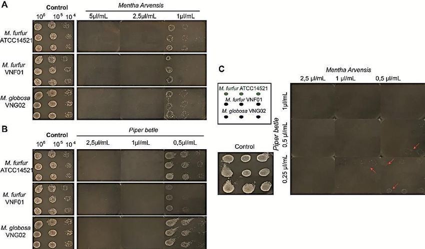
Introduction

        Malassezia spp. is a genus of lipid-dependent yeasts, which lacks the
ability to synthesize lipid, and hence, depends on lipid for their survivals.
Malassezia is the most abundance commensal yeasts of human and warm-
blood animal skins (Ashbee, 2007). Nonetheless, the yeasts can become
pathogenic agents under certain circumstances. For example, malassezia
yeasts have been found in various dermatological afflictions, including
pityriasis versicolor, malassezia folliculitis, seborrheic dermatitis, atopic
dermatitis, and psoriasis in immunologically competent hosts (Difonzo et
al., 2013). Additionally, the species have also associated with catheter-
related fungemia, sepsis, and a variety of deeply invasive infections in
immunocompromised patients, including patients with acquired immune-
deficiency syndrome (AIDS), immunohematological and oncological
patients, as well as solid organ and bone marrow transplant recipients
(Tragiannidis et al., 2010).
        Malassezia genus is classified as Malasseziomycetes (Wang et al.,
2014), which is further divided into three clusters; Cluster A consists of M.
furfur, M. japonica, M. obtusa, and M. yamatoensis; subcluster B1 contains
M. globosa and M. restricta, the most abundantly occurring human skin
inhabitants; subcluster B2 includes M. sympodialis, M. dermatis, M. caprae,
M. equina, M. nana, and M. pachydermatis; and cluster C embraces M.
cuniculi and M. slooffiae (Wu et al., 2015). M. globosa and M. restricta are
the species most commonly found on healthy and diseased human skin
(Crespo Erchiga et al., 1999). Nevertheless, other species, such as M.
sympodialis and M. furfur, have also linked to many human skin disorders
(Jagielski et al., 2014).
        In vitro studies demonstrated that different Malassezia species
respond dissimilarly to various antifungal agents. For instance, M. furfur, M.
globosa, and M. obtusa are more tolerant to terbinafine than other
Malassezia spp., whereas M. sympodialis is highly susceptible (Gupta et al.,
2000). Fluconazole is highly active against M. sympodialis and M. slooffiae,
but inactive against M. globosa and M. restricta (Rojas et al., 2014).
Itraconazole had high activity against M. globosa while ketoconazole is
more active against M. furfur than econazole and miconazole (Hammer et
al., 2000). Although these antifungal agents either alone or in combination
can be used for treating malassezia-associated disorders, the
hypersensitivity, toxicity, resistance to synthetic antimicrobials due to their
improper application concerned public health. Hence, identification of
antifungal agents with safer properties has been actively seeking.
        Plant essential oils (EO) are complex mixtures of natural
compounds, which have antiseptic and medicinal properties. EOs have been
used as folk medicine and agro-food preservers since ancient times and have
been shown to possess antimicrobial and antioxidant activities. Although

1620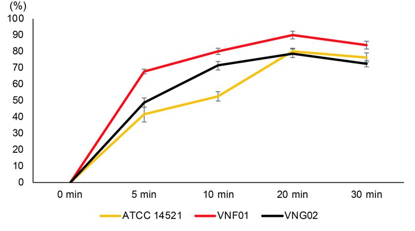
International Journal of Agricultural Technology 2021Vol. 17(4):1619-1630

chemical compositions of EOs differ among species, the main compounds
identified in EOs often belong to the family of terpenes, which are highly
lipophilic and low molecular weight, and thus, capable of disrupting the cell
membrane, causing cell death or inhibiting the sporulation and germination
of fungi. Chemical components of EOs are affected by factors such as the
geographical location, environment, the stage of maturity and method of
extraction. This chemical difference is directly correlated to dissimilarity in
biological activities. EOs can inactivate fungi by disrupting structure and
function of cell wall/membrane, mitochondria, biofilm, and mycotoxin
synthesis (Nazzaro et al., 2017). EOs and their components, therefore, has
been shown to inhibit the development of a variety of fungi (Karpiński,
2020; Abd-Rashed et al., 2021). For instance, EOs from Thymus, Artemisia,
Malaleuca, Cinnamomun, Ocimum, Zataria, Rosmarinus, Origanum,
Syzigium, Foenicolum, Thapsia, Tachyspermum, Myrtus have been
demonstrated to eliminate the growth of malassezia genus. Furthermore,
EOs from Cymbopogon citratus and C. flexuosus have been formulated in
shampoo, cream or lotion for the successful treatment of dandruff and
pityriasis versicolor in two clinical trials (Donato et al., 2020). EOs from
Mentha arvensis (M. arvensis) and Piper betle (P. betle) plants are widely
used in Vietnam, and capable of killing several fungi and bacteria.
However, the chemical compounds and inhibitory effects of these two EOs
on the development of malassezia spp. have not been characterized.
        In this study, chemical compositions of the essential oils (Eos) from
M. arvensis and P. betle was analyzed by Gas chromatography–mass
spectrometry (GC-MS) while inhibitory effects and minimum inhibitory
concentrations of the single or combined EOs against M. globosa and M.
furfur species were identified by agar diffusion and dilution assays. The
anti-malassezia kinetics curves of M. arvensis and P. betle EOs were
determined by kill-time analyses.

Materials and methods

Plant materials

       P. betle Leaves and whole M. arvensis plants (except roots) with
flowers were collected at the Hanam and Bacninh province. The materials
were washed thoroughly with tap water, and then, with sterilized distilled
water before EO isolation.

EO isolation

       Fresh materials (100 g each) were grinded to 0.5 mm to 1.5 mm and
subjected to hydro distillation in a conventional Clevenger type apparatus

                                                                                 1621
containing 300 ml of distilled water for 3 hours at 100oC. The EOs were
o
dried over anhydrous sodium sulfate and kept in sterile tubes at 4 C until use.
GC/MS analyses
GC/MS were analyzed according to the method described by
Sparkman (2005). Briefly, GC/MS data were obtained on an Agilent
Technologies HP 6890 mass spectrometer instrument using a HP-5MS
column (0.25 µm x 60 m x 0.25 mm). The carrier gas was helium;
temperature programming, 1 min at 40C, rising at 3C/min to 230C and
maintain at 230C for 10 min. Duplicate analysis was performed.
Quantitative results are mean data derived from GC analysis.
Fungal strains
Malassezia furfur VNF01 and Malassezia globosa VNG02 were
provided by the Center of Experimental Biology - National Center for
Technological Progress while the Malassezia furfur ATCC 14521 were
purchased from ATCC.
Agar diffusion assay
Agar diffusions assays were conducted as described by Hadacek and
Harald (2000). Briefly, 50 μl of each Malassezia strain (106 cells/ml) were
spread on mDixon agar plate (malt extract 36 g/l, desiccated oxbile 20 g/l,
tween 40 10 ml/l, peptone 6 g/l, glycerol 2 ml/l, oleic acid 2ml/l, pH 6). A 9
mm well was generated at the middle of each plate. 50 μl of a serial dilluted
EO was added to each well. 50 μl of DMSO (Sigma-Aldrich, cat # 67-68-5)
was added to the well of the negative control plate because EOs was dilluted
in DMSO (Makkar et al., 2018). The plates were kept at 4oC for 4 hours,
and then transferred to 30oC. The inhibition zones (plate circles without
visible fungi) were measured after 72 hours of incubation at 30oC. Each
experiment was repeated 3 times, and included 3 replicates for each
condition.
Agar dilution assay
Agar dilution methods were performed as demonstrated by Lambert
and Pearson (2000). Briefly, 75 μl of a serial dilluted EO (either alone or in
combination) was added to each plates containing 7.5 ml of mDixon agar
media. 75 μl of DMSO was added to the negative control plate. 5 μl of each
cell concentration (106, 105, 104 cells/ml) were dropped on each plate with 3
technical replecates. The plates were incubated at 30oC. The visible growth
was accessed at the third day. Each experiment was repeated 3 times.
1622International Journal of Agricultural Technology 2021Vol. 17(4):1619-1630
Kill-time analyses
The kill-time curve assay was modified from Joray et al. (2011).
Briefly, broth media with 106 yeast cells/ml were supplemented with either
DMSO or a mixture of M. arvensis (1 μl/ml) and P. betle (0.5 μl/ml) and
incubated at 5, 10, 20, and 30 minutes. At selected time intervals, samples
from the test culture were taken, serially diluted in the same broth media,
and plated in the agar plate media. All plates were then incubated at 30oC,
and CFU were counted after 72 hours of incubation. Percent of cell death
were calculated by 100% - % of living cells. % of living cells =
(concentration of cells at 5, 10, or 20 minutes divided to concentration of
cells at zero time point) x 100.
Results
Chemical profiles of M. arvensis and P. betle oils
The averaged EO yields from the M. arvensis and P. betle were
1.09% and 0.25%, respectively (data not shown). The components of the
two EOs. GC-MS detected 11 compounds, accounting for 99.87% of M.
arvensis EO, while GC-MS analyses of P. betle EO identified to be 4
compounds, which composed of 78.43% of the P. betle EO. Menthol and
menthone, the two major compounds of M. arvensis EO, comprised 68.19%
and 22.77% of the EO content, respectively while eugenol acetate and m-
eugenol, the two most abundant molecules in P. betle EO, constituted
38.66% and 30.28% of the EO, respectively (Tables 1 and 2).
Table 1. Chemical composition of M. arvensis EO
Compound Relative Compound Relative
composition ratio, composition
% ratio, %
α-pinene 1.45 α - bourbonene 0.12
α-Myrcene 1.35 Camphene 0.04
D-Limonene 1.73 Eucalyptol 0.53
I-menthone 22.77 α -santalol 0.33
dI-menthol 68.19 Bis(2-ethylhexyl)phthalate 0.55
Menthyl 2.81 Total 99.87
Acetate
Table 2. Chemical composition of P. betle EO
Compound Relative Compound Relative composition
composition ratio, ratio, %
%
Chavicol 3.71% Eugenol acetate 38.66%
m-Eugenol 30.28% Chavicol acetate 5.78%
Total 78.43%
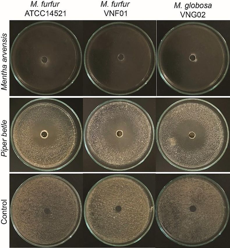
1623Inhibitory effects of M. arvensis and P. betle oils
The anti-malassezia activities of of M. arvensis or P. betle EOs was
evaluated against M. furfur ATCC 14521 and VNF01 strains, and M.
globosa VNG02 strain by using disc diffusion assays. M. arvensis EO
showed stronger inhibition than P. betle one (Figure 1). The fungi fully
covered the discs with 50 μl of DMSO (negative controls) after culturing for
three days (bottom panel). On other hand, no visible yeast cells were
appeared in the discs with 50 μl of M. arvensis 100% EO (top panel) while
the fungal growth were inhibited by 30-43% in the discs containing 50 μl of
P. betle 100% EO (middle panel) in the same experimental conditions as the
negative control.
Figure 1. Anti-malassezia properties of M. arvensis and P. betle EOs
against 106/ml of M. furfur (ATCC 14521 and VNF01), and M. globosa
(VNG02) cells
Minimum inhibitory concentration of M. arvensis and P. betle oils
The minimum inhibitory concentrations (MIC) of single or
combination of M. arvensis and P. betle EOs were identified by agar
dilution assays. As shown in Figure 2A, the visible growth of M. furfur
1624International Journal of Agricultural Technology 2021Vol. 17(4):1619-1630
ATCC 14521 and VNF01 strains, and M. globosa VNG02 strain was
completely absent when cultured with M. arvensis oil at the concentration of
5 µl/ml and 2.5 µl/ml, but the colonies were visible at 1 µl/ml, suggesting
that MIC of M. arvensis EO was 2.5 µl/ml. Similarly, MIC of P. betle oils
was identified at the concentration of 1 µl/ml (Figure 2B).
To evaluate the additive/synergic effects of M. arvensis and P. betle
EOs on the development of M. furfur ATCC 14521 and VNF01 strains, and
M. globosa VNG02 strain, different combinations of these two EOs were
added to agar medium plates. Among 9 combinations, the mixtures of M.
arvensis and P. betle essential oils with the ratios of 2.5 + 1 µl/ml; 2.5 + 0.5
µl/ml; 2.5 + 0.25 µl/ml; 1 + 1 µl/ml; 1 + 0.5 µl/ml; 0.5 + 1 µl/ml,
respectively; completely suppressed visible extension of all three fungal
strains (Figure 2C).
Figure 2. Minimum inhibitory concentrations of single or combination of
M. arvensis and P. betle EO. Inhibitory growth by M. arvensis EO (A), P.
betle EO (B), and their combination (C) at different dilution
Anti-malassezia kinetics of M. arvensis and P. betle EO
The anti-massezia kinetics curves of M. arvensis and P. betle EOs
were presented in Figure 3. The number of cells treated with DMSO did not
vary over 30 minutes of incubation. However, the fungi inoculated with a
mixture of 1 µl/ml M. arvensis and 0.5 µl/ml P. betle EOs increased
proportions of cell deaths gradually and was peaked at 20 minutes. The cell
death ratios started to drop after 30 minutes of incubation. After 20 minutes
1625of treatment, the M. furfur VNF01 strain had the highest percent of cell
deaths (90%), while those of M. furfur ATCC 14521 and M. globosa
VNG02 strains were 78% and 80%, respectively. However, the number of
cell deaths were slightly decreased after 30 minutes of incubation.
Figure 3. Anti-malassezia kinetics linears of M. arvensis and P. betle EOs
against 106 CFU/ml M. furfur (ATCC 14521, VNF01) and M. globosa
(VNG02) yeast cells
Discussion
EOs are complex mixtures of chemically distinct molecules. EOs
mainly contain teroenoidic compounds, but nitrogenous derivatives,
glucosinolates or isothiocyanate constituents, coumarins or furocoumarins
and short-chain aliphatic molecules also present in EOs of certain plants
(Friedrich, 1976). EOs usually compose of 2 – 3 main components, which
could be ascribed to their major biological activities. Nevertheless, the
additive or synergistic actions have also been evidenced (Franz, 2010).
Chemical components of EOs are affected by factors such as the
geographical location, environment, the stage of maturity and method of
extraction. This chemical difference is directly correlated to dissimilarity in
biological activities of different samples. EOs can inactivate fungi by
disrupting structure and function of cell wall/membrane, mitochondria,
biofilm, and mycotoxin synthesis (Nazzaro et al., 2017). EOs from M.
arvensis and P. betle plants are widely used in Vietnam, and capable of
killing several fungi and bacteria. However, their chemical components and
anti-malassezia properties have not been characterized. Here, we present the
chemical compositions and anti-malassezia attributes of EOs from M.
arvensis and P. betle cultivated in Vietnam.
1626International Journal of Agricultural Technology 2021Vol. 17(4):1619-1630
M. arvensis leaves contain 0.4 - 0.8% of EO, which is usually
dominated by menthol (above 60%) and menthone (4 - 18%) (Kalemba and
Agnieszka, 2020). Average EO yield from M. arvensis grown in Bacninh,
Vietnam were ranged from 0.9 – 1.09%, which were slightly higher than
that reviewed in Kalemba and Agnieszka, 2020. Menthol (68.19%)
identified in this study was in the range of literature review whereas
menthone (22.77%) were faintly elevated. These data indicate that the yield
and lead compound proportions of EO from M. arvensis cultured in
Bacninh, Vietnam were similar to that reported in literatures. Consistently,
the Vietnamese M. arvensis EO has the MIC values of 2.5 µl/ml for M.
furfur and M. globose species, which is in the MIC ranges (0.25 to 3 µl/ml)
of M. arvensis EO against a variety of bacteria and fungi (Kalemba and
Agnieszka, 2020).
P. betle plants consist of 0.1% to 0.15% of EO, whose chemical
components belong to the classes of monoterpenes, sesquiterpenes,
phenylpropanoids, and aldehydes. Chemical constituents of P. betle EO
vary depend on botanical origin, plant ages, and harvesting time, but
eugenol, carvacrol, chavicol, chavibetol (m-eugenol) are usually major
molecules identified in P. betle leaf EO (Nayaka et al., 2021). Our P. betle
plants contains 0.2% - 0.3% of EO, whose main components were eugenol
acetate, chavibetol, chavicol, chavicol acetate. Of these compounds, eugenol
acetate and chavibetol (m-eugenol) were the two dominant compounds and
accounted for 38.66% and 30.28%. Again, our EO yield was higher than,
but contains similar compound as that reported in literature. Accordingly,
our agar dilution assays showed that MIC of P. betle EOs were 1 µl/ml,
which is slightly higher than MIC values ( 0.3 µl/ml to 0.7 µl/ml) against 14
fungal species identified in (Prakash et al., 2010).
This study also revealed the additive effects of M. arvensis and P.
betle EOs against two malassezia species, that is a combination of a half
dose of the MIC value of each EOs can inhibit visible growth of all fungi
tested. Even though the mechanism(s) underlying this phenomenon remains
to be clarified, the additive property may depend on the combined actions of
the EOs’ main components. Eugenol has been reported to change or disrupt
fungal cell wall structure, causing cell deaths due to dysfunction of
membrane fluidity and permeability. Furthermore, eugenol acetate and
chavicol acetate were predicted to strongly interact to amino acid
constructing fungal protein structures, and therefore may reduce fungal
survivability (Nayaka et al., 2021). M. arvensis EO and its lead compounds
have been shown to kill Candida species by inhibiting ergosterol
biosynthesis and PM-ATPase activity (Samber et al., 2015). Altogether,
these data suggest that the combination of M. arvensis and P. betle EOs may
enhance killing capacity by targeting different cell organelles. Moreover,
kill-time assays revealed the mixture of M. arvensis and P. betle EOs can
1627eliminate up to 90% of M. furfur and 80% of M. globosa after 20 minutes of
treatment.
This is the first study to demonstrate that M. arvensis and P. betle
EOs can inhibit the development of members of malassezia genus with the
MIC of 2.5 µl/ml and 1 µl/ml, respectively. This work also showed that M.
arvensis and P. betle EOs additively eliminated M. furfur and M. globosa
species, probably by disrupting structures and/or functions of different
organelles. Furthermore, M. arvensis and P. betle EOs were capable of
killing up to 90% of M. furfur and 80% of M. globose species after 20
minutes of treatment. Altogether, this study suggested that M. arvensis and
P. betle EOs can be potential agents for formulating shampoo, cream or
lotion to treat malassezia-associated disease.
Acknowledgments
This work was funded by the National Center for Technological Progress, Ministry
of Science and Technology, Viertnan.
References
Abd-Rashed, A., Rathi, D. N. G., Ahmad Nasir, N. A. H. and Abd Rahman, A. Z. (2021).
Antifungal properties of essential oils and their compounds for application in skin
fungal infections: conventional and nonconventional
approaches. Molecules, 26:1093-1137.
Ashbee, H. R. (2007). Update on the Genus Malassezia. Medical Mycology, 45:287-303.
Crespo Erchiga, V., Ojeda Martos, A. A., Vera Casaño, A., Crespo Erchiga, A. and
Sánchez Fajardo, F. (1999). Isolation and identification of Malassezia spp. in
pytiriasis versicolor, seborrheic dermatitis and healthy skin. Revista Iberoamericana
De Micologia, 16:16-21.
Difonzo, E. M., Faggi, E., Bassi, A., Campisi, E., Arunachalam, M., Pini, G., Scarfì , F. and
Galeone, M. (2013). Malassezia skin diseases in Humans. Giornale Italiano Di
Dermatologia E Venereologia: Organo Ufficiale, Societa Italiana Di Dermatologia E
Sifilografia, 148:609-19.
Donato, R., Sacco, C., Pini, G. and Bilia, A. R. (2020). Antifungal activity of different
essential oils against Malassezia pathogenic species. Journal of Ethnopharmacology,
249:112376.
Franz, C. M. (2010). Essential oil research: Past, present and future. Flavour and Fragrance
Journal, 25:112-113.
Friedrich, H. (1976). Phenylpropanoid constituents of essential oils. Lloydia, 39:1-7.
Gupta, A. K., Kohli, Y., Li, A., Faergemann, J. and Summerbell, R. C. (2000). In vitro
susceptibility of the seven Malassezia species to ketoconazole, voriconazole,
itraconazole and terbinafine. The British Journal of Dermatology, 142:758-765.
Hadacek, F. and Harald, G. (2000). Testing of antifungal natural products: Methodologies,
comparability of results and assay ahoice. Phytochemical Analysis, 11:137-147.
1628International Journal of Agricultural Technology 2021Vol. 17(4):1619-1630
Hammer, K. A., Carson, C. F. and Riley, T. V. (2000). In vitro activities of ketoconazole,
econazole, miconazole, and melaleuca mlternifolia (Tea tree) oil against Malassezia
species. Antimicrobial Agents and Chemotherapy, 44:467-469.
Jagielski, T., Rup, E., Ziółkowska, A., Roeske, K., Macura, A. B. and Bielecki, J. (2014).
Distribution of Malassezia species on the skin of patients with atopic dermatitis,
psoriasis, and healthy volunteers assessed by conventional and molecular
identification methods. BMC Dermatology, 14:1-15.
Joray, M. B., del Rollán, M. R., Ruiz, G. M., Palacios, S. M. and Carpinella, M. C. (2011).
Antibacterial activity of extracts from plants of central argentina isolation of an
active principle from achyrocline satureioides. Planta Medica, 77:95-100.
Kalemba, D. and Agnieszka, S. (2020). Agrobiological interactions of essential oils of two
menthol mints: Mentha piperita and Mentha arvensis. Molecules, 25:59-91.
Karpiński, T. M. (2020). Essential oils of lamiaceae family plants as antifungals.
Biomolecules, 10:103-167.
Lambert, R. J. W. and Pearson, J. (2000). Susceptibility testing: accurate and reproducible
minimum inhibitory concentration (MIC) and non-inhibitory concentration (NIC)
values. Journal of Applied Microbiology, 88:784-790.
Makkar, M. K., Sharma, S. and Kaur, H. (2018). Evaluation of Mentha arvensis essential
oil and its major constituents for fungitoxicity. Journal of Food Science and
Technology, 55:3840-3844.
Nayaka, N. M. D. M. W., Sasadara, M. M. V., Sanjaya, D. A., Yuda, P. E. S. K., Dewi, N.
L. K. A. A., Cahyaningsih, E. and Hartati, R. (2021). Piper betle (L): Recent review
of antibacterial and antifungal properties, safety profiles, and commercial
applications.” Molecules, 26:2321-2341.
Nazzaro, F., Fratianni, F., Coppola, R. and Feo, V. D. (2017). Essential oils and antifungal
activity. Pharmaceuticals, 10:86-105.
Prakash, B., Shukla, R., Singh, P., Kumar, A., Mishra, P. K. and Dubey, N. K. (2010).
Efficacy of chemically characterized Piper betle L. essential oil against fungal and
aflatoxin contamination of some edible commodities and its antioxidant activity.
International Journal of Food Microbiology, 142:114-119.
Rojas, F. D., Sosa, M. D. L. A., Fernandez, M. S., Cattana, M. E., Cordoba, S. B. and
Giusiano, G. E. (2014). Antifungal susceptibility of Malassezia furfur, Malassezia
sympodialis, and Malassezia globosa to azole drugs and amphotericin B evaluated
using a broth microdilution method. Medical Mycology, 52:416-646.
Samber, N., Khan, A., Varma, A. and Manzoor, N. (2015). Synergistic anti-candidal
activity and mode of action of Mentha piperita essential oil and its major
components. Pharmaceutical Biology, 53:1496-1504.
Sparkman, O. D. (2005). Identification of essential oil components by gas
chromatography/quadrupole mass spectroscopy Robert P. Adams. Journal of the
American Society for Mass Spectrometry, 16:1902-1903.
Tragiannidis, A., Bisping, G., Koehler, G. and Groll, A. H. (2010). Minireview: Malassezia
infections in immunocompromised patients. Mycoses, 53:187-195.
Wang, Q. M., Theelen, B., Groenewald, M., Bai, F. Y. and Boekhout, T. (2014).
Moniliellomycetes and Malasseziomycetes, two new classes in
Ustilaginomycotina. Persoonia: Molecular Phylogeny and Evolution of Fungi,
33:41-47.
1629Wu, G., Zhao, H., Li, C., Rajapakse, M. P., Wong, W. C., Xu, J., Saunders, C. W, Reeder,
N. L., Reilman, R. A., Scheynius, A., Sun, S., Billmyre, B. R., Li, W., Averette,
F., Mieczkowski, P., Heitman, J., Theelen, B., Schröder, M. S., Sessions, P. F. D.,
Butler, G., Maurer-Stroh, S., Boekhout, T., Nagarajan, N. and Dawson Jr, T. L.
(2015). Genus-wide comparative genomics of Malassezia delineates its phylogeny,
Physiology, and niche adaptation on human skin. PLoS Genetics, 11:1-26.
(Received: 23 March 2021, accepted: 21 June 2021)
1630You can also read