Clinicomorphological Study of Leiomyoma Associated Endometrial Changes in Correlation with LMP: In a Tertiary Care Hospital in Rural Tamil Nadu
←
→
Page content transcription
If your browser does not render page correctly, please read the page content below
DOI: 10.7860/JCDR/2019/41034.12909
                                                                                                                                  Original Article

                         Clinicomorphological Study of Leiomyoma
 Pathology Section

                                Associated Endometrial Changes in
                                 Correlation with LMP: In a Tertiary
                                  Care Hospital in Rural Tamil Nadu
Anusha Babu Rajendran

Abstract                                                                        was observed as 41-50 years (56.50%). Multiparous women
Introduction: Leiomyomas are benign smooth muscle tumors.                       (94.86%) are more commonly affected. Menorrhagia was the
They are the single most common cause for hysterectomy in                       most common symptom seen in 46.53%. Majority of the cases
the reproductive and peri-menopausal age group associated                       had a single leiomyoma (59.82%). The most common location
with abnormal uterine bleeding. There are certain endometrial                   was intramural (49.85%). The most common degenerative
changes with leiomoyomas due to hormonal and mechanical                         change associated with a leiomyoma was hyaline change
influences of the leiomyomas.                                                   (23.87%). The most common type of endometrium was
                                                                                proliferative endometrium observed in 44.41%. The percentage
Aim: To study the incidence of leiomyomas of uterus in a rural
                                                                                of mailignacy associated with a leiomyoma was 0.60%.
area of Tamil Nadu and the endometrial changes associated
with it correlating it to the LMP, in hysterectomy specimens.                   Conclusion: Leiomyomas are the most common causes for
                                                                                hysterectomy. The associated endometrial change is proliferative
Materials and Methods: This is a retrospective study of three
                                                                                in nature suggesting the role of harmones such as oestrogen
hundred thirty one patients who underwent hysterectomy for
                                                                                and progesterone on the leiomyoma and simultaneously on the
uterine leiomyoma between 2013 and 2015, histopathological
                                                                                corresponding endometrium.
analysis of the cases for endometrial changes was done.
Results: A total of 331 hysterectomy specimens done for
leiomyomas were studied. The most common age group

                                                              Keywords: Degeneration, Endometrium, Oestrogen, Proliferative, Uterine leiomyoma

Introduction                                                                    Materials and Methods
Leiomyomas are benign smooth muscle tumours. The most                           A retrospective study was done on 331 hysterectomy specimens,
common site of origin of the leiomyomas (uterine fibroids) is the               done for leiomyomas during the period June 2013-May 2015.
myometrium of the uterus. They are the single most common                       Haematoxylin and eosin stained sections obtained from the
cause for hysterectomy in the reproductive and peri-menopausal                  331 hysterectomy specimens were used for the study. All
age group [1,2]. They are mostly asymptomatic but may present                   hysterectomy specimems done for leiomyomas were included
with menstrual disturbances [3]. These symptoms are mainly due                  and myomectomy specimens, endometrial biopsies, endocervical
to the hyper-oestrogenic status associated with these tumours.                  biopsies, endometrial currettings were excluded from the study.
Adenomyosis is a myometrial lesion characterised by the presence                Histopathological evaluation of the gross specimens were done
of ectopic endometrium in the myometrium. Furthermore, both                     and H&E stained slides were studied and the endometrium was
adenomyosis and leiomyomas commonly coexist in nearly 33%                       classified as proliferative, secretory, hyperplastic and atrophic,
                                                                                stromal changes and type of leiomyoma was also classified.
of the cases [4]. This shows the hormone dependency of the
leiomyomas. The pathogenesis of the fibroids is still unclear; however
recent evidences show that they are due to the steroid hormones,                Results
                                                                                In this study, out of 1463 hysterectomy specimens received in
oestrogen and progesterone [5]. The endometrium of the uterus is
                                                                                two years (June 2013-May 2015) 331 (22.62%) had leiomyomas.
also under the cyclical influence of these steroid hormones. There
                                                                                Most common age group affected was 41-50 years (56.5%) [Table/
are studies suggesting the association of endometrial changes with
                                                                                Fig-1]. Multiparous women (94.86%-314 cases) harboured more
leiomoyomas such as glandular hyperplasia in the endometrium,
                                                                                leiomyomas compared to primipara (3.63%-12 cases) and nullipara
this may be the expression of oestrogenic hyperactivity and atrophic
                                                                                (1.51%-5 cases). It was observed that menorrhagia was the
endometrium which could be the result of mechanical forces upon                 most common symptom seen in 46.53% (154 cases) followed by
the endometrium [6]. Distortion, elongation, and dilatation of the              abdominal pain in 26.89% (89 cases) of cases. The other symptoms
glands are said to be the result of pressure effect of the leiomyoma.           that were observed were post menopausal bleeding (9.67%-
Since the leiomyomas are hormone dependant tumours and the                      32 cases), dysmenorrhoea (3.93%-13 cases), mass descending
uterine endometrium is also under the influence of these hormones,              per vaginum (2.72%-9 cases), polymenorrhoea (2.42%-8 cases),
there could be an association between the hormone influence on                  dysuria (1.81%-6 cases) and urinary retention (0.91%-3 cases).
the leiomyoma and the endometrium changes associated with it. In                A 5.14% (17 cases) of the women were asymptomatic. It was
this study LMP is used as a guide to phase the endometrium and                  observed that adenomyosis was associated with leiomyoma in 110
there by identify the abnormal changes in the endometrium which                 cases (33.23%). It was most commonly seen in multiparous women
are not correlating with the phase and date of the endometrium.                 and in the age group of 30-50 years.
Journal of Clinical and Diagnostic Research. 2019 Jun, Vol-13(6): EC01-EC04                                                                          1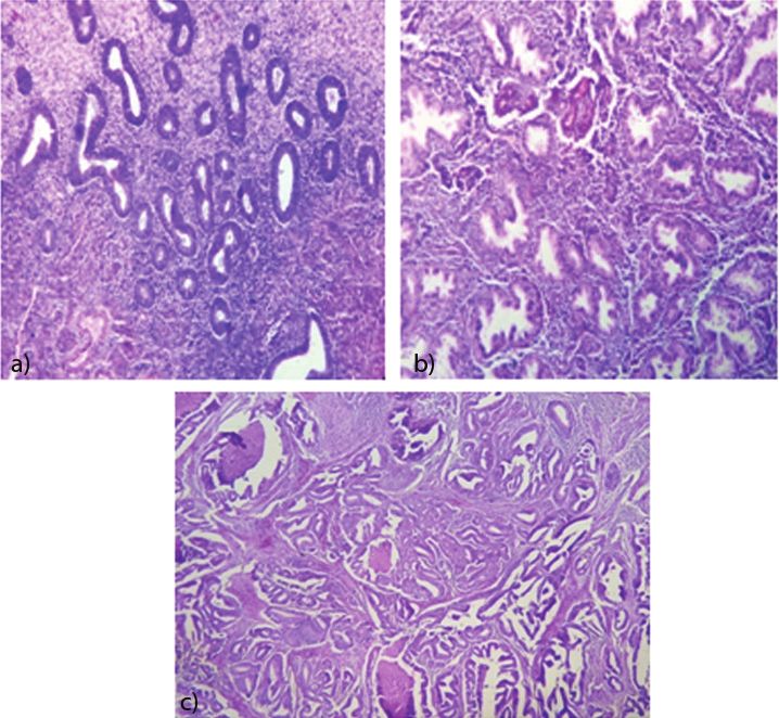
Anusha Babu Rajendran, Clinicomorphological Study of Leiomyoma Associated Endometrial Changes in Correlation with LMP www.jcdr.net
Age group Frequency Percentage Type of Leiomyoma
Total
21-30 2 0.60% endometrium Intra-Mural Multi-Ple Submucosal Sub serosal
31-40 92 27.79% Proliferative 78 19 18 32
147
41-50 187 56.50% endometrium 47.28% 46.34% 36% 42.67%
51-60 42 12.69% Secretory 24 12 11 10
57
61-70 7 2.11% endometrium 14.55% 29.27% 22% 13.34%
71-80 1 0.30% Disordered 16 1 5 8
proliferative 30
Total 331 100% endometrium 9.7% 2.44% 10% 10.67%
[Table/Fig-1]: Age distribution of leiomyoma.
Atrophic 15 2 5 8
30
endometrium 9.1% 4.88% 10% 10.67%
It was observed that majority of the cases of leiomyomas were
single in number (59.82%-198 cases) and multicentric was 40.18% Hyperplasia of 26 7 8 15
56
(133 cases). It was observed that the most common type of endometrium 15.76% 17.07% 16% 20%
leiomyoma encountered was intramural type in 49.85% (165 cases) 6 - 3 2
followed by subserosal and submucosal fibroid seen in 22.66% Others 11
3.64% - 6% 2.67%
(75 cases) and 15.11% (50 cases) respectively, leiomyomas in
165 41 50 75
multiple locations was observed in 12.39% (41 cases). One case Total 331
of broad ligament fibroid was observed. Secondary changes were 49.85% 12.39% 15.1% 22.7%
observed in 39.27% (130 cases) of the leiomyomas of which the [Table/Fig-3]: Correlation of type of leiomyoma with type of endometrium.
most commonest secondary change was found to be hyaline
change observed in 23.87% (79 cases) of the leiomyomas. The other LMP in days
Type of endometrium Total
secondary changes that were observed were cystic change (11 7-15 16-30 >30
cases), haemorrhage (12 cases), calcification (2 cases), lymphocytic 3 2 5 10
infiltration (4 cases), infarction (1 case), osseous metaplasia (1 case) Early proliferative endometrium
3.6% 1.7% 3.9% 3.0%
and lipoleiomyoma (5 cases). Red degeneration which occurs in
pregnancy was observed in one case. 52 34 51 137
Late proliferative endometrium
61.9% 28.8% 39.5% 41.4%
In this study of 331 cases, the most common type of endometrium
associated with a leiomyoma was proliferative endometrium 3 24 9 36
Early secretory endometrium
observed in 44.41% (147 cases). Hyperplasia without atypia was 3.6% 20.3% 7.0% 10.9%
seen in 16.92% (56 cases) [Table/Fig-2]. 2 15 4 21
Late secretory endometrium
2.4% 12.7% 3.1% 6.3%
Type of endometrium Frequency Percentage
Secretory endometrium with arias stella - 3 - 3
Early proliferative endometrium 10 3.02%
reaction - 2.5% - .9%
Late proliferative endometrium 137 41.39%
1 1 4 6
Early secretory endometrium 36 10.88% Pill endometrium
1.2% .8% 3.1% 1.8%
Late secretory endometrium 21 6.34%
3 5 22 30
Secretory endometrium with arias stella reaction 3 0.91% Atrophic endometrium
3.6% 4.2% 17.1% 9.1%
Pill endometrium 6 1.81%
6 11 13 30
Atrophic endometrium 30 9.06% Disordered proliferative endometrium
7.2% 9.3% 10.1% 9.1%
Disordered proliferative endometrium 30 9.06%
12 10 12 34
Simple hyperplasia without atypia 34 10.27% Simple hyperplasia without atypia
14.3% 8.5% 9.3% 10.3%
Complex hyperplasia without atypia 22 6.65%
2 13 7 22
Endometrioid adenocarcinoma 2 0.60% Complex hyperplasia without atypia
2.4% 11.0% 5.4% 6.6%
Total 331 100%
[Table/Fig-2]: Endometrial changes in leiomyoma. - 2 2
Endometrioid adenocarcinoma -
1.6% .6%
It was found that hyperplasia without atypia was most common [Table/Fig-4]: Last Menstrual Period (LMP) and endometrium.
in subserosal fibroids (20%) and atrophic endometrium was most
common in multiple leiomyoma [Table/Fig-3]. is of importance owing to the fact that they are the most common
The stromal changes were in concurrence to the corresponding cause for hysterectomies in the women in the reproductive age
type of endometrium observed i.e., for proliferative endometrium group. In the current study, out of 1463 hysterectomy specimens in
and hyperplasia the stroma appeared cellular and compact, for two years 331 (22.62%) hysterectomies had leiomyomas. Cramer
secretory endometrium the stroma was edematous, decidualised SF et al., observed 77% of hysterectomies are due to leiomyomas
stroma was seen in Pill endometrium and Arias-Stella reaction. and Velu ARK et al., observed it to be 28% and Jha R et al., found
it to be 28% [1,7,8].
In this study, Last Menstrual Period in days were divided into 3 groups
as 7 to 15 (84 cases), 16 to 30 (118) and more than 30 (129). Leiomyomas most commonly occur in the mid reproductive age
Late proliferative endometrium was the predominant type in all group women. Baird DD et al., in their study of randomly selected
3 categories of LMP. [Table/Fig-4] suggests proliferative changes women between 31-49 years found that the incidence of uterine
persisting in majority of cases of leiomyoma beyond 15 days of fibroids was 60% by age 35 years which increased to 80% by age
LMP giving light to a hormonal influence of the leiomyoma. 50 years in African-American women and for Caucasian women
by 35 years of age the incidence of uterine fibroids was 40%
Discussion and by age 50 years it was found to be 70% [9]. In this study,
The leiomyomas are the most common benign tumours of the it was observed that majority of the cases (56.50%) were seen
uterus in the reproductive and post menopausal age group. This in the age group of 41-50 years and 84.29% cases were seen
2 Journal of Clinical and Diagnostic Research. 2019 Jun, Vol-13(6): EC01-EC04www.jcdr.net Anusha Babu Rajendran, Clinicomorphological Study of Leiomyoma Associated Endometrial Changes in Correlation with LMP
in age group between 31-50 years. This is in concurrence with
the studies of Gowri M et al., Velu ARK et al., and Begum S et
al, [2,3,7]. In this study it was also observed that 20.54% cases
were post-menopausal, this is in contrast with the findings of Velu
ARK et al., who had only 4% of the women who were in the post-
menopausal age group [7].
Multiparous women (94.86%) were found to have more leiomyomas
compared to primipara and nullipara. This is explained by the fact
that multiparous women, acquire increased levels of oestrogen,
progesterone and their receptors, ER and PR [10,11].
Most of the women with leiomyomas are asymptomatic, however
they most commonly present with menorrhagia. In the current
study menorrhagia was seen in 46.53% followed by abdominal
pain in 26.89% of cases. These findings are in concurrence with
other studies [2,3]. The cause for the predominance of menstrual
symptoms could be attributed to the fact of increased vascularity,
endometrial surface, altered contractility of endometrium and the
hormonal influence of the tumor.
Majority of the cases of leiomyomas were single in number (59.82%).
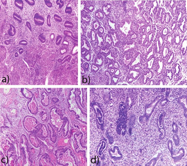
These findings are in agreement with studies of Gowri M et al., and [Table/Fig-5]: Proliferative endometrium (100x-H&E); b) Early secretory endometrium
Velu ARK et al., [3,7]. This finding is in contrast with Begum S et al., (100x-H&E); c) Late secretory endometrium (100x-H&E); d) Disordered proliferative
and Cramer SF et al. who found multiple leiomyomas to be more endometrium (100x-H&E).
frequent than single leiomyomas [1,2].
The most common type of leiomyoma encountered was intramural
type in 49.85%. These findings are in concurrence with Gowri M et
al., and Velu ARK et al., who found that the most common type of
leiomyoma to be of the intramural type [3,7].
Secondary changes were observed in 39.27% of the leiomyomas
of which the commonest secondary change was found to be
hyaline change observed in 23.87% of the leiomyomas. This is in
agreement with the observations of Gowri M et al., Begum S et al.,
and Persaud V et al., [2,3,12].
In the current study,110 (33.23 %) leiomyomas were associated
with adenomyosis. This is in contrast with Ben AN et al., who
reported 62 % of cases of leiomyoma with adenomyosis and Raju
GC et al., who reported 16 % of leiomyomas with associated
adenomyosis [12-14]. In the current study it was observed that
adenomyosis was most commonly seen in multiparous women
and in the age group of 30-50 years as observed by other Raju GC
et al., and Velu ARK et al., [7,13].
The most common type of endometrium associated with a
leiomyoma was proliferative endometrium observed in 44.41% of
which the late proliferative endometrium was the predominant type
[Table/Fig-6]: Simple hyperlasia without atypia (100X-H&E); b) Complex hyperlasia
in 41.38% and early proliferative in 3.02%. This is in concurrence without atypia (100x-H&E); c) Endometrioid adenocarcinoma (100x-H&E).
with Velu ARK et al., and Gowri M et al., [Table/Fig-5] [3,7].
Hyperplasia of endometrium was seen in 16.92% of the cases of in a hormonal context. The risk is higher in the perimenopausal
which 10.27% of the cases showed simple hyperplasia without period. The most frequent abnormality in endometrium is simple
atypia and 6.64% of the cases showed complex hyperplasia hyperplasia suggesting a rare progression to highest grades and a
without atypia. The findings of the current study is in contrast with possible protective role of leiomyoma as target tissue which capture
the studies of Velu ARK et al., Gowri M et al., and Chethana M et al., oestrogens [15].
who found hyperplastic changes in 25%, 22% and 24% respectively It was also observed that 159 cases (48.03%) of the total 331 cases
[3,7,11] [Table/Fig-6]. showed persistence of the proliferative phase beyond 15 days of
Atrophic endometrium was seen in 9.06% of the cases and disorderly the LMP. This shows that leiomyomas are also associated with
proliferating endometrium in 9.06% of cases of leiomyoma. This is in a persistent proliferative endometrium, which is due to a hyper-
concurrence with the studies of Velu ARK et al. whereas Gowri M et oestrogenic effect. This hyper-oestrogenic effect could be a common
al., and Chethana M et al., had observed more percentage of cases factor contributing to the leiomyoma and the endometrial changes
with atrophic changes probably due to more cases of submucosal as noted by Velu ARK et al., [7].
fibroids in both the studies which cause pressure effects on the The probable cause for the endometrial changes could be oestrogen,
endometrium leading to atrophy [3,7,11]. Atrophic endometrial progesterone and pressure effects, in cases of submucosal
changes are not only due to pressure effects but also due to post fibroids. Deligdish L et al., concluded that there is high oestrogen
menopausal hormone insufficiency [6]. level in women with fibroids and it is hypothesised that oestrogen
In this study of the 331 cases, 88 cases (26.58%), showed is synthesised by the endometrium which is responsible for the
abnormal endometrial changes. The most frequent abnormality growth of the fibroid [6]. Oestrogen and progesterone together
was simple hyperplasia without atypia. Teleman S et al., who play a role in fibroid growth [7]. The oestrogen up regulates both
proposed that leiomyoma and endometrial hyperplasia, develop oestrogen receptors and progesterone receptors in the fibroids
Journal of Clinical and Diagnostic Research. 2019 Jun, Vol-13(6): EC01-EC04 3Anusha Babu Rajendran, Clinicomorphological Study of Leiomyoma Associated Endometrial Changes in Correlation with LMP www.jcdr.net
during proliferative phase which is followed by the progesterone Thus, a proliferative endometrium beyond 16 days of LMP in a
induced mitogenesis during the luteal phase [16]. endometrial curettage sample could also be considered to be due
Study by Gull B et al., has shown that the tissue concentration of to a leiomyoma and thereby can be treated with harmone therapy
oestrogen receptor and progesterone receptor were more in the or simple myomectomy, thereby a hysterectomy can be avoided in
leiomyoma when compared with the normal myometrium [16]. these patients if detected early.
The endometrium is under the cyclical influence of the steroid
References
hormones. There is an entity called the sub-endometrial myometrium [1] Cramer SF, Patel A. The frequency of uterine leiomyomas. Am J Clin Pathol.
which also undergoes cyclical steroid receptor changes in 1990;94(4):435-38.
[2] Begum S, Khan S. Audit of leiomyoma uterus at Khyber Teaching Hospital,
concurrence with the endometrium during the menstrual cycle
Peshawar. J Ayub Md Coll. 2004;16(2):46-49.
[17]. There is growing evidence that the leiomyomas are benign [3] Gowri M, Mala G, Murthy S, Nayak V. Clinico-pathological study of uterine
tumours arising from this sub-endometrial myometrium Hence the leiomyomas in hysterectomy specimens. Journal of Evolution of Medical and
Dental Sciences. 2013;2(46):9002-09.
leiomyomas, arising from the sub-endometrial myometrium, also
[4] Taran FA, Weaver AL, Coddington CC, Stewart EA. Characteristics indicating
show cyclical changes in the oestrogen and progesterone receptors, adenomyosis coexisting with leiomyomas: A case-control study. Hum Reprod.
during the menstrual cycle, which adds to the evidence that the 2010;25(5):1177-82.
[5] Nissole M, Gillerot S, Casanas-Roux F, Squifflet J, Berliere M, Donnez J.
leiomyomas are not only steroid dependant for their growth but
Immunohistochemical study ofthe proliferation index, oestrogen receptors and
occur in concurrence with the changes in the endometrium [5,18]. progesterone receptors Aand B in leiomyomata and normal myometrium during
So, it could be said that, oestrogen and progesterone are common the menstrual cycle and under gonadotropin-relasing hormone agonist therapy.
Hum Reprod. 1999;14(11):2844-50.
factors affecting the leiomyoma and the endometrium.
[6] Deligdish L, Loewenthal M. Endometrial changes associated with myomata of
the uterus. J Clin Pathol. 1970;23(8):676-80.
Limitation and Recommendations [7] Velu ARK, Srinivasamurthy BC, Sundaram A. Uterine fibroid tumours and
associated changes in endometrium and myometrium: A three year prospective
The limitations of this study is the lack of IHC for ER, PR to
study in a tertiary care hospital. J Pharm Biomed Sci. 2014; 4(9):797-805.
demonstrate the hormonal dependency of the leiomyoma. This [8] Jha R, Pant AD, Jha A, Adhikari RC, Sayami G. Histopathological analysis of
could be carried out in the future to ascertain the fact of leiomyoma hysterectomy specimens. JNMA J Nepal Med Assoc. 2006;45(163):283-90.
[9] Baird DD, Dunson DB, Hill MC, Cousins D, Schectman JM. High cumulative
consuming the steroid hormones and thereby protecting
incidence of uterine leiomyoma in black and white women: Ultrasound evidence.
the endometrial influence of these hormones. A study of the Am J Obstet Gynecol. 2003;188(1):100-07.
subendometrial myometrium could also throw more light on the [10] Derek LJ. Benign enlargement of uterus. In: Fundamentals of Obstetrics and
Gynaecology. 5th Ed. London: Mosby;1990. p.193.
pathogenesis of a leiomyoma.
[11] Chethana M, Kumar HML, Munikrishna M. Endometrial changes in uterine
leiomyomas. J Clin Biomed Sci. 2013;3(2):72-79.
Conclusion [12] Persaud V, Arjoon PD. Uterine leiomyoma. Incidence of degenerative change and
Leiomyoma (22.62%) was the commonest condition noted a correlation of associated symptoms. Obstet Gynecol.1970;35(3):432-36.
[13] Raju GC, Naraynsingh V, Woo J, Jankey N. Adenomyosis uteri: A study of 416
in hysterectomy specimens and 33.23% of leiomyomas had Cases. Aust NZ J Obstet Gynaecol. 1988;28(1):72-73.
associated adenomyosis which suggests the hormonal dependency [14] Ben AN, Berriri H, Gara F. Adenomyosis: analysis of 35 cases. Tunis Med.
of these tumours. Abnormal endometrial changes do tend to occur 2001;79(8-9):447-51.
[15] Teleman S, Mihailovici MS. Morphological correlations between endometrial
in concurrence with a leiomyoma, as they constituted nearly one-
hyperplasia, uterine leiomyoma and ovarian associated lesions. Rev Med Chir
fourth of the cases. Most of these endometrial changes occur due to Soc Med Nat Iasi. 2003;107(2):379-82.
a hyper-oestrogenic status which is substantiated by the persistent [16] Gull B, Karlson B, Milsom I, Gramberg S. Factors associated with endometrial
proliferative phase seen beyond 15 days of LMP in 48.03% of cases, thickness and uterine size in random samples of postmenopausal women. Am J
Obstet Gynaecol. 2001;185(2):386-91.
which also explains the various menstural disturbances that the [17] Noe M, Kunz G, Herbertz M, Mall G, Leyendecker G. The cyclic pattern of the
patients present with in a leiomyoma. A 10.3% of the cases showed immunocytochemical expression of oestrogen and progesterone receptors in
simple hyperplasia without atypia and only 6.66 % showed complex human myometrial and endometrial layers: characterization of the endometrial-
sub-endometrial unit. Hum Reprod. 1999;14(1):190-97.
hyperplasia without atypia of endometrium. There were two cases
[18] Ciavattini A, Di-Giuseppe J, Stortoni P, Montik N, Giannubilo SR, Litta P, et
of leiomyoma with endometrial carcinoma. This study thus shows al. Uterine fibroids: pathogenesis and interactions with endometrium and
that the leiomyomas have a protective role on the endometrium. endomyometrial junction. Obstet Gynecol Int. 2013;2013:17384.
PARTICULARS OF CONTRIBUTORS:
1. Assistant Professor, Department of Pathology, Melmaruvathur Athiparasakthi Institute of Medical Sciences and Research, Chennai, Tamil Nadu, India.
NAME, ADDRESS, E-MAIL ID OF THE CORRESPONDING AUTHOR:
Dr. Anusha Babu Rajendran,
202, 2nd Floor, Jaag Homes Achyutha Square, No. 3, MTH Road, Villivakkam, Chennai-600049, Tamil Nadu, India. Date of Submission: Jan 01, 2019
E-mail: anusharajendran@yahoo.com Date of Peer Review: Feb 20, 2019
Date of Acceptance: Mar 07, 2019
Financial OR OTHER COMPETING INTERESTS: None. Date of Publishing: Jun 01, 2019
4 Journal of Clinical and Diagnostic Research. 2019 Jun, Vol-13(6): EC01-EC04You can also read