Co-Infection of Mucormycosis and Actinomycosis in COVID-19 Infection
←
→
Page content transcription
If your browser does not render page correctly, please read the page content below
International Journal of Health Sciences and Research
                                                                             DOI: https://doi.org/10.52403/ijhsr.20210818
                                                                                             Vol.11; Issue: 8; August 2021
                                                                                                   Website: www.ijhsr.org
Case Report                                                                                              ISSN: 2249-9571

Co-Infection of Mucormycosis and Actinomycosis in
                COVID-19 Infection
             Sunil V Jagtap1, Atul Hulwan2, Snigdha Vartak3, Ramnik Singh4,
                                     Swati S. Jagtap5
 1
     Professor, Department of Pathology, Krishna Institute of Medical Sciences Deemed University, Karad, India.
       2
         Assistant Professor, Department of Pathology, Krishna Institute of Medical Sciences Deemed University,
                                                     Karad, India.
      3,4
          Assistant Lecturer, Department of Pathology, Krishna Institute of Medical Sciences Deemed University,
                                                     Karad, India.
     5
       Associate Professor, Department of Physiology, Krishna Institute of Medical Sciences Deemed University,
                                                     Karad, India.
                                         Corresponding Author: Swati Sunil Jagtap

ABSTRACT

Coronavirus disease 2019 (COVID-19) is an infection caused by severe acute respiratory syndrome
coronavirus-2 (SARS-CoV-2). COVID-19 infection may be associated with a wide range of bacterial
and fungal co-infections.
Herewith a case of 46 year-old male patient of post COVID-19 developed co-infection. He had
received steroid treatment and improved in last month. He is known case of diabetes type II since last
one year and was on treatment. Now presented to our hospital having fever, facial pain, and swelling
mid-face region. His RT-PCR test was positive. The CT scan of the nasal septum, medial walls of
bilateral maxillary, ethmoid, sphenoid and frontal sinuses extending into bilateral nasal cavities.
Features sugges tive of infective pathology invasive fungal rhinosinusitis On clinical, radio imaging
and on histopathological findings diagnosed as maxillary mucormycosis with actinomycosis.
Conclusion: We are presenting this rare case of COVID-19 associated with co-infection of
mucormycosis and actinomycosis for its clinical, radio imaging, and on histopathological findings.

Key Words: Coronavirus Disease 2019 (COVID-19), Mucormycosis, Actinomycosis, Co-infections.

INTRODUCTION                                                    particularly reports about secondary fungal
        Coronavirus disease 2019 (COVID-                        disease mucormycosis are on rise. [2] Only a
19) has become a pandemic. Covid 19 is                          few cases have been reported in the
associated with a significant incidence of                      literature related to Covid 19 and
secondary infections, both bacterial and                        mucormycosis associated with actino-
fungal. [1] Mucormycosis is an opportunistic                    mycosis.
fungal infection caused by fungi belonging
to Mucormycetes (Mucor, Rhizopus,                               CASE REPORT
Lichtheimia, Cunninghamella). Mucormy-                                 A 46 year old male patient,
cosis are usually seen in the soil and water.                   diagnosed as COVID-19 infection month
Secondary infections are seen in Covid 19                       back, also a known case of type 2 diabetes
infected patients, mostly in individuals with                   mellitus, presented with complaints of
pre-existing immunosuppression. There are                       headache, bilateral nasal obstruction,
increased in the super infections which were                    swelling mid-face region, foul smelling
rarely reported in the beginning of the                         nasal discharge and fever. The patient RT –
current pandemic of COVID 19.There is                           PCR test was positive for Covid 19 one

                      International Journal of Health Sciences and Research (www.ijhsr.org)                           127
                                            Vol.11; Issue: 8; August 2021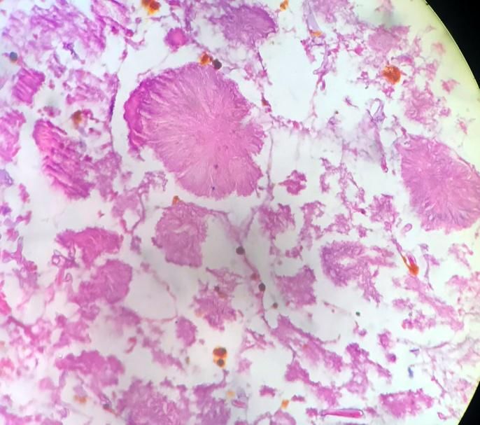
Sunil V Jagtap et.al. Co-infection of mucormycosis and actinomycosis in COVID-19 infection.
month back. CT scan - PNS (plain + contrast) nodes seen on both sides. Bilateral orbits
showed residual/recurrent ill-defined including extraocular m uscles and retro
h eterogeneously enhancing soft tissue orbital fat appear normal. Cribriform plate
thickening noted in the parts of nasal and critsa galli appear normal. Rest of the
septum, medial walls of bilateral maxillary, visu alised bones appea r n ormal. Visualised
ethmoid, sphenoid and fron tal sinuses brain parenchyma appears normal.
extending into bilateral nasal cavities. Patient underwent right
Features suggestive of infective pathology hemimaxillectomy with bilateral sinus
of invasive fungal rhinosinusitis. (Figure 1 curettage; debridement of the devitalized
A,B) There is evidence of bone defect in areas in the nasal floor was done. Necrotic
an terior wall of left maxillary sinus maxillary and palatal bone with teeth 18-21
extending to the alveolar process of maxilla were removed.
on left side. Few enlarged cervical lymph
Figure-1A,B: CT scan - PNS h eterogeneously enhancing soft tissue thickening of nasal septum, walls of maxillary, ethmoid,
sphenoid and fron tal sinuses extending in to bilateral nasal cavities.
tissue showed extensive areas necrosis and
inflammation predominantly of neutrophils
along with scattered lymphocytes, plasma
cells, macrophages and few giant cells.
Areas of inflamed granulation tissue,
hemorrhage and congestion was noted.
Necrotic areas showed numerous colonies
of mucormycosis consisting of broad, non-
septate hyphae.(Figure -2, Figure 3) The
PAS, Grocott’s methenamine silver stain
showed fungal hyphae of mucormycosis and
actinomycosis. On histopathological
examination the patient was diagnosed with
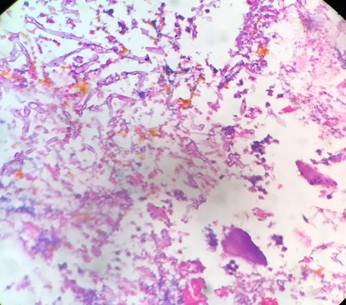
Figure-2: On microscopy areas of inflamed granulation tissue mucormycosis and actinomycosis. The
and necrosis showing colonies of mucormycosis consisting of specific treatment guide followed for
broad, non-septate hyphae and actinomycosis.(H&E Stain.40x)
COVID-19 and co-infection .After
treatment with Amphotericin-B -antifungal
On microscopy revealed as tissue
therapy showed improvement. Patient is
lined by columnar epithelium showing
advised regular follow up.
ulceration and underlying subepithelial
International Journal of Health Sciences and Research (www.ijhsr.org) 128
Vol.11; Issue: 8; August 2021Sunil V Jagtap et.al. Co-infection of mucormycosis and actinomycosis in COVID-19 infection.
neutropenia, corticosteroid use, diabetes
mellitus, HIV etc.[5,6]
This fungus usually induces a
pyogenic inflammatory reaction which is
characterised by abscess formation. The
hyphae are characteristically broad, usually
aseptate, thin walled. The size range in
width from 3 - 25µm and in length upto 200
µm. They are empty, having focal bulbous
dilatation or irregular branching mostly at
right angles. They take its invasive form
resulting in vascular invasion, thrombosis
and necrosis.
Figure-3: On microscopy high power view of mucormycosis Actinomycosis is commensal in the
consisting of broad, non-septate hyphae and actinomycosis.
(H&E Stain.100x) oral cavity. Actinomycosis is an infrequent
invasive bacterial disease that has been
DISCUSSION recognized for over a century.
Co-infection of mucormycosis, Actinomyces spp. are, anaerobic, Gram-
actinomycosis and COVID-19 of these three positive, filamentous bacteria. It causes
pathogens in a same host is rare. The cases chronic suppurative inflammation.
of mucormycosis have been reported in Actinomycosis commonly involves
patients those recently recovered from cervicofacial, thoracic, abdomino-pelvic
COVID-19 in many regions of India. The region and central nervous system.[7,8]
COVID 19 pandemic has thrown up yet Within the cervicofacial region
another challenge for the healthcare sector, actinomycosis has been reported in oral
with isolated cases of an unusual fungal cavity, mandible, maxilla, paranasal sinuses,
infection being reported with also showing salivary glands, and eye, ear and neck areas.
rise in mortality. In recent studies observed Invasion of Actinomyces is precipitated by
that, in COVID-19 infected patient who factors such as dental caries, trauma,
treated with widespread use of broad immunosuppression and periodontitis.
spectrum antibiotic or steroid and recovered Beyond the pathogenesis of SARS-
developed secondary bacterial or fungal CoV-2, microbial coinfection plays an
infection in 8%. [3] important role in the occurrence and
The critically ill-patients, patients on development of increasing the disease
mechanical ventilation and those with symptom and mortality. Among COVID-19
longer hospital stay are more likely to patients the various co-pathogens included
develop fungal co-infection Mucormycosis bacteria, such as Streptococcus pneumoniae,
are seen in the nature widely distributed. Staphylococcus aureus, Klebsiella
The primary sites of invasion are nasal pneumoniae, Mycoplasma pneumoniae,
sinuses which spread as rhino-maxillary and Chlamydia pneumonia, Legionella
rhino-cerebral-orbital mucormycosis.[4] The pneumophila and Acinetobacter baumannii;
common clinical features are mild fever, Candida species and Aspergillus flavus; and
headache, swelling, facial pain, also other viruses such as influenza, rhinovirus/
sites for mucormycosis invasion are enterovirus, parainfluenza, metapneumo-
pulmonary, cutaneous, gastrointestinal tract virus, influenza B virus, and human
or disseminated. They infect immunodeficiency virus were noted. 8
immunosuppressed individuals and the Combined orofacial aspergillosis and
major predisposing factors are mucormycosis was reported by Chermetz M
malignancies, organ transplants, et al.[9] Jagtap SV, et al reported case of
mucormycosis in post COVID-19 infection
International Journal of Health Sciences and Research (www.ijhsr.org) 129
Vol.11; Issue: 8; August 2021Sunil V Jagtap et.al. Co-infection of mucormycosis and actinomycosis in COVID-19 infection.
who received steroid treatment and also a 5. Singh AK, Singh R, Joshi SR, Misra A.
case of HIV/AIDS.[10] Lin et al reported a Mucormycosis in COVID-19: A systematic
case of a cavitary pulmonary lesion with review of cases reported worldwide and in
aspergillosis, mucormycosis, and India. Diabetes Metab Syndr. 2021 May
actinomycosis co-infection.[11] The specific 21;15(4):102146.
6. Garg D, Muthu V, Sehgal IS,
treatment guide lines should be follow for Ramachandran R, Kaur H, Bhalla A, Puri
COVID-19 and co-infection.[12] The GD, Chakrabarti A, Agarwal R.
appropriate antimicrobial agents treatment is Coronavirus Disease (Covid-19) Associated
essential to reduce the mortality. Mucormycosis (CAM): Case Report and
Systematic Review of Literature.
CONCLUSION Mycopathologia. 2021;186(2):289-298.
Among COVID-19 patients, 7. Valour F, Sénéchal A, Dupieux C, et al.
recognizing the possible pathogens causing Actinomycosis: etiology, clinical features,
co-infection is important. We are presenting diagnosis, treatment, and
this rare case of COVID-19 associated with management. Infect Drug Resist. 2014;7:
co-infection of mucormycosis and 183-197.
8. Khatib WM, Jagtap SV, Patel PM et al.
actinomycosis for its clinical, radio imaging, Tonsillar actinomycosis – A case report. Int
and on histopathological findings. The J Health Sci Res. 2015; 5(9):607-609.
appropriate antimicrobial agents treatment is 9. Chermetz M, Gobbo M, Rupel K, Ottaviani
essential to reduce the mortality. G, Tirelli G, Bussani R, et al. Combined
orofacial aspergillosis and mucormycosis:
Acknowledgement: None fatal complication of a recurrent paediatric
glioma-case report and review of
Conflict of Interest: None literature. Mycopathologia 2016; 181:723–
733.
Source of Funding: None 10. Jagtap SV, Jagtap SS, Nagar V, Varshney
K. Invasive mucormycosis in post COVID-
19 infection: Case report with review. IP
REFERENCES Arch Cytol Histopathology Res 2021;6(2):
1. Zhou P, Liu Z, Chen Y, Xiao Y, Huang X,
135-139.
Fan XG. Bacterial and fungal infections in
11. Lin, Lan, Xue, Dan; Lin, et al .Pulmonary
COVID-19 patients: A matter of
aspergillosis, mucormycosis, and
concern. Infect Control Hosp Epidemiol.
actinomycosis co-infection presenting as a
2020;41(9):1124-1125.
cavitary lesion in a patient with diabetes,
2. Mehta S, Pandey A – Rhino-Orbital
Chinese Medical Journal: 2019; 132, 20;
Mucormycosis Associated With COVID-19.
2512-2513.
Cureus. 2020;12(9):e10726.
12. Lai CC, Wang CY, Hsueh PR. Co-
3. Rawson TM, Moore LSP, Zhu N,
infections among patients with COVID-19:
Ranganathan N, et al . Bacterial and fungal
The need for combination therapy with non-
coinfection in individuals with coronavirus:
anti-SARS-CoV-2 agents? J Microbiol
a rapid review to support COVID-19
Immunol Infect. 2020;53(4):505-512.
antimicrobial prescribing. Clin Infect Dis.
2020 ;3;71(9):2459-2468.
How to cite this article: Jagtap SV, Hulwan A,
4. Gupta S, Goyal R, Kaore NM. Rhino-
Vartak S et.al. Co-infection of mucormycosis
Orbital-Cerebral Mucormycosis: Battle with
and actinomycosis in COVID-19 infection. Int J
the Deadly Enemy. Indian J Otolaryngol
Health Sci Res. 2021; 11(8): 127-130. DOI:
Head Neck Surg. 2020;72(1):104–11.
https://doi.org/10.52403/ijhsr.20210818
******
International Journal of Health Sciences and Research (www.ijhsr.org) 130
Vol.11; Issue: 8; August 2021You can also read