Annals of Cardiology and Cardiovascular Medicine
←
→
Page content transcription
If your browser does not render page correctly, please read the page content below
Annals of Cardiology and Cardiovascular Medicine Case Report
Published: 19 Apr, 2019
Ebstein’s Anomaly with Severe Tricuspid Valve Stenosis
Presenting as Acute Embolic Stroke Secondary to Deep
Vein Thrombosis
Veena Nanjappa, Hema Raveesh*, Sadanand KS and Manjunath CN
Department of Cardiology, Sri Jayadeva Institute of Cardiovascular Sciences and Research, India
Abstract
Ebstein’s malformation is an uncommon congenital heart disease with great variability in the
morphology of the tricuspid valve. Its prevalence is 0.3% to 0.5% and less than 10% cases are
associated with severe tricuspid valve stenosis. Patients with Ebstein’s anomaly have 10% incidence
of pre-excitation. We hereby report a similar case of Ebstein’s anomaly with asymptomatic cardiac
status presenting for the first time at adult age with embolic stroke as a result of paradoxical
embolism via ostium secundum atrial septal defect secondary to left leg deep vein thrombosis with
pre-excitation syndrome on ECG.
Keywords: Ebstein’s anomaly; Atrial septal defect; Tricuspid valve stenosis; DVT
Introduction
Ebstein’s malformation is an uncommon congenital heart disease with great variability in
the morphology of the tricuspid valve. In the majority of cases, dysplasia and displacement of the
tricuspid valve leaflets result in varying degrees of tricuspid regurgitation. In some, however, there
may be linear attachments of the antero-superior and mural leaflets of tricuspid valve to the junction
between the inlet and trabecular components of the right ventricle, resulting in imperforate type of
tricuspid valve or embryologically may arrest later on in development resulting in severe tricuspid
OPEN ACCESS
stenosis [1].
*Correspondence:
We hereby report a case of Ebstein’s anomaly with severe tricuspid valve stenosis with NYHA
Hema Raveesh, Department of
functional class II status presenting for the first time at adult age with left hemiparesis as a result of
Cardiology, Sri Jayadeva Institute of paradoxical embolism via ostium secundum Atrial Septal Defect (ASD) secondary to left leg Deep
Cardiovascular Sciences and Research, Vein Thrombosis (DVT) with pre-excitation syndrome on ECG.
Mysore, India, Tel: 919886512210;
E-mail: hemaraveesh@yahoo.com Case Presentation
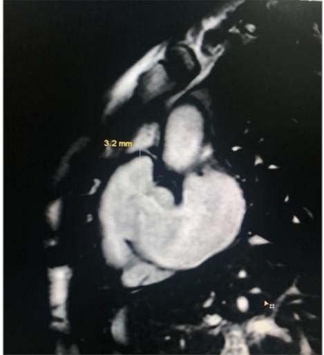
Received Date: 14 Mar 2019 A 32 year old young male presented to the neurologist with transient left hemiparesis and
Accepted Date: 12 Apr 2019 dysarthria and was diagnosed with stroke. He also gave history of painful swelling of both lower
Published Date: 19 Apr 2019 limbs of two weeks duration. MRI brain revealed acute infarcts in right basal ganglia and corona
Citation: radiate (Figure 1); subacute infarcts in right posterior temporal lobe and also chronic infarcts in
Nanjappa V, Raveesh H, Sadanand bilateral periventricular and centrum semi ovale regions. MR angiogram revealed distal middle
KS, Manjunath CN. Ebstein’s Anomaly cerebral artery embolus.
with Severe Tricuspid Valve Stenosis On clinical examination he had bilateral tender swelling of both lower limbs (left > right) and
Presenting as Acute Embolic Stroke vitals were stable with no perceptible murmur on cardiac examination. He had central cyanosis
Secondary to Deep Vein Thrombosis. with saturation of 88% at rest and prior to his CVA he was in functional class II and was working
Ann Cardiol Cardiovasc Med. 2019; as a farmer. There was no differential cyanosis. He had pan digital grade II clubbing. On further
3(1): 1020. questioning, he gave no history of cyanosis, palpitations or dyspnea in the past. He was born of
Copyright © 2019 Hema Raveesh. a non-consanguineous marriage and had uneventful birth history. There was no history of any
This is an open access article congenital heart disease in his siblings. His ECG revealed pre-excitation syndrome with short PR
distributed under the Creative
and delta wave suggestive of a possible right postero-septal pathway; his ECHO work up revealed
Ebstein’s anomaly with 4.5 mm apical displacement of hinge point of tricuspid valve; he also
Commons Attribution License, which
had severe tricuspid valve stenosis with dilated right atrium with trickle of flow seen across the
permits unrestricted use, distribution,
tricuspid valve. Linear attachments of the anterior and the mural leaflets resulted in almost complete
and reproduction in any medium,
separation of the inlet portion from the apical trabecular and outlet portions of the right ventricle.
provided the original work is properly
He had a large bidirectional ostium secundum ASD 4.2 cm in size (Figure 2). He had LV global
cited.
Remedy Publications LLC. 1 2019 | Volume 3 | Issue 1 | Article 1020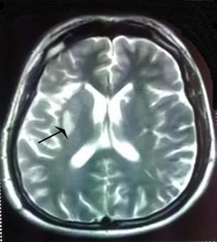
Hema Raveesh, et al.,                                                                                      Annals of Cardiology and Cardiovascular Medicine

 Figure 1: MRI brain: Image of acute infarcts in right basal ganglia and             Figure 3: Cardiac MRI shows severe tricuspid valve stenosis-Annular
 corona radiata; subacute infarcts were seen in right posterior temporal lobe        diameter of tricuspid valve 3.2 mm. Atrialization of right ventricle seen.
 and chronic infarcts were seen in bilateral periventricular and centrum semi
 ovale regions. MR angiogram revealed distal MCA embolus.
                                                                                    prevalence of 0.3% to 0.5% [2], occurring in 1 per 200,000 live births.
                                                                                    The following anomalies have been reported that may be associated
                                                                                    with Ebstein’s anomaly: Atrial septal defect (90%), anatomic or
                                                                                    functional pulmonary atresia (30%) and ventricular septal defect (less
                                                                                    common). Its association with an imperforate tricuspid valve and
                                                                                    tricuspid valve stenosis is seen in only less than 10% of cases [3,4].
                                                                                    This imperforation or stenosis occurs due to the excessive redundancy
                                                                                    of the anterior leaflet, with fusion of the commissures. Partial fusion
                                                                                    of developing tricuspid valve leaflet components results in tricuspid
                                                                                    stenosis and complete fusion results in tricuspid valve atresia [1,5].
                                                                                        Tethering and displacement of the tricuspid leaflets result in
                                                                                    a very peculiar echocardiographic appearance, with an “empty”
                                                                                    tricuspid annulus and membranous separation of the inflow and the
                                                                                    outflow portions of the right ventricle. This anatomic condition has
 Figure 2: Transthoracic 2D images: Ebstein’s anomaly in four chamber view;
 apical displacement of tricuspid valve seen, with large ostium secundum            been and continues to be the subject of discussion, as functionally it
 ASD.                                                                               may be classified as a subtype of tricuspid atresia, but embryologically
                                                                                    it belongs to the spectrum of Ebstein’s anomaly [6]. The first cases
hypokinesia with EF 40%. There was no Doppler gradient across                       were described in anatomic specimens by Van Praagh et al. [7] in
the right ventricular outflow tract and the pulmonary valve. Right                  1971.
ventricle appeared morphologically normal, although small. There                        Patients with Ebstein‘s anomaly have 10% incidence of pre-
was no demonstrable VSD or PDA on transthoracic echo. Since he                      excitation. Our patient had asymptomatic pre-excitation syndrome
gave no history of palpitations or syncope and no family history of                 with demonstrable delta wave in resting ECG. He was however not
heart disease he was not considered for electrophysiological study.                 subjected to stress test in view of his acuity of illness.
    His cardiac MRI revealed severe tricuspid valve stenosis with                       Celermajer et al. [6] reviewed 220 cases of Ebstein’s anomaly
atrialization of right ventricle suggestive of Ebstein’s anomaly (Figure            with 1 to 34 years of follow-up. Actuarial survival for all live-born
3). Annular diameter of tricuspid valve was 3 mm (Normal is: 2.8 cm                 patients was 67% at 1 year and 59% at 10 years. Predictors of death
to 3.1 cm) suggesting severe tricuspid valve stenosis. His RV was very              were echocardiographic grade of severity at presentation (relative risk
small.                                                                              increased by 2.7 for each increase in grade), fetal presentation, and
    He also had contrast induced nephropathy following MR                           right ventricular outflow tract obstruction.
angiogram. Venous Doppler of lower limb revealed tibial vein DVT                        Observation alone is advised for asymptomatic patients with no
extending till great saphenous vein. His thrombophilia work up                      right-to-left shunting and only mild cardiomegaly. Children who
and Antinuclear Antibodies (ANA) were negative. His hemoglobin                      have survived infancy generally do well for several years, and surgery
was 17% with hematocrit of 59%. He was started on parenteral and                    can be postponed until symptoms appear, cyanosis becomes evident,
overlap-oral anticoagulation till therapeutic INR was achieved.                     or paradoxical emboli occur. Deliberations about an operation should
Cardiac catheterisation study was not considered in view of his                     begin if evidence of deterioration exists, such as progressive increase
extensive DVT. His Celermajer score was not conducive for intra                     in right heart size, reduction in systolic function, or appearance of
cardiac repair.                                                                     ventricular or atrial tachyarrhythmias. Once symptoms progress
Discussion                                                                          to NYHA functional class III or IV, medical management has little
                                                                                    to offer, surgical risks increase, and operation is clearly indicated.
    Ebstein’s anomaly is an uncommon congenital heart defect with a                 A biventricular reconstruction is feasible for most patients in an

Remedy Publications LLC.                                                        2                                          2019 | Volume 3 | Issue 1 | Article 1020
Hema Raveesh, et al.,                                                                                     Annals of Cardiology and Cardiovascular Medicine

experienced cardiac centre. However, because of the wide spectrum                   malformation and related lesions of the tricuspid valve. Pediatr Cardio.
of anatomic variations in the tricuspid valve, the surgical approach to             1987:721-36.
patients with Ebstein’s anomaly needs to be individualized according             3. Maron BJ, Towbin JA, Thiene G, Antzelevitch C, Corrado D, Arnett D, et
to the specific morphology found at operation. A one and half                       al. Contemporary definitions and classification of the cardiomyopathies:
ventricle repair can be applied to the failing right ventricle. Heart               An American heart association scientific statement from the council on
transplantation is reserved for patients with severe biventricular                  clinical cardiology, heart failure and transplantation committee; quality
                                                                                    of care and outcomes research and functional genomics and translational
dysfunction. There are no surgical series data of Ebstein’s in India.
                                                                                    biology interdisciplinary working groups; and council on epidemiology
In the society of thoracic surgeons’ congenital heart surgery database              and prevention. Circulation. 2006;113(14):1807-16.
data [8], in hospital operative mortality was found to be 3.3% in
adults and late mortality of 10%.                                                4. Attenhofer Jost CH, Connolly HM, O’Leary PW, Warnes CA, Tajik AJ,
                                                                                    Seward JB. Left heart lesions in patients with Ebstein’s anomaly. Mayo Clin
    Bidirectional Glenn with closure of atrial septal defect was                    Proc. 2005;80(3):361-8.
contemplated for this patient in concordance with surgeon at later               5. Rao PS. Tricuspid atresia. In: Long WA, editor. Fetal and Neonatal
date.                                                                               Cardiology. Xth ed. Philadelphia: WB Saunders: 1990.p. 525-40.
Conclusion                                                                       6. Celermajer DS, Bull C, Till JA, Cullen S, Vassillikos VP, Sullivan ID, et al.
                                                                                    Ebstein’s anomaly: Presentation and outcome from fetus to adult. J Am
    This case highlights the importance of cardiac evaluation                       Coll Cardiol. 1994;23(1):170-6.
especially in stroke. Congenital heart diseases as a causal pathology
need to be kept in mind in the evaluation of stroke in young.                    7. Van Praagh R, Ongley PA, Swan HJC. Anatomic types of single or common
                                                                                    ventricle in man. Morphologic and geometric aspects of sixty necropsied
References                                                                          cases. Am J Cardiol. 1964;13:367-86.

1. Wilson AD, Rao PS. Embryology. In: Kambam J, editor. Cardiac Anesthesia       8. Davies RR, Pasquali SK, Jacobs ML, Jacobs JJ, Wallace AS, Pizarro
   for Infants and Children. St Louis: MoMosby; 1994. p. 3-9.                       C. Current spectrum of surgical procedures performed for Ebstein’s
                                                                                    malformation: An analysis of the society of thoracic surgeons’ congenital
2. Anderson RH, Shinebourne EA, Macartney FJ, Tynan M. Ebstein’s                    heart surgery database. Ann Thorac Surg. 2013;96(5):1703-9.

Remedy Publications LLC.                                                     3                                            2019 | Volume 3 | Issue 1 | Article 1020
You can also read