Comparative in vitro study of the cleaning efficacy of AirFloss ultra and I Prox Sulcus brushes in an orthodontic phantom model - Nature
←
→
Page content transcription
If your browser does not render page correctly, please read the page content below
www.nature.com/scientificreports
OPEN Comparative in vitro study
of the cleaning efficacy of AirFloss
ultra and I‑Prox Sulcus brushes
in an orthodontic phantom model
Hanna Boes1*, Sören Brüstle1, Gholamreza Danesh2, Stefan Zimmer1 & Mozhgan Bizhang1
Preventing biofilm-related risks such as gingivitis and white spot lesions during orthodontic
treatments is very challenging. The cleaning efficiencies of AirFloss Ultra and I-Prox P sulcus brushes
were evaluated using an orthodontic phantom model. After attaching brackets onto black-coated
maxillary KaVo teeth, a plaque substitute was applied. The evaluated tooth surfaces were divided
into two areas. Cleaning was performed with an AirFloss Ultra with two (A-2) or four (A-4) sprays or an
I-Prox P for two (I-2) or four (I-4) seconds. Images before and after cleaning were digitally subtracted,
and the percentage of fully cleaned surfaces was determined (Adobe Photoshop CS5, ImageJ).
Statistical analysis was performed by ANOVA and post hoc tests with Bonferroni correction (SPSS 25,
p < 0.05). The mean values of total cleaning efficacy were 26.87% for I-2, 43.73% for I-4, 34.93%, for
A-2 and 56.78% for A-4. The efficacy was significantly higher for A-4 than for A-2, I-4, and I-2. There
were significant differences between the four groups. Repeated cleaning led to an improved result.
Within the study limitations, the AirFloss Ultra with four sprays proved to be more efficient than the
sulcus brush I-Prox P for cleaning.
Effective oral hygiene plays a key role in preventing caries and periodontal diseases during orthodontic treat-
ment. The attachment of brackets, bands, and arches creates additional surfaces that differ from the oral tissue
structures. These retention sites provide ideal conditions for an increased accumulation of food residue and
bacterial plaque by also limiting the natural cleansing mechanisms of saliva and the oral muscles1–3. In addition,
the physiological germination pattern changes.
The presence of Streptococcus mutans and Lactobacillus represents an increased risk for the development of
carious lesions4–6. The study of Corbett, Brown et al. showed there was an increased prevalence of S. mutans
during orthodontic treatment. Before treatment, patients showed a lower concentration of this bacteria in the
plaque than patients during t reatment7. The concentration of S. mutans increased fourfold during orthodontic
treatment than before treatment and decreased after treatment to the initial value8. As a result, initial deminer-
alization of the enamel can be observed in this group of patients within a very short time after insertion of the
orthodontic device9.
The incidence of developing white spot lesions during orthodontic treatment varies between 2 and 96%,
and not every patient is affected s imilarly3,10–13. To avoid these side effects, preventive procedures need to be
performed to reduce the proliferation of plaques and to efficiently remove them. In addition to proper nutrition
and the use of fluoride, dental and oral hygiene are important.
The studies published thus far have mainly compared the effectiveness of different toothbrushes for plaque
removal in multibracket patients and have reported different r esults14–18. Rosema, Slot et al. showed that the use
of a toothbrush reduces plaque by 46%19. In particular, the hard-to-reach circular surfaces around brackets and
proximal areas are not cleaned sufficiently with a toothbrush, leading to the demineralization of the enamel and
periodontal diseases20.
Alternative cleaning procedures are therefore necessary to make the cleaning process more efficient for
patients with fixed orthodontic appliances. The literature recommends using proximal cleaning devices to remove
plaque in these barely accessible areas21,22. However, studies reviewing these proximal cleaning procedures usually
consider only participants without fixed orthodontic appliances23,24.
1
Department of Operative and Preventive Dentistry, Faculty of Health, Witten/Herdecke University,
Alfred‑Herrhausen‑Str. 50, 58455 Witten, Germany. 2Department of Orthodontics, Faculty of Health,
Witten/Herdecke University, Witten, Germany. *email: Hanna.Boes@uni‑wh.de
Scientific Reports | (2021) 11:1921 | https://doi.org/10.1038/s41598-021-81603-y 1
Vol.:(0123456789)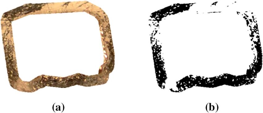
www.nature.com/scientificreports/
In 2015, Philips presented the Sonicare AirFloss Ultra device, promising efficient plaque removal of approxi-
mal areas (the tooth surface facing an adjacent tooth) and easy use. Previous studies have shown meaningful
results when applying the AirFloss25,26. Another possibility is to use a mono-tuft brush27. The v-shaped Miradent
mono-tuft brush ensures gentle and efficient cleaning of hard to access areas. Both devices have the advantage
that no proximal contact point needs to be bypassed during cleaning. This eases cleaning tremendously. One
study showed that an oral hygiene procedure with an electric toothbrush combined with AirFloss was more
effective than a manual toothbrush combined with flossing in reducing plaque and gingivitis in orthodontic
subjects following 3 weeks of use28.
A very important parameter is the compliance of the patient11. A previous study proved the high accept-
ance and easy handling of the AirFloss. Compared to the use of dental floss, 78% of the patients preferred the
AirFloss29. To date, no studies have described the cleaning abilities of the I-Prox P brush. However, it can be
assumed that accidental sliding of the brush on the gingiva might lead to pain and therefore reduce patient
compliance.
To date, no studies have published the results of the cleaning efficiency of the AirFloss Ultra compared to that
of the I-Prox P brush in multibracket patients using each a single time. Thus, the aim of this prospective in vitro
study was to investigate the cleaning efficiency of the AirFloss Ultra and the I-Prox P brushes on an orthodontic
phantom model under standardized conditions.
Materials and methods
The cleaning process was performed on a maxillary (upper jaw) model with full dentition of plastic teeth (KaVo
Dental GmbH, Ulm, Germany).
Preparation. First, the buccal (tooth area facing towards the cheek) and the approximal surfaces of the plas-
tic teeth were sandblasted with 110 µm aluminium oxide Korox at 2 bars (Bego, Bremer Goldschlägerein Wilh.
Herbst GmbH & Co KG, Bremen, Germany; device: Renfert Classic Plus 3, Renfert GmbH, Hilzingen, Ger-
many). A labelled diagram explaining the dental anatomy of the maxilla can be found online in Supplementary
Figure S2. This process led to the adequate adhesion of the black two-component varnish applied to the plastic
teeth (DuPont Refinish, Willich, Germany). After drying for 24 h, a second coat was applied. The black varnish
provided an ideal contrast to white artificial plaque. Then, brackets (Mini 2000, Straight-Wire, Ormco, Orange,
California, USA) were attached to the teeth by superglue (Henry Schein, Instant Fix, Melville, USA), and a wire
(Niti-Wire round 0.012 inch, Orthana, Recklinghausen, Germany) was ligated with orthodontic ligatures (Den-
talastics Personal, Dentauraum, Ispringen, Germany).
To ensure standardized and repositionable fixation of the teeth for photography, the teeth were placed into a
plaster block after each cleaning stage. A total of three blocks were produced for the buccal, mesial (approximal
surface directed towards the midline of the face) and distal (approximal surface directed away from the midline
of the face) areas (Dento-stone 220, Dentona AG, special superhard plaster type 4, Dortmund, Germany). To
make the blocks, the root of each tooth needed to be covered with wax to prevent areas divergence (Modelling
wax, Henry Schein, Melville, USA). The teeth were set into plaster 2 cm apart from each other. To ensure an even
height and distance, the crown of the tooth was fixed in A-silicone (polysiloxane, Henry Schein, Melville, USA).
Twenty millilitres of water was added to the plaster (100 g) and it was stirred with a vacuum mixer (Motova
SLA, BEGO AG, Bremen, Germany). Then, the root of each blocked tooth was set into the plaster. To enable easy
insertion and removal of the teeth from the block, cavities were milled into the plaster block. Then, the blocks
were trimmed to their final shape (Renfert GmbH, Hilzingen, Germany).
To standardize the evaluation, it was important that the artificial plaque did not peel off, to ensure high
contrast with the black tooth surface, and that its composition did not change during the trial. A suspension of
titanium dioxide (13 g) and isopropyl alcohol (28 millilitres) (VWR International GmbH, Langenfeld, Germany)
was used to simulate the plaque. With a magnetic stirrer (IKA-Werke GmbH & Co. KG, Staufen, Germany), the
two ingredients were mixed for 30 min, turning them into a viscous compound. Standardized conditions were
maintained by using mechanical stirring and constant ingredient mixing ratio. After a resting time of 24 h, the
plaque was applied manually to the teeth by one brushstroke with a clean brush. By tapping the tooth on the edge
of the table, the plaque spread uniformly over the tooth surface. It was allowed to dry for 4 h. For each cleaning
trial, one layer was applied.
Cleaning process. Cleaning was accomplished by the Sonicare AirFloss Ultra HX8431 (Philips GmbH,
Amsterdam, Netherlands) or by the sulcus brush I-Prox P (Miradent, Hager & Werken GmbH & Co. KG, Duis-
burg, Germany) (Fig. 1). Both devices were characterized by the fact that no proximal contact can be bypassed.
The spray nozzle of the AirFloss device faces the buccal surface of the approximal area. By pressing a button,
water sprays at a speed of 72 km per hour from the buccal to the oral surface facing the mouth cavity. Depending
on the mode, one, two, or three spray bursts are applied. The sulcus brush consists of an approximately 14-cm-
long handle with a tapered v-shaped brush. A new brush was used for each cleaning process. Cleaning was car-
ried out by rotating movements in the buccal and approximal areas.
To standardize the cleaning processes, the AirFloss Ultra was always applied in contact with the tooth surface
at a constant angle. The use of the devices was performed as recommended in the respective instruction manuals
(see Supplementary Document S3). In contrast to a standard toothbrush, the I-Prox P brush is held at a right
angle in relation to the buccal and approximal tooth surfaces. Because of the tilted head of the brush (120° angle),
this cleaning position allows for gentle cleaning without irritating the gingiva. The brush was used with a contact
pressure of two bars, and the application time was managed with a timer.
Scientific Reports | (2021) 11:1921 | https://doi.org/10.1038/s41598-021-81603-y 2
Vol:.(1234567890)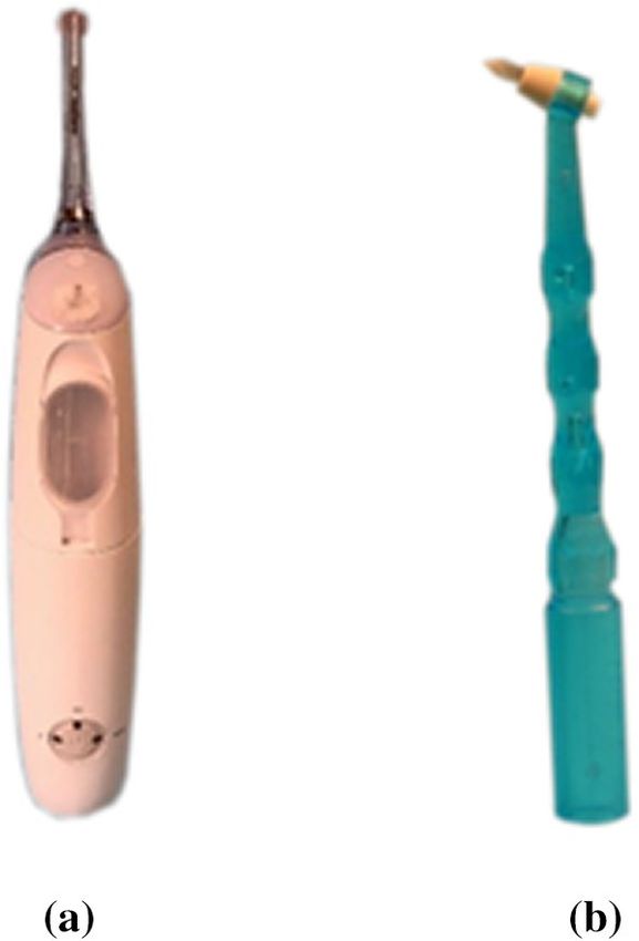
www.nature.com/scientificreports/

                                  Figure 1.  The Sonicare AirFloss Ultra HX8431 (a) and the sulcus brush I-Prox P (b) were used for cleaning.

                                  Figure 2.  A specially built appliance was attached to the KaVo model to ensure standardized cleaning.

                                      First, the buccal area was cleaned followed by the mesial and distal approximal surfaces. After each cleaning
                                  procedure, the brush of the I-Prox P was replaced to maintain a constant hardness of the cleaning filaments.
                                  The AirFloss pressure of the sprays was pre-set and did not change during the trial. Depending on the cleaning
                                  routine, the double burst mode was accomplished one (two spray bursts per area) and two times (four spray
                                  bursts per area). The nozzle tip of the AirFloss was placed horizontally between two teeth at the gum line while
                                  the activation button was pressed to deliver two and four bursts, respectively.
                                      To achieve cleaning as close to reality as possible, the KaVo model was fixed to a special appliance. This setup
                                  allowed simulated cleaning of the upper teeth at eye level and from the front as if they were the teeth of the trial
                                  operator. Additionally, it prevented the model from moving during the process (Fig. 2).
                                      Every cleaning stage was divided into four groups. Each group was repeated four times leading to a total of
                                  16 cleaning procedures.

                                  •   The use of the I-Prox P brush for two seconds per area (I-2).
                                  •   The use of the I-Prox P brush for four seconds per area (I-4).
                                  •   The use of the AirFloss Ultra with two spray bursts per area (A-2).
                                  •   The use of the AirFloss Ultra with four spray bursts per area (A-4).

Scientific Reports |   (2021) 11:1921 |                https://doi.org/10.1038/s41598-021-81603-y                                                   3

                                                                                                                                               Vol.:(0123456789)
www.nature.com/scientificreports/
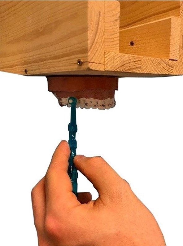
Figure 3. (a) shows the extracted photograph. (b) presents the black-and-white picture of the cleaned, white
areas and the uncleaned, black areas.
Each cleaning routine (I-2, I-4, A-2, A-4) included the cleaning of four incisors, two canines, four premolars,
and four molars (see Supplementary Figure S2). Since only two canines were included in the KaVo model, an
extra cleaning stage was carried out by cleaning only the two canine. Cleaning a total of 16 teeth in each clean-
ing routine and repeating each group four times led to an equal number of 64 evaluated teeth of each tooth type
(incisors, canines, premolars, molars). All the cleaned teeth in all four groups totalled 256 teeth.
Photograph editing. After each completed cleaning procedure, the teeth were removed from the KaVo
model and placed into the plaster blocks. In a photobooth, photographs were taken by a Canon EOS 60D with
an EF-S 18–25 mm IS II lens (Canon GmbH, Krefeld, Germany) at 62.2 cm.
Before applying titanium oxide to the teeth, a total of 12 masks of the buccal and approximal surfaces were
produced, defining the areas considered for evaluation (Adobe Photoshop CS5, Adobe Systems Software Ireland
Limited, Dublin, Ireland).
After putting the teeth in the plaster blocks, the masks produced in advance were layered on the cleaned
photographed areas. The extraction was conducted using Adobe Photoshop CS5. The generated cut-outs were
evaluated with the ImageJ software program (National Institutes of Health, Bethesda, USA). By turning them into
black-and-white pictures, the cleaned areas (black) were distinguishable from the uncleaned surfaces (white).
The ratio of white pixels to the total number of pixels of the mask corresponded to the cleaning efficacy (Fig. 3).
Outcome measures. The working hypothesis assumed that the AirFloss Ultra would achieve a higher
cleaning efficacy than the sulcus brush I-Prox P.
Statistical analysis. To produce significant results, the sample size was estimated with the G* Power 3.1.9.2
program30. A sample size of 32 teeth (16 teeth per group) was analysed with a statistical power of 0.8, an alpha
error of 0.05, and an effect size of 1.04.
All data were analysed with the SPSS 25.0 software program (IBM Germany GmbH, Ehningen, Germany).
All 256 teeth served as the bases for the statistical analysis. Each area of each tooth (mesial, distal, and buccal)
was evaluated, leading to a total of 768 single values. For the descriptive statistics, the minimum, maximum,
median, mean, and standard deviation were used. Kolmogorov–Smirnov test identified a normal distribution
with an asymptomatic significance value of p = 0.2 for the mesial, distal, and buccal areas in all the cleaning
groups. Within groups I-2, A-2, and A-4, a normal distribution with an asymptomatic significance value of p = 0.2
was found. The values of cleaning groups I-4 showed an asymptomatic significance value of p = 0.01. Subsequent
ANOVAs and post hoc tests with Bonferroni correction were used to determine the significance between the
cleaning groups at a significance level of p = 0.05.
Results
The cleaning efficacy was based on the evaluation of 256 teeth. The mesial, distal, and buccal surface of every
tooth was observed. Each cleaning procedure provided 14 buccal, 14 distal and 14 mesial photos. A total of 768
single pictures were obtained by conducting the two cleaning stages. The I-Prox brush achieved an overall clean-
ing efficacy of 26.87% (SD: 11.37) with a two-second application (I-2) and 43.73% (SD: 10.74) with a four-second
application (I-4). The AirFloss managed to clean 34.93% (SD: 9.54) of the evaluated tooth surfaces with two
sprays (A-2) and 56.78% (SD: 10.12) with four sprays (A-4). The results of the cleaning efficacy of the Sonicare
AirFloss Ultra and the I-Prox P brush are described in Table 1.
With a mean cleaned area of 56.78% (SD: 10.12%), the overall cleaning efficacy of the AirFloss Ultra (A-4)
was significantly higher. The least efficient result was achieved by the I-Prox P brush (I-2), with a mean cleaned
area of 26.87% (SD: 11.37%). The application of the AirFloss Ultra with two sprays (A-2) cleaned 34.93% (SD:
9.54%) of the tooth surface and the I-Prox P brush (I-4) for 4 s cleaned 43.73% of the tooth surface (SD: 10.74%).
Significant differences existed between all groups, with p < 0.001 and p < 0.01 (Fig. 4).
Significant differences between cleaning groups were not observed based on the mean cleaning efficacy of the
cleaned area (buccal, mesial, distal) (Fig. 5). The corresponding mean and standard deviation of each application
in relation to the tooth area are presented in Table 1.
Scientific Reports | (2021) 11:1921 | https://doi.org/10.1038/s41598-021-81603-y 4
Vol:.(1234567890)www.nature.com/scientificreports/
Area of tooth Type of tooth
Group Total Buccal Mesial Distal Incisor Canine Premolar Molar
I-2 (n = 14) 26.87 (11.37) 39.48 (5.45) 17.51 (5.73) 23.62 (8.27) 29.74 (10.51) 29.09 (7.08) 23.86 (14.07) 25.89 (11.33)
I-4 (n = 14) 43.73 (10.74) 53.84 (5.67) 37.01 (9.07) 40.35 (8.84) 48.51 (5.61) 45.29 (8.57) 38.01 (12.38) 43.89 (12.22)
A-2 (n = 14) 34.93 (9.54) 31.98 (6.34) 37.24 (12.43) 35.57 (8.76) 30.80 (6.34) 29.41 (8.92) 35.69 (7.99) 41.05 (11.05)
A-4 (n = 14) 56.78 (10.12) 53.18 (8.93) 59.71 (10.88) 57.44 (10.07) 49.47 (6.20) 56.50 (11.20) 60.87 (7.86) 60.13 (11.66)
Table 1. The means (in percent) and standard deviation (SD) of the cleaned areas of each cleaning group
(I-Prox P brush for 2 s (I-2) and four seconds (I-4) and the Airflow for 2 sprays (A-2) and four sprays (A-4)).
Figure 4. The mean total cleaning efficacy (in percent) with the standard deviation of the I-Prox P brush
for two seconds (I-2) and four seconds (I-4) and the AirFloss Ultra with 2 sprays (A-2) and 4 sprays (A-4).
Horizontal lines with “***” show significant differences between the devices with p < 0.001, and “**” indicates a
significant difference with p < 0.01.
Figure 5. The means and standard deviation of the cleaning efficacy of the buccal (a), mesial (b) and distal (c)
areas. Horizontal lines with “***” describe significant differences with p < 0.001, “**” with p < 0.01, and “*” with
p < 0.05 between the I-Prox P brush applied for two (I-2) and four seconds (I-4) and the AirFloss with two (A-2)
and four sprays (A-4). “n.s.” indicates no significant differences.
Further significant differences in the mean cleaning efficacy between the applied devices can be seen with
respect to the type of tooth (incisor, canine, premolar, and molar) (Fig. 6). The corresponding mean and standard
deviation of each application with respect to tooth type are presented in Table 2.
Table 2 presents an overview of the maximum and minimum cleaning efficacy of each tooth surface and
tooth type achieved by the use of the I-Prox P brush for two (I-2) or four (I-4) seconds or the application of the
AirFloss Ultra with two (A-2) or four (A-4) sprays.
Scientific Reports | (2021) 11:1921 | https://doi.org/10.1038/s41598-021-81603-y 5
Vol.:(0123456789)www.nature.com/scientificreports/
Figure 6. The means and standard deviation of the cleaning efficacy of the incisors (a), canines (b), premolars
(c), and molars (d). Horizontal lines with “***” describe significant differences with p < 0.001, “**” with p < 0.01,
and “*” with p < 0.05 between the I-Prox brush applied for two (I-2) and four seconds (I-4) as well as the
AirFloss with two (A-2) and four sprays (A-4). “n.s.” indicates no significant differences.
Area of tooth Type of tooth
Total Buccal Mesial Distal Incisor Canine Premolar Molar
Maximum A-4 A-4/I-4 A-4 A-4 A-4/I-4 A-4/I-4 A-4 A-4
Minimum I-2 A-2 I-2 I-2 A-2/I-2 A-2/I-2 A-2/I-2 I-2
Table 2. Maximum and minimum cleaning efficacy of the I-Prox P brush and the AirFloss Ultra cleaning
devices at different application frequencies (two seconds (I-2)/sprays (A-2) vs. four seconds (I-4)/sprays (A-4)).
Discussion
The long duration of orthodontic therapy with fixed appliances usually causes patients to lose compliance with
adequate oral h ygiene9,31. This in vitro study compared the sulcus brush I-Prox P by Miradent to the Philips
AirFloss Ultra device, which shows potential for easy and effective cleaning.
This study shows the advantages and disadvantages regarding their cleaning efficacy. However, uncertainty
parameters associated with in vivo models, such as the dependency of compliance, cleaning routine, manual
skills, and saliva composition, were eliminated. On the other hand, the plaque substitute and the teeth themselves
do not correspond exactly to real conditions. To measure the cleaning efficacy more precisely, in vivo studies
are required using measurable parameters such as the approximal plaque or the stage of inflammation of the
interdental papilla32. Because of the greater plaque formation on the buccal sites of maxillary teeth compared to
mandibular teeth, maxillary teeth are thought to be more difficult to clean than lower t eeth33. For this reason,
the maxillary model was chosen for the cleaning procedures.
During the cleaning process, the approximal areas are not visible to the person conducting the study. This
study used plaster blocks to create standardized and reproducible conditions for the trial process. In addition,
this setup enabled a precise digital analysis of the buccal and approximal areas. The evaluation and analysis of the
percent of cleaning efficacy were performed by ImageJ software. Previous studies used this program to evaluate
photographs, describing it as a reliable measurement t ool34,35.
Scientific Reports | (2021) 11:1921 | https://doi.org/10.1038/s41598-021-81603-y 6
Vol:.(1234567890)www.nature.com/scientificreports/
The simulation of dental plaque is one of the challenges of clinical studies. To standardize laboratory condi-
tions, a well-established test method for the cleaning effectiveness of different devices was used36. Schätzle et al.
also used titanium oxide to simulate dental b iofilm37. Titanium oxide does not peel off during the cleaning
procedure and shows high contrast to black-coloured teeth38. This method enables a precise evaluation of the
cleaned areas. Although the plaque substitute does not show any other similarities to natural plaque, significant
findings can be a chieved36,37,39. However, in vivo studies are required to verify the results.
Previous studies have proven that cleaning devices remove plaque reliably in areas with direct contact between
the device and the tooth surface40. This situation leads to the assumption that the cleaning result is dependent
on the dimensions of the contact area of the cleaning device and the tooth surface. Within this study, the aim
was not only to measure the efficacy of cleaning but also to illustrate all areas reached by the application of the
tested devices.
Statement 1: The overall cleaning performance of the AirFloss ultra is superior to that of the
I‑Prox P brush. The overall cleaning performance is a summary of the cleaning effects on the individual
tooth surfaces (buccal, mesial, and distal) and tooth types (incisors, canines, premolars, and molars). The I-Prox
P brush with a four-second application (I-4) achieved a comparable cleaning efficiency on the buccal surfaces
and on the incisor and canine teeth compared to the AirFloss Ultra with four applications (A-4), but it per-
formed significantly worse in the proximal and posterior areas. Since the overall cleaning effect is based on the
summation of all tooth surfaces and tooth types, the overall cleaning efficacy of the AirFloss Ultra is significantly
higher when used four times. The reasons for the significantly better cleaning effect of the AirFloss Ultra com-
pared to the I-Prox P brush are mainly due to the structures and types of the cleaning devices. Key statements
two and three provide a detailed statement about the results achieved.
Statement 2: I‑Prox P cleans more efficiently on buccal surfaces than on proximal sur‑
faces. No significant difference was found regarding the cleaning efficacy of the I-Prox P brush (I-4) and
AirFloss Ultra (A-4) when applied for four seconds and four sprays on the buccal surface, respectively. The
slightly better cleaning result of the I-Prox P sulcus brush on the buccal surface can be attributed to its bristle
design. Due to the conically tapered V-shaped bristle tufts, the brush tips also clean difficult-to-reach surfaces
underneath the bracket-arch area. This reach results in a larger contact area between the tooth and bristle tips,
which is crucial for efficient plaque r emoval40. In contrast, the wall shear stress of the AirFloss Ultra spray jet is
not powerful enough at the margins to remove plaque adequately. In summary, the buccal area of the teeth can
be cleaned as adequately by using the sulcus brush as by using the AirFloss Ultra.
However, the limitations of the I-Prox P brush appear when considering the cleaning of the proximal areas. In
the present study, the proximal region was divided into mesial and distal areas. The evaluation showed a signifi-
cant superiority of the AirFloss Ultra with four sprays (A-4) on both surfaces. The inferior result of the I-Prox P
brush in the proximal areas is due to the special brush architecture of the sulcus brush. In contrast to its effect on
buccal surfaces, the V-shaped design of the bristle tufts is unsuitable for cleaning the proximal surfaces because
it does not conform to the shape of the interdental area. When the bristles are inserted, the incongruity between
the bristle design and the interdental space causes them to lie closely on top of each other, preventing the cleaning
of the orally located areas of the proximal space. This phenomenon is called the “umbrella effect”40. Additionally,
the increase in insertion resistance leads to a drift of the brush tip towards the gingiva, which may cause injuries.
Statement 3: the AirFloss ultra cleans premolars and molars more efficiently than incisors and
canines. Table 2 illustrates that the AirFloss Ultra is significantly superior to the I-Prox P in cleaning premo-
lars and molars. However, when cleaning incisors and canines, there was no significant difference between the
two cleaning devices when applied four times and four seconds. The AirFloss Ultra cleans slightly less efficiently
on the incisors and canines than in the premolar and molar areas. This outcome is due to the different morphol-
ogy of the proximal region of the teeth. Compared to premolars and molars, the proximal region of the anterior
and canine teeth is characterized by a larger apical and incisal area and mesial and distal extension. In contrast,
the posterior teeth have greater extension in the vestibular and oral directions.
When cleaning with the AirFloss Ultra, only the areas reached by the spray are cleaned. The radius of the
spray jet is determined by the spray head. Because of the greater expansion in the apical and incisal, mesial,
and distal directions of the proximal surfaces of the anterior and canine teeth, areas that are not directly in the
radius of the spray remain uncleaned. Since the proximal surfaces in the premolar and molar regions tend to
have the greatest extension in the vestibular and oral directions, the cleaning power increases. The AirFloss Ultra
is therefore most efficient in the premolar region.
A previous study used a calculation model to determine the force per area generated by a spray from the
AirFloss and called it wall shear s tress41. A spray of 1.7 kPa (kilopascal) was adequate to dissolve the adhesion of
most of the bacterial colonies. Because of the tooth morphology, the wall shear stress decreases from the buccal
to oral direction. The fluid flow generated by the AirFloss moves straight, without following the tooth anatomy.
A wall shear stress of 2.7 kPa was determined at the vestibular site of the proximal region, which decreased to
1.7 kPa in the middle area and measured 0.3 kPa in the oral area. The minor cleaning effect of the AirFloss in the
oral region can be confirmed by this study. The small spray radius of the AirFloss spray head justifies the minor
cleaning efficacy in the anterior and canine regions compared to the posterior region. A larger spray head on the
AirFloss Ultra might lead to superior cleaning performance in the anterior region. This hypothesis is confirmed
by the results of the present study.
Scientific Reports | (2021) 11:1921 | https://doi.org/10.1038/s41598-021-81603-y 7
Vol.:(0123456789)www.nature.com/scientificreports/
Statement 4: repeated use of the cleaning device leads to increased cleaning efficacy. The
lowest cleaning efficiency was achieved with a two-second (I-2) and two-spray (A-2) application of the clean-
ing device, contrary to the maximum cleaning efficiency achieved by four second (I-4) and four spray (A-4)
repetitions. Thus, the percentage of the plaque removed can be improved by increasing the cleaning time or the
number of sprays. The cleaning performance of the AirFloss Ultra is based on the wall shear stress developed
by the spray. If this stress is greater than the adhesive strength of the plaque substitute, then the tooth surface is
cleaned. Additionally, if the spray of the AirFloss device is applied several times, then the wall shear stress acts
more frequently on the examined tooth surface. As a result, plaque particles that were not completely detached
by the two sprays seemed to be removed by reapplying the wall shear stress. The reason for this outcome may be
the reduction in the adhesive strength of the artificial plaque. A comparative study also assessed higher plaque
removal when increasing the brushing t ime42. The cleaning performance of the I-Prox P brush behaves in a simi-
lar way. This confirms the generally accepted statement in the literature that cleaning is improved by extending
the cleaning time43,44.
Conclusion
Within the limitations of an in vitro study with plaque substitutes, the results of this study have shown that Air-
Floss Ultra and I-Prox P brushes achieve a reduction in plaque substitution in all cleaning cycles. They enable
a cleaning process without bypassing the approximal contact point of the teeth. This is an important aspect for
patients wearing fixed orthodontic appliances and helps simplify their daily oral hygiene. The significantly higher
overall cleaning performance of the AirFloss Ultra compared to the I-Prox P brush found in this study provides
an indication for recommending the AirFloss Ultra for oral hygiene during multibracket therapy. The extent to
which the I-Prox P brush is suitable for plaque reduction in multibracket patients cannot be conclusively deter-
mined by the present study. Although the test method proved to be practicable and effective, additional studies
must follow to clinically verify the results; specifically the cleaning efficiency of the I-Prox P brush in patients
undergoing orthodontic treatment needs to be evaluated and the issue of compliance needs to be addressed. Our
results indicated effective cleaning with different devices but the result may be very different than those of true
teeth cleaning. Additionally, the cleaning performance was improved by a longer cleaning process or multiple
applications. Considering this finding, when applying the tested devices, patients should be instructed to use the
respective devices for a longer period or several times during each cleaning procedure.
Data availability
The authors confirm that the data supporting the findings of this study are available within the article and its
supporting materials.
Received: 24 August 2020; Accepted: 7 January 2021
References
1. Chang, H. S., Walsh, L. J. & Freer, T. J. Enamel demineralization during orthodontic treatment. Aetiology and prevention. Aust.
Dent. J. 42(5), 322–327 (1997).
2. Zimmer, B. W. & Rottwinkel, Y. Assessing patient-specific decalcification risk in fixed orthodontic treatment and its impact on
prophylactic procedures. Am. J. Orthod. Dentofac. 126(3), 318–324 (2004).
3. Hadler-Olsen, S., Sandvik, K., El-Agroudi, M. A. & Ogaard, B. The incidence of caries and white spot lesions in orthodontically
treated adolescents with a comprehensive caries prophylactic regimen—a prospective study. Eur. J. Orthod. 34(5), 633–639 (2012).
4. Mitchell, L. Decalcification during orthodontic treatment with fixed appliances—an overview. Br. J. Orhtod. 19(3), 199–205 (1992).
5. Ahn, S. J., Lim, B. S. & Leem, S. J. Prevalence of cariogenic streptococci on incisor brackets detected by polymerase chain reaction.
Am. J. Orthod. Dentofac. 131(6), 736–741 (2007).
6. Al Mulla, A. H., Kharsa, S. A., Kjellberg, H. & Birkhed, D. Caries risk profiles in orthodontic patients at follow-up using Cariogram.
Angle Orthod. 79(2), 323–330 (2009).
7. Corbett, J. A., Brown, L. R., Keene, H. J. & Horton, I. M. Comparison of Streptococcus mutans concentrations in non-banded and
banded orthodontic patients. J. Dent. Res. 60(12), 1936–1942 (1981).
8. Rosenbloom, R. G. & Tinanoff, N. Salivary Streptococcus mutans levels in patients before, during, and after orthodontic treatment.
Am. J. Orthod. Dentofac. 100(1), 35–37 (1991).
9. Gorton, J. & Featherstone, J. D. In vivo inhibition of demineralization around orthodontic brackets. Am. J. Orthod. Dentofac.
123(1), 10–14 (2003).
10. Boersma, J. G., van der Veen, M. H., Lagerweij, M. D., Bokhout, B. & Prahl-Andersen, B. Caries prevalence measured with QLF
after treatment with fixed orthodontic appliances: Influencing factors. Caries Res. 39(1), 41–47 (2005).
11. Chapman, J. A., Roberts, W. E., Eckertm, G. J., Kulam, K. S. & González-Cabezas, C. Risk factors for incidence and severity of
white spot lesions during treatment with fixed orthodontic appliances. Am. J. Orthod. DEntofac. 138(2), 188–194 (2010).
12. Julien, K. C., Buschang, P. H. & Campbell, P. M. Prevalence of white spot lesion formation during orthodontic treatment. Angle
Orthod. 83(4), 641–647 (2013).
13. Brown, M. D., Campbell, P. M., Schneiderman, E. D. & Buschang, P. H. A practice-based evaluation of the prevalence and predis-
posing etiology of white spot lesions. Angle Orthod. 86(2), 181–186 (2016).
14. Trimpeneers, L. M., Wijgaerts, I. A., Grognard, N. A., Dermaut, L. R. & Adriaens, P. A. Effect of electric toothbrushes versus manual
toothbrushes on removal of plaque and periodontal status during orthodontic treatment. Am. J. Orthod. Dentofac. 111(5), 492–497
(1997).
15. Heasman, P., Wilson, Z., Macgregor, I. & Kelly, P. Comparative study of electric and manual toothbrushes in patients with fixed
orthodontic appliances. Am. J. Orthod. Dentofac. 114(1), 45–49 (1998).
16. Thienpont, V., Dermaut, L. R. & Van Maele, G. Comparative study of 2 electric and 2 manual toothbrushes in patients with fixed
orthodontic appliances. Am. J. Orhtod. Dentofac. 120(4), 353–360 (2001).
17. Hickman, J., Millett, D. T., Sander, L., Brown, E. & Love, J. Powered vs manual tooth brushing in fixed appliance patients: a short
term randomized clinical trial. Angle Orthod. 72(2), 135–140 (2002).
Scientific Reports | (2021) 11:1921 | https://doi.org/10.1038/s41598-021-81603-y 8
Vol:.(1234567890)www.nature.com/scientificreports/
18. Costa, M. R. et al. Efficacy of ultrasonic, electric and manual toothbrushes in patients with fixed orthodontic appliances. Angle
Orthod. 77(2), 361–366 (2007).
19. Rosema, N., Slot, D. E., van Palenstein Helderman, W. H., Wiggelinkhuizen, L. & Van der Weijden, G. A. The efficacy of powered
toothbrushes following a brushing exercise: A systematic review. Int. J. Dent. Hyg. 14(1), 29–41 (2016).
20. Arici, S., Alkan, A. & Arici, N. Comparison of different toothbrushing protocols in poor-toothbrushing orthodontic patients. Eur.
J. Orthod. 29(5), 488–492 (2007).
21. Kossack, C. & Jost-Brinkmann, P. G. Plaque and gingivitis reduction in patients undergoing orthodontic treatment with fixed
appliances-comparison of toothbrushes and interdental cleaning aids. A 6-month clinical single-blind trial. J. Orofac. Orthop. 66,
20–38 (2005).
22. Sudjalim, T. R., Woods, M. G. & Manton, D. J. Prevention of white spot lesions in orthodontic practice: A contemporary review.
Aust. Dent. J. 51(4), 284–289 (2006).
23. Jackson, M. A., Kellett, M., Worthington, H. V. & Clerehugh, V. Comparison of interdental cleaning methods: A randomized
controlled trial. J. Periodontol. 77(8), 1421–1429 (2006).
24. Rösing, C. K., Daudt, F. A., Festugatto, F. E. & Oppermann, R. V. Efficacy of interdental plaque control aids in periodontal main-
tenance patients: A comparative study. Oral Health Prev. Dent. 4(2), 99–103 (2006).
25. Stauff, I. et al. Efficacy and acceptance of a high-velocity microdroplet device for interdental cleaning in gingivitis patients—a
monitored, randomized controlled trial. Int. J. Dent. Hyg. 16(2), e31–e37 (2018).
26. Mwatha, A. et al. Gingival health and plaque regrowth response following a four-week interdental hygiene intervention. J. Clin.
Dent. 28(1), A36-44 (2017).
27. Bock, N. C., von Bremen, J., Kraft, M. & Ruf, S. Plaque control effectiveness and handling of interdental brushes during multibracket
treatment—a randomized clinical trial. Eur. J. Orthod. 32(4), 408–413 (2010).
28. Nammi, K. et al. The effects of use of a powered and a manual home oral hygiene regimen on plaque and gum health in an ortho-
dontic population. J. Clin. Dent. 30, A1-8 (2019).
29. Bruce, J.D. Effect of Philips Sonicare AirFloss on plaque index, gingival index, and bleeding index in patients with fixed orthodontic
appliances. Preprint at https://studyres.com/doc/8615351/effect-of-philips-sonicare-airfloss-on-plaque-index-ging... (2013).
30. Faul, F., Erdfelder, E., Buchner, A. & Lang, A.-G. Statistiacal power analysis using G*Power 3.1: Test for correlation and regression
analyses. Behav. Res. Methods. 41, 1149–1160 (2009).
31. Ogaard, B. Prevalence of white spot lesions in 19-year-olds: A study on untreated and orthodontically treated persons 5 years after
treatment. Am. J. Orthod. Dentofac. 96(5), 423–427 (1989).
32. Sjögren, K., Lundberg, A. B., Birkhed, D., Dudgeon, D. J. & Johnson, M. R. Interproximal plaque mass and fluoride retention after
brushing and flossing–a comparative study of powered toothbrushing, manual toothbrushing and flossing. Oral Health Prev. Dent.
2(2), 119–124 (2004).
33. Addy, M., Griffiths, G., Dummer, P., Kingdom, A. & Shaw, W. C. The distribution of plaque and gingivitis and the influence of
toothbrushing hand in a group of South Wales 11–12 year-old children. J. Clin. Periodontol. 14(10), 564–572 (1987).
34. Rajyalakshmi, R., Prakash, W. D., Ali, M. J. & Naik, M. N. Periorbital biometric measurements using ImageJ software: Standardisa-
tion of technique and assessment of intra- and interobserver variability. J. Cutan Aesthet Surg. 10(3), 130–135 (2017).
35. Otsuka, R. et al. Properties of manual toothbrush that influence on plaque removal of interproximal surface in vitro. J. Dent. Sci.
15, 14–21 (2020).
36. Schätzle, M., Imfeld, T., Sener, B. & Schmidlin, P. R. In vitro tooth cleaning efficacy of manual toothbrushes around brackets. Eur.
J. Orthod. 31(1), 103–107 (2009).
37. Schätzle, M., Sener, B., Schmidlin, P. R., Imfeldm, T. & Attin, T. In vitro tooth cleaning efficacy of electric toothbrushes around
brackets. Eur. J. Orhtod. 32(5), 481–489 (2010).
38. Imfeld, T., Sener, B. & Simonovic, I. In-vitro-Untersuchung der mechanischen Wirkung von handelsüblichen Handzahnbürsten.
Acta Med. Dent. Helv. 5, 37–47 (2000).
39. Vogel, M., Sener, B., Roos, M., Attin, T. & Schmidlinm, P. R. Interdental cleaning and gingival injury potential of interdental
toothbrushes. Swiss Dent. J. 124(12), 1290–1301 (2014).
40. Wolff, D., Joerss, D., Rau, P. & Dörfer, C. E. In vitro cleaning efficacy and resistance to insertion test of interdental brushes. Clin.
Oral Investig. 10(4), 297–304 (2006).
41. Rmaile, A. et al. Removal of interproximal dental biofilms by high-velocity water microdrops. J. Dent. Res. 93, 68–73 (2014).
42. Gallagher, A. et al. The effect of brushing time and dentifrice on dental plaque removal in vivo. J. Dent. Hyg. 83(3), 111–116 (2009).
43. Mc Cracken, G. I. et al. Effect of brushing force and time on plaque removal using a powered toothbrush. J. Clin. Periodontol. 30,
409–413 (2003).
44. Williams, K. et al. One- and 3-minute plaque removal by a battery-powered versus a manual toothbrush. J. Periodontol. 75(8),
1107–1113 (2004).
Author contributions
Conceptualization: M.B. Methodology: M.B., S.B., G.D. Formal analysis: M.B., H.B. Investigation: S.B. Writing
(original draft preparation): H.B., S.B., M.B. Writing (review and editing): S.Z., G.D. Supervision: M.B., S.Z., G.D.
Competing interests
The authors declare no competing interests.
Additional information
Supplementary Information The online version contains supplementary material available at https://doi.
org/10.1038/s41598-021-81603-y.
Correspondence and requests for materials should be addressed to H.B.
Reprints and permissions information is available at www.nature.com/reprints.
Publisher’s note Springer Nature remains neutral with regard to jurisdictional claims in published maps and
institutional affiliations.
Scientific Reports | (2021) 11:1921 | https://doi.org/10.1038/s41598-021-81603-y 9
Vol.:(0123456789)www.nature.com/scientificreports/
Open Access This article is licensed under a Creative Commons Attribution 4.0 International
License, which permits use, sharing, adaptation, distribution and reproduction in any medium or
format, as long as you give appropriate credit to the original author(s) and the source, provide a link to the
Creative Commons licence, and indicate if changes were made. The images or other third party material in this
article are included in the article’s Creative Commons licence, unless indicated otherwise in a credit line to the
material. If material is not included in the article’s Creative Commons licence and your intended use is not
permitted by statutory regulation or exceeds the permitted use, you will need to obtain permission directly from
the copyright holder. To view a copy of this licence, visit http://creativecommons.org/licenses/by/4.0/.
© The Author(s) 2021
Scientific Reports | (2021) 11:1921 | https://doi.org/10.1038/s41598-021-81603-y 10
Vol:.(1234567890)You can also read