Adjustable Rigid Interspinous Process Fixation: A Biomechanical Study of Segmental Lordosis and Interbody Loading in the Lumbar Spine - Semantic ...
←
→
Page content transcription
If your browser does not render page correctly, please read the page content below
Open Access Original
Article DOI: 10.7759/cureus.4317
Adjustable Rigid Interspinous Process
Fixation: A Biomechanical Study of
Segmental Lordosis and Interbody Loading
in the Lumbar Spine
Anup Gandhi 1 , Chris Ferry 2 , Jason A. Inzana 3 , Steve W. Chang 4 , Ryan DenHaese 5
1. Orthopaedics, Zimmer Biomet Spine, Westminster, USA 2. Orthopaedics, Cooper Medical School of
Rowan University, Camden, USA 3. Orthopaedics, Telos Partners, LLC, Denver, USA 4. Neurosurgery,
Barrow Neurological Institute, St. Joseph's Hospital and Medical Center, Phoenix, USA 5. Neurosurgery,
AXIS Neurosurgery and Spine, Buffalo, USA
Corresponding author: Chris Ferry, ferry.chris.m@gmail.com
Disclosures can be found in Additional Information at the end of the article
Abstract
Background
Rigid interspinous process fixation (ISPF) may serve as a minimally disruptive adjunct to
lumbar interbody fusion. Previous biomechanical assessments of ISPF have demonstrated
particularly advantageous outcomes in stabilizing the sagittal plane. However, ISPF has not
been well characterized in regard to its impact on interbody load, which has implications for
the risk of cage migration or subsidence, and sagittal alignment. The purpose of this study was
to biomechanically assess in vitro the interbody load (IBL), focal lordosis (FL), and spinous
process loading generated by in situ compression/distraction with a novel ISPF device capable
of incremental in situ shortening/extension. Bilateral pedicle screw fixation (BPSF) was used as
a control.
Methods
Two fresh frozen human lumbar spines were thawed and musculature was removed, leaving
ligaments intact. Seven functional spinal units were iteratively tested, which involved a
standard lateral discectomy, placement of a modified lateral cage possessing two load cells, and
posterior fixation. BPSF and ISPF were performed at each level, with order of fixation was
randomized. BPSF was first performed with maximum compressive exertion followed by 75%
exertion to represent clinical application. The ISPF device was implanted at a neutral height
and incrementally shortened/extended in situ in 1-mm increments. IBL and FL were measured
under each condition. Loads on the spinous processes were estimated through bench-top
Received 12/26/2018 mechanical calibration.
Review began 12/29/2018
Review ended 03/19/2019
Published 03/25/2019 Results
© Copyright 2019
No significant differences in IBL were observed, but the ISPF device produced a significantly
Gandhi et al. This is an open access
greater change in FL compared to the clinically relevant BPSF compression. IBL, as a function of
article distributed under the terms of
the Creative Commons Attribution ISPF device height, expressed linear behavior during compression and exponential behavior
License CC-BY 3.0., which permits during distraction.
unrestricted use, distribution, and
reproduction in any medium, provided
the original author and source are Conclusions
credited.
The novel ISPF device produced clinically effective IBL and FL, performing well in comparison
How to cite this article
Gandhi A, Ferry C, Inzana J A, et al. (March 25, 2019) Adjustable Rigid Interspinous Process Fixation: A
Biomechanical Study of Segmental Lordosis and Interbody Loading in the Lumbar Spine. Cureus 11(3):
e4317. DOI 10.7759/cureus.4317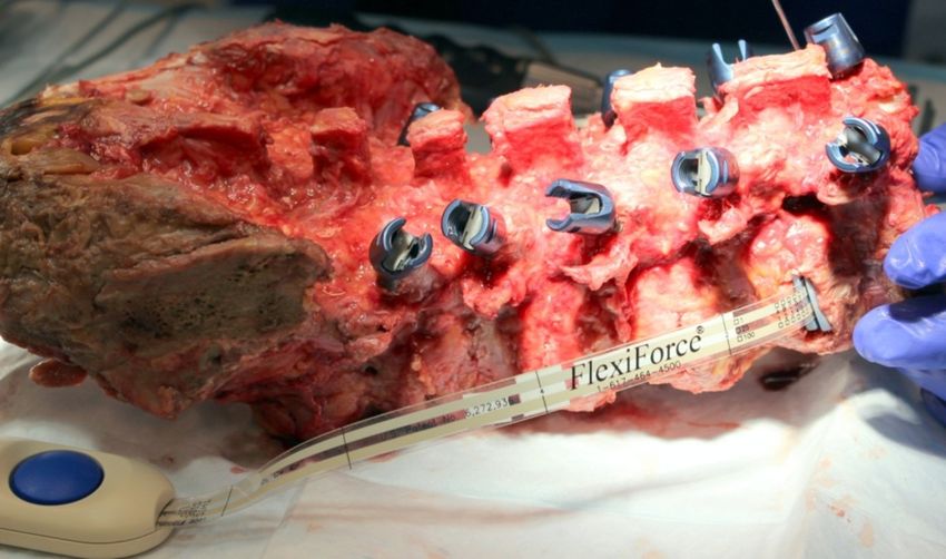
to BPSF. Additionally, incremental ISPF device manipulation demonstrated predictable and
                                   clinically safe trends regarding loading of the interbody space and spinous processes.

                                   Categories: Orthopedics, Other
                                   Keywords: interspinous process fixation, posterior fixation, lumbar, cadaveric, spine biomechanics,
                                   lateral lumbar interbody fusion, pedicle screw fixation, spine surgery, lordosis

                                   Introduction
                                   Rigid interspinous process fixation (ISPF) has been proposed as a less invasive alternative to
                                   pedicle screw fixation (PSF) for supplemental use in circumferential lumbar fusion [1-2]. While
                                   the body of literature evaluating the mechanical efficacy of ISPF continues to grow, particularly
                                   with respect to understanding segmental rigidity, the mechanisms through which sagittal
                                   correction and interbody (IB) loading are achieved and maintained with ISPF are not well
                                   characterized [3-10]. Given the previously demonstrated intraoperative benefits of ISPF, which
                                   include diminished bone invasion and limited midline paraspinal disruption, a greater
                                   biomechanical understanding of the ISPF technology would be highly advantageous in further
                                   defining its role as a minimally invasive adjunct in spine surgery [1-2,11-12].

                                   With ISPF, compression or distraction is applied through the spinous processes and leverages a
                                   larger posterior moment arm about the IB space compared to manipulations through PSF. The
                                   larger moment arm achieved by ISPF may translate to substantial variations in the focal
                                   lordosis and IB loading, even with small adjustments. The option to apply compression or
                                   distraction in a controlled manner enables the surgeon to carefully modulate focal lordosis to
                                   achieve optimal sagittal balance. Simultaneously, the resulting compressive loading of the IB
                                   cage between the vertebral endplates may help resist migration, but should also be moderated
                                   to minimize the risk of subsidence [13-15]. Therefore, it is essential to characterize the focal
                                   lordosis and IB loading during spinous process manipulation via ISPF in comparison with the
                                   standard clinical technique of bilateral PSF (BPSF).

                                   Accordingly, the objective of this study was to evaluate the translated effects of spinous process
                                   manipulation, via a novel ISPF device, on IB loading and sagittal correction. The novel ISPF
                                   device, which can be incrementally shortened/extended in situ, provides an ideal mechanism to
                                   achieve precise compression/distraction of the spinous processes without the need for device
                                   substitution or additional instrumentation. The IB loading and focal lordosis induced through
                                   the ISPF device was compared with BPSF - the gold standard for posterior fixation. In addition,
                                   the loads applied to the spinous processes were characterized to compare with previously
                                   measured failure loads.

                                   Materials And Methods
                                   Cadaveric specimen preparation
                                   Two fresh-frozen human cadaveric spines were used in this study (age-sex: 57 years, male; 61
                                   years, male). Each spine was thawed at room temperature and the lumbosacral specimens (L1-
                                   S1) were dissected out. Ligamentous structures were maintained. Residual musculature and
                                   adipose tissue were removed. Osseous structural integrity was confirmed via standard
                                   anteroposterior and lateral radiographs. Any specimens exhibiting previous lumbosacral
                                   surgery, excessive degeneration, or anatomical discrepancy were excluded. No structural
                                   failures or abnormalities were observed during testing.

                                   One level was excluded due to damage incurred at L1 during specimen dissection/preparation,
                                   leaving a combined total of seven (n = 7) functional spinal units (FSUs) that were utilized in the

2019 Gandhi et al. Cureus 11(3): e4317. DOI 10.7759/cureus.4317                                                                      2 of 16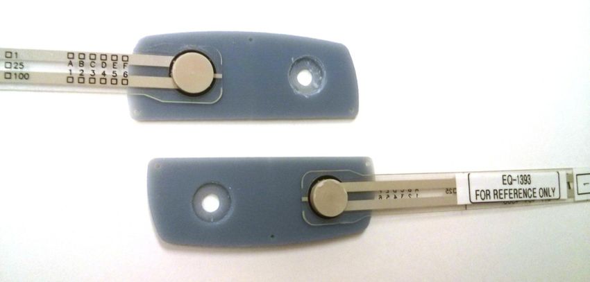
study. The specimens were preemptively instrumented with bilateral pedicle screws
                                   (Silverton® Spinal Fixation System; Zimmer Biomet Spine, Westminster, CO USA) at levels L1-
                                   L5 (Figure 1). Doing so ensured the consistent composition of the vertebral bodies throughout
                                   all testing and reduced the amount of manipulation to the vertebral column once testing had
                                   commenced. The pedicle screws did not contact any adjacent vertebrae or screws; hence, the IB
                                   loads and spinal motion were not affected by the preemptive screw insertion. Connecting rods
                                   were not inserted during this initial instrumentation phase. Prior to both ISPF and BPSF
                                   instrumentation, the interspinous and supraspinous ligaments were preemptively removed to
                                   accommodate the ISPF device. This does not alter the results of BPSF fixation, considering that
                                   distraction with BPSF was not measured in this study and those ligaments do not provide
                                   notable compressive resistance.

                                     FIGURE 1: Test specimen with bilateral pedicle screw
                                     instrumentation
                                     Note that a modified, force-sensing lateral interbody cage has been inserted at the most superior
                                     level.

                                   Following pedicle screw placement, a partial discectomy was performed, through a standard
                                   lateral approach to accommodate the placement of a modified lateral lumbar interbody fusion
                                   (LLIF) cage. The cage (22 mm (W) × 60 mm (L)) was modified such that it possessed two load
                                   cells (FlexiForce®; Tekscan Inc., Boston, MA USA; Figure 2). The load cells acted as a bridge
                                   between the inferior/superior endplates of the implant, and so all loading transduced through
                                   the cage was assumed by the load cells. The two load cell forces were summed to determine the
                                   total IB cage load at each treated level. The cage footprint, profile, and surface design were
                                   consistent with a commercially available lateral cage (Timberline® Lateral Fusion System;
                                   Zimmer Biomet Spine, Westminster, CO USA). An appropriate cage height was determined
                                   specific to each affected level and was accounted for in the thickness of the cage endplates. The
                                   sequence of testing was randomized such that four levels were tested with BPSF first and three
                                   with the ISPF device first.

2019 Gandhi et al. Cureus 11(3): e4317. DOI 10.7759/cureus.4317                                                                          3 of 16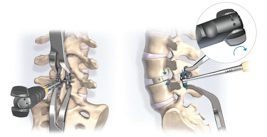
FIGURE 2: Modified, force-sensing lateral interbody cage
                                     Note the two load cells situated between the superior and inferior end-plates of the cage.

                                   BPSF testing
                                   BPSF testing was performed in two stages at each treated level. Following preparation of the
                                   disc space, the modified lateral cage of an appropriate height was placed. The baseline IB cage
                                   load was recorded, and lateral fluoroscopic images were taken prior to each loading stage.

                                   Following baseline parameter measurement, bilateral connecting rods were fixed to the pedicle
                                   screws across a single FSU at a time under maximum attainable compression. The maximum
                                   compressive force was recorded using a fixed load cell on the compressor handles. Final set
                                   screw tightening was then performed and the IB cage load was recorded and lateral
                                   fluoroscopic images were taken. Next, the set screws were loosened from the connecting rods
                                   and the vertebral segment was allowed to relax to a neutral state. The pedicle screws were then
                                   recompressed to 75% of the previously measured maximum compressive force, which was
                                   determined by the surgeon authors to be representative of the exertion during clinical
                                   application. Since the force applied by each surgeon may be subjective and variable, the
                                   maximum compression was applied by a single individual to minimize inter-specimen
                                   variability and the load values were measured to ensure consistency. The set screws were then
                                   retightened to secure the connecting rods and the IB cage load and the lateral fluoroscopic
                                   images were collected (Figure 3A). The set screws and connecting rods were then removed and
                                   the vertebral segment was allowed to return to a neutral state.

2019 Gandhi et al. Cureus 11(3): e4317. DOI 10.7759/cureus.4317                                                                   4 of 16
FIGURE 3: Lateral fluoroscopic images
                                     Bilateral pedicle screw fixation (A) and interspinous process fixation (B) constructs with the
                                     modified, force-sensing lateral cage placed at the index level.

                                   ISPF testing
                                   The ISPF device (Alpine XCTM Adjustable Fusion System; Zimmer Biomet Spine, Westminster,
                                   CO USA) was implanted with a device post height of 14 mm (Figures 4, 5). The post is
                                   considered the adjustable (axial) portion of the device that sits within the interspinous space
                                   when the device is implanted. To maintain consistency between specimens, the spinous
                                   processes were trimmed when necessary to ensure that a post height of 14 mm was
                                   consistently an appropriate fit for the neutral loading state. During clinical application,
                                   trimming of the spinous process is not necessary considering that the post height can be
                                   adjusted to fit each patient and level. Sagittal compression of the spinous processes by the ISPF
                                   device was achieved such that the device spikes were seated with good bone apposition.
                                   Additionally, device placement was as far anterior and as close to the laminar junction as
                                   possible. Once in place, the post height of the device can be compressed downward from 14 mm
                                   to 6 mm or expanded upward from 14 mm to 18 mm.

                                     FIGURE 4: Novel adjustable interspinous process fixation
                                     device
                                     Interspinous process fixation device demonstrating various post heights for maximal compression
                                     (A) and maximal distraction (C) relative to the center height (B).

                                     Image source: Zimmer Biomet Spine, Westminister, CO USA

2019 Gandhi et al. Cureus 11(3): e4317. DOI 10.7759/cureus.4317                                                                       5 of 16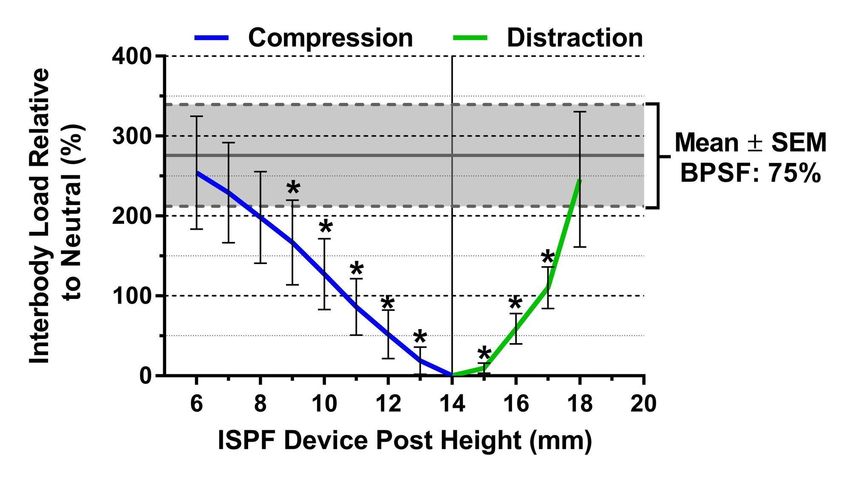
FIGURE 5: Schematic representation of the adjuster tool
Note the schematic (right) demonstrates device compression; however, expansion occurs through
the same mechanism by reversing the dial.
Image source: Zimmer Biomet Spine, Westminister, CO USA
Following device fixation to the spinous processes, the device post height was decreased in 1-
mm increments to the minimum post height of 6 mm. This 8 mm of compression was
determined by the surgeon authors to be clinically appropriate for both specimens given their
respective anatomy. However, the authors emphasize that 8 mm of compression is not to be
considered a standard value and that the degree of compression or distraction is dependent
upon patient specific anatomy.
IB cage load measurements were collected at each 1-mm increment during compression. The
device was then expanded to its maximum post height of 18 mm. Similarly, IB cage load was
recorded at each 1-mm increment. The torque applied to the height adjustment instrument was
also recorded at each 1 mm increment during compression and distraction. Torque values were
used in subsequent calculations to estimate the force exerted by the ISPF device on the spinous
processes. Lateral fluoroscopic images were collected at neutral, fully compressed, and fully
distracted states (Figure 3B). Subsequent measurement of focal lordosis for both fixation types
was performed using Cobb’s method.
Spinous process force calculation
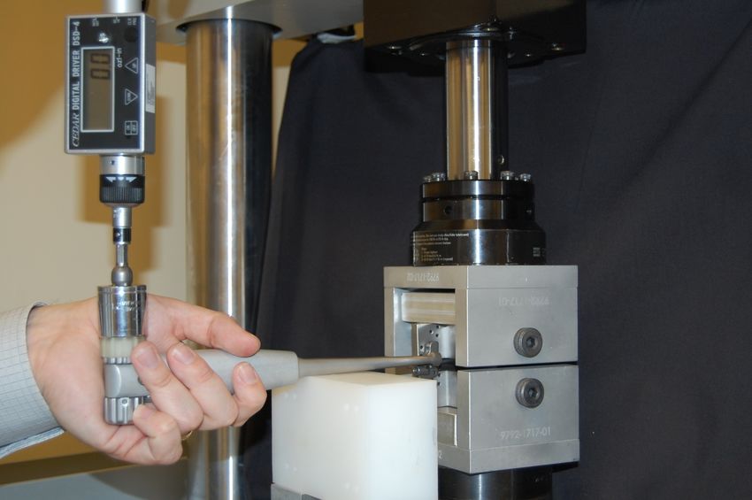
Independent of the cadaveric testing phase, a calibration plot was developed to estimate the
forces applied by the ISPF device on the spinous processes while in compression/distraction.
The ISPF device was first assembled to two modified ASTM (American Society for Testing and
Materials) F1717 stainless steel test blocks and then placed within a test frame possessing a
load cell (MTS Bionix® Tabletop Test Systems; MTS Systems Corporation, Eden Praire, MN
USA; Figure 6). The ISPF device was then compressed by applying known torque values to the
adjuster tool. The corresponding force transmitted by the ISPF device on the test blocks was
measured by the load cell. The applied torque values were plotted against their corresponding
loads. A best-fit linear equation was then fit to the data. This linear standard curve was utilized
2019 Gandhi et al. Cureus 11(3): e4317. DOI 10.7759/cureus.4317 6 of 16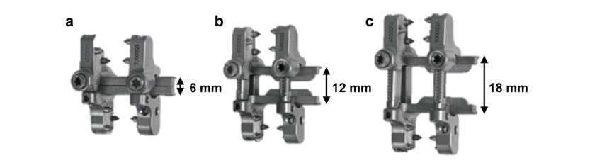
to convert the torque values that were measured during cadaveric testing to the estimated
                                   forces exerted by the ISPF device on the spinous processes.

                                     FIGURE 6: Test assembly to determine device force as the
                                     function of height
                                     Note the interspinous process fixation device assembled to two modified ASTM (American Society
                                     for Testing and Materials) F1717 stainless steel test blocks, within a load cell testing frame, with
                                     adjuster tool attached.

                                   Statistical methods
                                   The change (%) in IB load, relative to baseline, and change in focal lordosis (deg.) were
                                   compared between the ISPF device at its in situ compressed state (post height = 6 mm), the ISPF
                                   device at its in situ distracted state (post height =18 mm), BPSF under 75% exertion, and BPSF
                                   under 100% exertion by Friedman’s test (non-parametric repeated measures ANOVA) with
                                   Dunn’s test for multiple post hoc comparisons. Multiplicity-adjusted p-values less than 0.05
                                   were considered statistically significant. Non-parametric tests were used for statistical testing
                                   due to the small sample size. To ensure consistency between the specimens, the BPSF
                                   compression loads were checked for outliers using the robust regression and outlier removal
                                   (ROUT) method with a Q coefficient equal to 5%. All statistical analyses were performed using
                                   GraphPad Prism 7.01 (GraphPad Software, San Diego, CA USA).

                                   Results
                                   BPSF compression
                                   The maximum load (100% exertion) applied to the instrument handles during BPSF
                                   compression was 204 ± 8 N (range: 194-218 N) and the clinically relevant loading (75% of
                                   maximum) was 157 ± 11 N (range: 148-178 N). There were zero statistical outliers according to
                                   the ROUT method with a Q coefficient equal to 5%, suggesting good inter-specimen
                                   repeatability.

2019 Gandhi et al. Cureus 11(3): e4317. DOI 10.7759/cureus.4317                                                                         7 of 16
Interbody loading
No significant differences in IB load relative to baseline (%) were observed between the four
testing conditions. ISPF in maximal compression (6 mm) or distraction (18 mm) and the
clinically relevant BPSF compression produced similar IB load values (Figure 7). Applying
maximum compression on the BPSF construct tended to produce the greatest IB loads, but were
not significantly different from the other 3 test conditions (p = 0.068). The difference in IB load
was significantly different between BPSF at 75% effort and the ISPF device when it was adjusted
less than 6 mm in compression or 4 mm in distraction (Figure 8). The mean compressive IB load
ranged from 101 to 329 N over the four test conditions.
FIGURE 7: Interbody load versus fixation condition
No significant differences in the interbody load, relative to neutral, were observed between the four
posterior fixation/manipulation techniques. Note that bilateral pedicle screw fixation under the
extreme case of 100% compressive exertion tended to produce the greatest load relative to
bilateral pedicle screw fixation at 75% exertion (clinically relevant loading) and the interspinous
process fixation techniques (p = 0.068). Values represent the mean and error bars are standard
error measurements.
ISPF, interspinous process fixation; BPSF, bilateral pedicle screw fixation.
The mean IB load as a function of ISPF device post height demonstrated linear behavior (R 2=
0.995) in the compression phase, and non-linear, second-order behavior in distraction (R 2 =
0.998; Figure 8).
2019 Gandhi et al. Cureus 11(3): e4317. DOI 10.7759/cureus.4317 8 of 16FIGURE 8: Interbody load versus interspinous fixation device
post height
The compressive interbody cage load increased regardless of whether the interspinous process
fixation device is compressed or distracted. During compression, the interbody cage load tends to
increase linearly, while distraction produced second-order or exponential increases in interbody
cage load. * denotes pFIGURE 9: Change in lordosis versus fixation condition
Interspinous process fixation at 8 mm of compression produces significantly greater focal lordosis
compared to clinically relevant compression with bilateral pedicle screw fixation. Note that the
distraction of the interspinous process fixation successfully reduces focal lordosis. Values represent
the mean and error bars are SEM. * denotes pFIGURE 10: Approximate load on spinous processes versus
interspinous fixation device post height
A post height of 14 mm was the neutral height used during device insertion. Lines represent the
mean and error bars are standard error measurements. Note that no fractures were observed in this
study and that the estimated forces on the spinous processes are below the range of mean failure
values reported in the literature [16].
SP, spinous process; ISPF, interspinous process fixation
Discussion
Biomechanical outcomes
While considerable literature exists characterizing the biomechanical effectiveness of various
posterior lumbar fixation constructs, the focus has almost exclusively been segmental
rigidity/range-of-motion (ROM). IB loading and focal lordosis are rarely assessed in a cadaveric
model [17-19]. However, given the continued incidence of cage subsidence and cage migration
at noteworthy rates, improved understanding of such parameters is warranted [20-24]. When
evaluating rigid ISPF, it is critical to consider IB loading and focal lordosis since the ISPF device
leverages a larger moment arm compared to traditional BPSF, which may translate to greater
biomechanical effects within the IB space. Indeed, significantly greater focal lordosis was
observed during compression with the ISPF device compared to the clinically relevant BPSF
compression, yet the compressive IB loads were similar.
The ideal compressive loading on an IB cage should be sufficient to prevent migration or
expulsion, yet low enough to mitigate the risk of subsidence. Of course, the relevant load for
each of these cases largely depends on the areal profile in contact with the vertebral endplates
and is therefore specific to individual cage designs. Kwon and colleagues compressed a lateral
cage footprint (18 mm × 60 mm) into vertebral endplates following cage insertion and observed
compressive strength values of 1764 ± 966 N, which far exceed the IB loads observed herein
[22]. On the other hand, resistance to migration or expulsion is more complicated and depends
2019 Gandhi et al. Cureus 11(3): e4317. DOI 10.7759/cureus.4317 11 of 16largely on the static friction coefficient between the cage and endplates, the stability of the
spinal motion segment, as well as the areal contact. The IB loading induced by the clinically
relevant levels of BPSF compression and ISPF compression or distraction were similar in this
study, which suggests a similar level of resistance to migration in the neutral state. Although IB
loading was not measured during kinematic analyses herein, previous studies have
demonstrated similar rigidity between ISPF and BPSF in flexion-extension, but ISPF does tend
to be less rigid in lateral bending and axial rotation [3,6]. Future studies that investigate the IB
load during kinematic analysis may be useful to better understand the implications of
supplemental ISPF or PSF for the risk of potential cage migration.
In contrast to an interspinous spacer, which acts more passively as a blocking device, the ISPF
device can be utilized to actively increase focal lordosis through compression or focal kyphosis
through distraction. Additionally, distraction can serve to relieve facet loads and open the
spinal canal and neural foramens [25-27]. Interestingly, the distraction of the spinous processes
using the ISPF device not only increased the compressive IB load but did so in exponential
fashion. A 245% increase was achieved after just 4 mm of distraction, which was similar to that
achieved with 8 mm of compression. This finding underscores the importance of the fulcrum
that is leveraged during compression versus distraction of the posterior elements and has
important implications for the effects on the anterior column. During ISPF compression, the IB
cage itself likely served as the fulcrum considering that IB cages typically provide some
distraction to the disc space and an increased compressive IB load was observed. In contrast,
the effects on the anterior column from spinous process distraction are likely influenced by
tensioning structures, such as the posterior longitudinal ligament, which act as a middle
column tensile fulcrum to increase the anterior compressive load [27]. This observation of
increased load at the anterior column following posterior distraction is consistent with a
previous study by Zheng et al, where the pressure distribution was mapped across the disc space
[27].
In regard to sagittal correction, compression of the ISPF device (8 mm in situ) produced a
significantly greater change in focal lordosis compared with BPSF under 75% compression.
Furthermore, distraction with the ISPF device (4 mm in situ) produced an increase in local
kyphosis of 2.0o , demonstrating an effective range of sagittal correction of 7.0 o (compressed-
to-distracted states). Such range, achieved in a calculable manor, allows the surgeon to
consider optimal sagittal balance while not compromising the load placed on the IB cage. This
robust ability to manipulate the sagittal plane is consistent with previous ROM studies that
have demonstrated an inherent ability of ISPF to resist flexion-extension [3-10].
A final important consideration of this study was whether or not the novel ISPF device exerts
excessive forces on the spinous processes during compression or distraction. The estimated
mean load on the spinous processes was 292 N under 8 mm of ISPF device compression and 212
N under 4 mm of distraction. Although the failure strength depends strongly on the bone size
and quality of each individual, the applied loads were well below the range of mean failure
strength values from previous studies (339-493 N) [16, 28]. Accordingly, no spinous process
fractures were observed during this study. It is also important to reiterate that clinical
application of this device requires appropriate selections of post height and
compression/distraction for each patient to mitigate any risk of spinous process failure.
In this study, a novel ISPF device, which affords incremental compression/distraction in situ,
was compared with traditional BPSF under varied degrees of compression. While the semi-
quantitative approach taken with BPSF was an inherent limitation to this study, it is consistent
with the subjective nature of PSF in general. Compression/distraction with PSF is almost
always achieved by surgeon feel alone, making calculable manipulation and characterization
extremely challenging. Although this technique is naturally subjective, it is an important
2019 Gandhi et al. Cureus 11(3): e4317. DOI 10.7759/cureus.4317 12 of 16parameter to quantitatively evaluate in controlled studies, considering its potential impact on
multiple outcomes. Therefore, the loads applied during BPSF compression were recorded to
ensure repeatability at the maximal compressive effort and at a more clinically relevant level of
compression (75% effort). The 75% effort level was deemed by the surgeon authors to be
representative of clinical application. While the ISPF device can be precisely adjusted during
compression/distraction, the extent of manipulation is still a subjective choice that depends on
patient anatomy. In this study, 8 mm of compression (reduction in post height from 14 mm to 6
mm) was deemed to be clinically appropriate for the specific anatomies. However, since IB load
was measured at 1 mm increments, additional comparisons with BPSF at 75% compression can
be made (Figure 8). Further comparisons with BPSF at 100% effort were not made, considering
such extreme compression is not reflective of clinical scenarios and is often avoided, as
excessive stress at the screw-to-bone interface and at the rod-to-set screw juncture predisposes
the screw to pull-out and construct failure [29].
Clinical implications
While the amount of evidence regarding rigid ISPF is still limited, early clinical and
biomechanical assessments have demonstrated good utility, particularly in anterior lumbar
interbody fusion (ALIF) and LLIF application [1-12]. Given the ability for anterior and lateral
cages alone to provide significant reduction and stability, notably in the axial and coronal
planes, extensive and invasive posterior fixation may be replaced by less invasive supplemental
fixation strategies in many cases [3-4, 6-7]. ISPF is a minimally invasive strategy that provides a
robust mechanism for locking the sagittal plane while largely preserving the midline structures,
making it a particularly well-suited adjunct to a large anterior or lateral IB cage.
Despite such an influential stabilization mechanism with ISPF, the ability to manipulate the
spinous processes has traditionally been a challenge. Drilling and pinning of the spinous
processes have been the predominant technique to achieve the leverage required to compress
the spinous processes and provide focal sagittal correction. Such a technique not only places an
additional burden on the surgeon but can also predispose the spinous processes to the
fracturing. Furthermore, similar to PSF, the technique is entirely subjective. No opportunity
exists for precise, controlled manipulation. The novel adjustable ISPF device explored in this
study may provide a solution to the challenges of this intervention. The in situ manipulation
capabilities of the device avoid drilling and pinning while enabling incremental adjustments
without the need for device substitution or repositioning. Additionally, the ability to compress
or distract the segment post-implantation allows for a more favorable loading environment of
the IB space.
Limitations
The authors acknowledge several inherent limitations of this study. Of previous note, the BPSF
compression technique was somewhat subjective, but the protocol provided a reproducible
mechanism through which consistent inter-specimen testing may be performed. Likewise, the 8
mm of compression with the ISPF device is a subjective surgeon choice that must be matched
to the anatomy as best as possible. The extent to which the spinous processes can be
manipulated without predisposing the bone mass to the fracturing is largely dependent upon
patient anatomy and bone quality. The specimens utilized in this study were selected and, in
some cases, the spinous processes were trimmed slightly to ensure consistency in the neutral
post height and range of allowable compression/distraction.
The authors also acknowledge the limitation in performing several iterative tests on each
motion segment. This limitation requires the assumption that each test iteration is
independent of any previous testing condition, which may not be accurate considering the
viscoelastic nature of the soft tissue structures. However, this an inherent limitation of any
2019 Gandhi et al. Cureus 11(3): e4317. DOI 10.7759/cureus.4317 13 of 16biomechanical assessment of spinal motion segments in which iterative testing is performed at
the same level. The use of a standardized protocol that included specimen relaxation, a
randomized testing order, and outcomes normalized to baseline values was employed to
mitigate this limitation as much as possible.
Conclusions
The novel ISPF device demonstrated IB loading that is similar to that of BPSF under clinically
relevant compression. Additionally, the ISPF device produced a greater increase in focal
lordosis than BPSF, demonstrating the mechanical advantage of ISPF to readily provide sagittal
correction through the extended lever arm of the spinous process. The ISPF device also
demonstrated an ability to produce compressive loading of the IB space during distraction,
resulting in increased focal kyphosis. Such a phenomenon shows that the novel ISPF device can
afford a range of sagittal angulation that does not compromise IB loading. Given the less
invasive nature and technical feasibility of the ISPF approach, such characteristics may present
the novel ISPF device as a viable alternative to PSF in circumferential lumbar fusion.
Additional Information
Disclosures
Human subjects: All authors have confirmed that this study did not involve human
participants or tissue. Animal subjects: All authors have confirmed that this study did not
involve animal subjects or tissue. Conflicts of interest: In compliance with the ICMJE uniform
disclosure form, all authors declare the following: Payment/services info: This study was
funded by Zimmer Biomet Spine. Anup Gandhi, Chris Ferry, and Jason Inzana are/were
employees (salary) of Zimmer Biomet Spine at the time of study execution. Ryan DenHaese and
Steve Change have received consulting fees and royalties from Zimmer Biomet Spine. .
Financial relationships: Anup Gandhi, Chris Ferry, Jason Inzana declare(s) employment from
Zimmer Biomet Spine. Steven Chang, Ryan DenHaese declare(s) personal fees from Zimmer
Biomet Spine. Steven Chang, Ryan DenHaese declare(s) royalties from Zimmer Biomet Spine.
Ryan DenHaese declare(s) personal fees from Corelink Surgical. Other relationships: All
authors have declared that there are no other relationships or activities that could appear to
have influenced the submitted work.
References
1. Villavicencio AT, Serxner BJ, Mason A, Nelson EL, Rajpal S, Faes N, Burneikiene S: Unilateral
and bilateral pedicle screw fixation in transforaminal lumbar interbody fusion: radiographic
and clinical analysis. World Neurosurg. 2015, 83:553-9. 10.1016/j.wneu.2014.12.012
2. Wang JC, Haid RW, Miller JS, Robinson JC: Comparison of CD HORIZON SPIRE spinous
process plate stabilization and pedicle screw fixation after anterior lumbar interbody fusion.
Invited submission from the Joint Section Meeting On Disorders of the Spine and Peripheral
Nerves, March 2005. J Neurosurg Spine. 2006, 4:132-6. 10.3171/spi.2006.4.2.132
3. Doulgeris JJ, Aghayev K, Gonzalez-blohm SA, Lee WE, Vrionis FD: Biomechanical comparison
of an interspinous fusion device and bilateral pedicle screw system as additional fixation for
lateral lumbar interbody fusion. Clin Biomech Avon. 2015, 205-10.
10.1016/j.clinbiomech.2014.10.003
4. Fogel GR, Parikh RD, Ryu SI, Turner AW: Biomechanics of lateral lumbar interbody fusion
constructs with lateral and posterior plate fixation: laboratory investigation. J Neurosurg
Spine. 2014, 20:291-7. 10.3171/2013.11.SPINE13617
5. Fogel GR, Turner AW, Dooley ZA, Cornwall GB: Biomechanical stability of lateral interbody
implants and supplemental fixation in a cadaveric degenerative spondylolisthesis model.
Spine. 2014, 39:E1138-E1146. 10.1097/BRS.0000000000000485
6. Gonzalez-blohm SA, Doulgeris JJ, Aghayev K, Lee WE, Volkov A, Vrionis FD: Biomechanical
analysis of an interspinous fusion device as a stand-alone and as supplemental fixation to
2019 Gandhi et al. Cureus 11(3): e4317. DOI 10.7759/cureus.4317 14 of 16posterior expandable interbody cages in the lumbar spine. J Neurosurg Spine. 2014, 20:209-19.
10.3171/2013.10.SPINE13612
7. Kaibara T, Karahalios DG, Porter RW, et al.: Biomechanics of a lumbar interspinous anchor
with transforaminal lumbar interbody fixation. World Neurosurg. 2010, 73:572-7.
10.1016/j.wneu.2010.02.025
8. Karahalios DG, Kaibara T, Porter RW, et al.: Biomechanics of a lumbar interspinous anchor
with anterior lumbar interbody fusion. J Neurosurg Spine. 2010, 12:372-80.
10.3171/2009.10.SPINE09305
9. Techy F, Mageswaran P, Colbrunn RW, Bonner TF, Mclain RF: Properties of an interspinous
fixation device (ISD) in lumbar fusion constructs: a biomechanical study. Spine J. 2013, 572-9.
10.1016/j.spinee.2013.01.042
10. Yu X, Zhu L, Su Q: Lumbar spine stability after combined application of interspinous fastener
and modified posterior lumbar interbody fusion: a biomechanical study. Arch Orthop Trauma
Surg. 2014, 134:623-9. 10.1007/s00402-014-1977-9
11. Sclafani JA, Liang K, Ohnmeiss DD, Gordon C: Clinical outcomes of a polyaxial interspinous
fusion system. Int J Spine. 2014, 8: 10.14444/1035
12. Vokshoor A, Khurana S, Wilson D, Filsinger P: Clinical and radiographic outcomes after
spinous process fixation and posterior fusion in an elderly cohort. Surg Technol Int. 2014,
271-6.
13. Drazin D, Hussain M, Harris J, et al.: The role of sacral slope in lumbosacral fusion: a
biomechanical study. J Neurosurg Spine. 2015, 23:754-62. 10.3171/2015.3.SPINE14557
14. Zhao FD, Yang W, Shan Z, et al.: Cage migration after transforaminal lumbar interbody fusion
and factors related to it. Orthop Surg. 2012, 4:227-32. 10.1111/os.12004
15. Kwon B, Kim DH: Lateral lumbar interbody fusion indications, outcomes, and complications . J
Am Acad Orthop Surg. 2016, 24:96-105. 10.5435/JAAOS-D-14-00208
16. Sun X, Murgatroyd AA, Mullinix KP, Cunningham BW, Ma X, McAfee PC: Biomechanical and
anatomical considerations in lumbar spinous process fixation-an in vitro human cadaveric
model. Spine J. 2014, 14:2208-15. 10.1016/j.spinee.2014.03.002
17. Demetropoulos CK, Morgan CR, Sengupta DK, Herkowitz HN: Development of a 4-axis load
cell used for lumbar interbody load measurements. Med Eng Phys. 2009, 31:846-51.
10.1016/j.medengphy.2009.04.002
18. Ledet EH, Tymeson MP, Dirisio DJ, Cohen B, Uhl RL: Direct real-time measurement of in vivo
forces in the lumbar spine. Spine J. 2005, 85-94. 10.1016/j.spinee.2004.06.017
19. Lavoie S, Lindsey RW, Gugala Z, Kirking B, Hipp JA: Load sharing and kinematics of threaded
cages for lumbar interbody fusion. Clin Orthop Relat Res. 2003, 408:174-9.
20. Abbushi A, Cabraja M, Thomale UW, Woiciechowsky C, Kroppenstedt SN: The influence of
cage positioning and cage type on cage migration and fusion rates in patients with
monosegmental posterior lumbar interbody fusion and posterior fixation. Eur Spine J. 2009,
18:1621-8. 10.1007/s00586-009-1036-3
21. Duncan JW, Bailey RA: An analysis of fusion cage migration in unilateral and bilateral fixation
with transforaminal lumbar interbody fusion. Eur Spine J. 2013, 22:439-45. 10.1007/s00586-
012-2458-x
22. Joseph JR, Smith BW, La marca F, Park P: Comparison of complication rates of minimally
invasive transforaminal lumbar interbody fusion and lateral lumbar interbody fusion: a
systematic review of the literature. Neurosurg Focus. 2015, 39:10.3171/2015.7.FOCUS15278
23. Kim MC, Chung HT, Cho JL, Kim DJ, Chung NS: Subsidence of polyetheretherketone cage
after minimally invasive transforaminal lumbar interbody fusion. J Spinal Disord Tech. 2013,
26:87-92. 10.1097/BSD.0b013e318237b9b1
24. Wiseman CM, Lindsey DP, Fredrick AD, Yerby SA: The effect of an interspinous process
implant on facet loading during extension. Spine. 2005, 30:903-7.
25. Richards JC, Majumdar S, Lindsey DP, Beaupré GS, Yerby SA: The treatment mechanism of an
interspinous process implant for lumbar neurogenic intermittent claudication. Spine. 2005,
30:744-9.
26. Bonaldi G, Brembilla C, Cianfoni A: Minimally-invasive posterior lumbar stabilization for
degenerative low back pain and sciatica. A review. Eur J Radiol. 2014, 84:789-98.
10.1016/j.ejrad.2014.04.012
27. Zheng S, Yao Q, Cheng L, et al.: The effects of a new shape-memory alloy interspinous process
device on the distribution of intervertebral disc pressures in vitro. J Biomed Res. 2010, 24:115-
2019 Gandhi et al. Cureus 11(3): e4317. DOI 10.7759/cureus.4317 15 of 1623. 10.1016/S1674-8301(10)60019-X
28. Shepherd DE, Leahy JC, Mathias KJ, Wilkinson SJ, Hukins DW: Spinous process strength.
Spine. 2000, 25:319-23.
29. Chen CS, Chen WJ, Cheng CK, Jao SH, Chueh SC, Wang CC: Failure analysis of broken pedicle
screws on spinal instrumentation. Med Eng Phys. 2005, 27:487-96.
10.1016/j.medengphy.2004.12.007
2019 Gandhi et al. Cureus 11(3): e4317. DOI 10.7759/cureus.4317 16 of 16You can also read