Comparison of Titanium Vascular Closure Staples With Suture Repair of the Thoracic Aorta in Swine
←
→
Page content transcription
If your browser does not render page correctly, please read the page content below
Comparison of Titanium Vascular
           Closure Staples With Suture Repair of
           the Thoracic Aorta in Swine*
           Subrato Deb, MD; Barry Martin, MD; Leon Sun, MD; David Burris, MD;
           David Wherry, MD; Emmanuel Pikoulis, MD; and Peter Rhee, MD, MPH

                   Objective: Devices that reduce technical difficulty and anastigmatic time when repairing large
                   vessels such as the thoracic aorta would be beneficial. The aim of this study was to determine if
                   titanium vascular closure staples (3 mm) could be safely and quickly applied in the repair of large
                   vessels such as the thoracic aorta.
                   Design: Through a left thoracotomy in 10 female swine (110 to 130 lb), an interposition graft (14
                   to 16 mm textile) was placed into the aorta distal to the left subclavian artery. Animals were
                   randomized at the time of repair to either running sutures (n ⴝ 5; 6 – 0 polypropylene) or vascular
                   closure staples (n ⴝ 5; 3 mm). The anastomosis was evaluated after 2 months with aortograms,
                   and the aorta was harvested to evaluate healing.
                   Results: The clamp times (mean ⴞ SD) were 30.8 ⴞ 8.2 min for suture repair and 24.8 ⴞ 5.1 min
                   for vascular closure staple repair (p ⴝ 0.2). Anastomosis times were 20.0 ⴞ 6.2 min for the suture
                   group and 16.4 ⴞ 6.4 min for the vascular closure staple group (p ⴝ 0.4). Arch aortograms at 2
                   months revealed no significant difference in luminal narrowing between the two groups. Gross
                   and microscopic examination revealed no thrombosis, well-healed wounds with a continuous
                   intimal layer, and no differences in intimal thickness or inflammation between the two groups.
                   Conclusion: Vascular closure staples were equivalent to sutures in terms of durability, graft
                   patency, and wound healing at 2 months. Vascular closure staples may offer the trauma surgeon
                   a quick and easy alternative when repairing large vessels such as the thoracic aorta.
                                                                                      (CHEST 2000; 118:1762–1768)

                   Key words: anastomosis; aorta; healing; graft; suture; swine; thorax; thrombosis; trauma; vascular closure staples

                   Abbreviation: ANOVA ⫽ analysis of variance

           T horacic aortic injury as a result of blunt force
             trauma is highly lethal and accounts for 17% of
                                                                                    The single most important factor determining
                                                                                 outcome in survivors is the duration of the thoracic
           all motor vehicle-related deaths.1 Of those who                       aorta occlusion during the surgical repair. Ischemic
           survive the initial injury to reach the hospital, over                spinal cord injury as a consequence of prolonged
           half will die in the next 24 to 48 h if expedient                     crossclamping can result in postoperative paraplegia,
           surgical repair is not undertaken.2,3                                 and is the most debilitating complication of this
                                                                                 surgery. A prospective multicenter trial has demon-
                                                                                 strated that prolonged aortic crossclamp times are
           *From the Department of Surgery (Drs. Deb and Rhee), Na-              associated with paraplegia.4 Although the use of
           tional Naval Medical Center, Bethesda, MD; Walter Reed Army           bypass techniques, which provide distal aortic perfu-
           Medical Center (Drs. Martin and Burris), Washington, DC;
           Uniformed Services University of the Health Sciences (Drs. Sun,       sion, may reduce paraplegia rates, the key is to
           Wherry, and Pikoulis), Bethesda, MD.                                  improve the speed of the aortic repair. Improve-
           The opinions expressed herein are those of the authors and are        ments in surgical techniques that reduce clamp time
           not to be construed as reflecting the views of the Uniformed
           Services University of the Health Services, the Department of the     and make the procedure technically easier would be
           Navy, Army, or the Department of Defense.                             of benefit. Improved instruments that allow stapling
           Supported by a grant from the United States Surgical Corpora-         of large vessels may offer this possibility.
           tion, Norwalk, CT, and the Office of Naval Research.
           Manuscript received December 15, 1999; revision accepted May             Advancements in technology have resulted in the
           23, 2000.                                                             development of titanium vascular closure staples,
           Correspondence to: Peter Rhee, MD, MPH, Department of                 which allow vessel approximation in a rapid and
           Surgery, Uniformed Services University of the Health Sciences,
           4301 Jones Bridge Rd, Bethesda, MD 20814; e-mail: prhee@              accurate fashion. The clips are placed to evert the
           usuhs.mil                                                             vessel wall without transmural penetration of the

           1762                                                                                                  Laboratory and Animal Investigations

Downloaded From: http://journal.publications.chestnet.org/ on 05/08/2015
endothelium, reducing vessel trauma and subse- incision. Animals were placed in right lateral decubitus position,
quent tissue reaction.5 This sutureless technique, and a muscle-sparing thoracotomy through the fifth intercostal
space was performed. Proximal and distal vascular control of the
described by Kirsch et al,6 involved vascular recon-
descending thoracic aorta was obtained with vessel loops followed
struction with nonpenetrating titanium arcuate- by the establishment of a left subclavian to distal thoracic aorta
legged clips. Although vascular closure staples were shunt using 9F cordis catheter for spinal cord perfusion during
originally designed for microvascular surgery, the clamping. This type of shunting procedure is one of the recom-
development of larger sizes has widened their appli- mended methods to help prevent paralysis during thoracic aortic
cability. Currently the clips are used in transplant, repair. Following IV heparin (5,000 U), the aorta was cross-
cardiac, and trauma vascular surgery.7–9 To the clamped just distal to the left subclavian artery and the aorta was
transected. One liter of whole blood was withdrawn prior to
original three sizes of vascular closure staples (Fig 1),
aortic crossclamping to avoid hypertension associated with the
a larger clip (extra large, 3 mm) has been developed crossclamping and for use during postoperative resuscitation.
for use in large vessels. The method of withdrawing blood to avoid proximal hypertension
The main advantages of vascular closure staples is unique to this experiment and not typical during repair in
are better intimal healing and faster anastomosis trauma scenarios.
times than the standard suture technique.10 –12 Faster At this point, the animals were randomized into either a
anastomotic times might be advantageous in treating vascular closure staple anastomosis group (clip group) or suture
anastomosis group (suture group). Polypropylene monofilament
traumatic thoracic aorta in which repair times are so 5– 0 vascular suture was utilized to repair the aorta in a running
crucial. We therefore undertook this study to deter- fashion for the suture group. In the clip group, the clip placement
mine if extra large vascular closure staples could be was as close as possible, with ⱕ 0.5 mm between the clips as
used to repair large vascular structures such as the recommended by the manufacturer. Fourteen- to 16-mm aortic
thoracic aorta in a safe fashion with acceptable long- interposition grafts (Hemashield; Meadox Medicals; Oakland,
NJ) were placed in the descending aorta.
term outcome. A diagram of the use of the forceps and clip applier is shown in
Figure 3. Two stay sutures were first placed to the proximal end
of the vessel and graft at the dorsal and cephalad aspect. Because
Materials and Methods the distal end was free at this point, the placement of the clips to
the proximal anastomosis was relatively easy. The distal approx-
Ten female pigs weighing 110 to 130 lb were used for this imation was also performed by placing two stay sutures, which aid
study. Animal care complied with the standards described by in the rotation of the vessel in order to place the clips. After
Joubert13 and the Guide for the Care and Use of Laboratory placement of the clips, the stay sutures were cut and removed.
Animals.14 All surgery was performed in designated veterinary The procedures were performed by an attending trauma surgeon
surgical suites with sterilized instruments and procedure. (P.R.) and two third-year surgical residents (S.D., B.M.). The
Vascular closure staples (Autosuture VCS; United States Sur- sutures and clips were applied by the surgical residents who had
gical Corporation; Norwalk, CT) are currently available in four no prior experience with the clips prior to the experiments. Four
sizes, based on the clip span between the legs at the tip. The four animals were used to develop the model. Two of the animals were
sizes are extra large (3.0 mm), large (2.0 mm), medium (1.4 mm), randomized into the suture group and two into the clip group. It
and small (0.9 mm). The extra-large clips were used and placed was during the development of the model that it was found
with a clip applier that contains 25 clips and comes individually necessary to provide shunting of the distal aorta, as crossclamping
wrapped in sterilized packs. Specially designed forceps for caused excessive hypertension proximal to the aortic crossclamp.
aligning the vessel edge with intimal eversion and clip removers Thus the 10 animals who had the procedure performed with the
were also used (Fig 2). distal shunting were used for the data analysis.
The animals were sedated with IM ketamine and intubated. Hypotension following the removal of aortic clamps was
Anesthesia was maintained with inhaled isoflurane. A first- treated with whole blood, IV crystalloid, and norepinephrine to
generation cephalosporin was administered prior to the skin maintain a mean arterial pressure ⬎ 70 mm Hg. Anastomotic line
Figure 1. Titanium vascular closure staple applier and the available four sizes.
CHEST / 118 / 6 / DECEMBER, 2000 1763
Downloaded From: http://journal.publications.chestnet.org/ on 05/08/2015Figure 2. Top, A: Vascular closure staple forceps. Bottom, B: Clip remover.
bleeding following the release of clamps was treated with addi- Intraoperative data collection included clamp time, anastomo-
tional sutures or clips as needed for hemostasis. The chest was sis time, and total operative blood loss. All animals survived for 8
closed in layers, and the dermis was approximated in subcuticular weeks after surgery. At the end of this period, the aortic repair
fashion with absorbable suture. A thoracostomy tube was placed was evaluated with arch aortogram, followed by euthanasia and
prior to closure of the thorax and was removed prior to extubation. harvest of the descending aorta. Measurements of the internal
diameter at the proximal and distal aortic anastomosis were taken
with calipers, followed by tissue fixation in 10% buffered form-
aldehyde. Histologic evaluation included light microscopy of
hematoxylin-eosin-stained tissue sections to evaluate the endo-
thelium at the suture line, the degree of fibrosis, and inflamma-
tory infiltrates. Each section was graded on the extent of fibrosis
and given a grade between 1 and 4. Statistical analysis was
performed using one way analysis of variance (ANOVA).
Results
All 10 swine survived the full 8 weeks following
aortic repair. Two animals, one from each group,
developed wound seromas that were treated with
needle aspiration without recurrence. There were no
wound infections or spinal cord injuries in either
group.
Clamp times and anastomosis times were both
faster in the clip group in comparison to the suture
group, although this difference did not reach statis-
tical significance (Table 1). Intraoperative blood loss
was higher in the clip group than in the sutured
animals but was not significant (Table 2). At the end
of 8 weeks, grafts in both groups were patent without
evidence of thrombosis or dissection as judged by
aortography (Fig 4). Intraluminal vessel diameter at
the proximal and distal anastomotic sites did not
differ significantly between the two groups (Table 3).
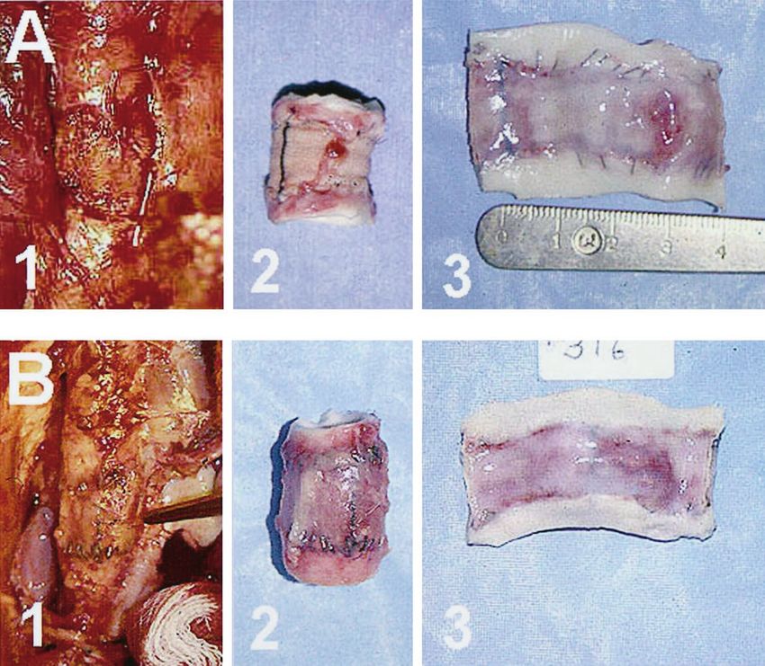
Figure 3. Illustration of vascular closure staples being applied to Gross pathologic examination revealed a smooth
the descending thoracic aorta with the aid of clip applier forceps. endothelial surfaced graft in both groups, with no
1764 Laboratory and Animal Investigations
Downloaded From: http://journal.publications.chestnet.org/ on 05/08/2015Table 1—Clamp and Anastomosis Times a significant risk of postoperative paraplegia, even if
Groups/Swine No. Clamp Time, min Anastomosis Time, mm
distal flow is augmented with bypass technique.4
There are additional adverse consequences of pro-
Suture group longed ischemia, such as reperfusion injury, which
1 41 22
2 31 27
can lead to adverse sequelae in distant organ sys-
3 35 23 tems. The US military also has interest in developing
4 28 11 methods to augment the skill of the surgeon in
5 19 17 combat and decrease the time required for vessel
Mean ⫾ SD 30.8 ⫾ 8.2 20.0 ⫾ 6.2 repair. Austere environments may make cardiopul-
p Value* 0.2 0.4
Clip group
monary bypass or shunts not available, making clamp
6 20 16 times more crucial.
7 21 13 This study found that vascular closure staples
8 23 16 provide a reliable method of aortic anastomosis, with
9 28 10 results comparable to conventional suture repair in
10 32 27
Mean ⫾ SD 24.8 ⫾ 5.1 16.4 ⫾ 6.4
terms of vessel patency, degree of narrowing, and
tissue reaction. The durability of vascular closure
*One-way ANOVA between suture group and clip group.
staple repair was demonstrated by the survival of all
animals for 8 weeks without evidence of dissection,
aneurysm, or anastomotic leak. Histologic examina-
evidence of leak or thrombosis (Fig 5). Histologi- tion (degree of fibrosis and intimal reaction) demon-
cally, there were no significant differences in fibrosis strated that the healing was comparable to suture
score or inflammatory infiltrates between the two repair.
groups. Both clamp time and anastomosis time were com-
parable to suture repair, although there was a trend
toward shorter times in the clip group. This may
Discussion afford the surgeon the potential for a faster repair
and decreased risk of ischemic injury. Bleeding after
The standard approach to the repair of the injured vascular closure staple repair tended to be higher
thoracic aorta after trauma involves a thoracotomy than suture repair; however, we believe that this was
through the left side of the chest with proximal and due to two factors. One, the clips were not large
distal aortic occlusion followed by repair. Although enough in one animal, as swine aorta can be thicker
distal aortic flow can be maintained by cardiopulmo- and softer than humans. Second, we tried to use a
nary bypass or heparinized shunts to minimize spinal completely sutureless technique with the vascular
cord ischemia, prolonged clamp times are not de- closure staples. The addition of suture as “stay
sired. When clamp time is prolonged, the patient has sutures” and for triangulation and for repair of
anastomotic leaks may have helped reduce the blood
loss. The repairs were performed by surgical resi-
Table 2—Intraoperative Blood Loss dents who had no prior experience with vascular
Intraoperative closure staples, with the aid of an attending trauma
Groups/Swine No. Blood Loss, mL surgeon. This demonstrates the relatively short
learning curve and the ease of use of the instru-
Suture group
1 150 ments.
2 250 Thoracic aortic injuries most often occur at the
3 980 aortic isthmus in the majority of the cases. Although
4 200 an injury to this area was not duplicated in this
5 200
animal model, the most common method of repair is
Mean ⫾ SD 356 ⫾ 351
Median 200 to resect the area of injury and place a graft, which
p Value* 0.48 was done in this model. Occasionally, the injury can
Clip group be directly repaired or other complex maneuvers can
6 200 be made; however, this is usually the exception. The
7 475
findings from this study would be relevant only to
8 650
9 200 those cases in which the injury is repaired with
10 1,100 resection and graft placement. The use of vascular
Mean ⫾ SD 525 ⫾ 374 closure staples would not be applicable in instances
Median 475 in which direct repair is needed.
*One-way ANOVA between suture group and clip group. Progress in vascular surgery has been enormous
CHEST / 118 / 6 / DECEMBER, 2000 1765
Downloaded From: http://journal.publications.chestnet.org/ on 05/08/2015Figure 4. Left, A: Aortogram of suture repair, with arrows pointing to proximal and distal anastomosis.
Right, B: Aortogram of vascular closure staple repair, with arrows pointing to proximal and distal
anastomosis; clips are visible around edges.
since Alexander Carrel15 first described the triangu- This has been postulated to be due to the clips
lation technique for vascular anastomosis in 1902. causing less vessel trauma due to their nonpenetrat-
Although vascular closure staples were originally ing nature.
designed for use in microvascular surgery, their use Clinically, vascular closure staples are used in
has been broadened and tested in multiple labora- various situations. Nataf et al8 used them in the
tory settings as well as clinically. Boeckx et al16 performance of coronary anastomosis in 10 patients
analyzed microvascular anastomosis in the rabbit with a marked reduction in anastomosis time. In
carotid artery under electron microscopy and found other applications, Mital et al7 used vascular closure
no histomorphologic differences between sutured staples to perform renal vascular anastomosis be-
and stapled anastomoses. In dogs, Dal Ponte et al17 tween cadaveric renal vessels and the external iliac
found that the use of the clips resulted in a more artery and vein of the recipient, with no postopera-
streamlined anastomosis, with decreased vessel wall tive complication. Vascular closure staples have also
damage, immediate hemostasis, and a trend toward been used for carotid endarteractomy, and vascular
shorter procedure times. Similar findings were re- access grafts for dialysis.21 The use of vascular clo-
ported in swine when used on the portal triad.18 sure staples in trauma was recently demonstrated by
Leppäniemi et al19,20 found a comparable degree of Rhee et al,9 who successfully used the clips to
narrowing, with similar tissue reaction using vascular perform repair and bypass of three vascular injuries
closure staples in the common bile duct and ureters, involving the brachial, axillary, and superficial fem-
compared to suture repair in swine, but with faster oral arteries, with excellent postoperative results.
repair times when using vascular closure staples. The use of vascular closure staples, however, is not
Previous studies have confirmed the ease of use, without concerns. Findlay and Megyesi22 raised the
better histologic healing, and faster repair times.7–9 question regarding the strength of clip closure, in
We found similar results in terms of tissue reaction. which the authors closed carotid endarterectomies
with either suture or clips. These authors found a
much faster closure time with clips; however, hemo-
Table 3—Mean (ⴞ SD) Proximal and Distal Suture stasis problems occurred in only two patients, which
Lines included clip failure in one patient with arterial
hypertension. The experience of vascular closure
Variables Suture Group Clip Group p Values*
staples in arteriosclerotic vessels is currently limited,
Proximal 13.6 ⫾ 1.9 13.7 ⫾ 2.0 1.0 and their use in chronically diseased vessels needs
Distal 13.9 ⫾ 2.2 13.7 ⫾ 2.5 0.62 further investigation. The vessels in patients with
*One-way ANOVA between suture group and clip group. traumatic aortic tears, however, are usually not dis-
1766 Laboratory and Animal Investigations
Downloaded From: http://journal.publications.chestnet.org/ on 05/08/2015Figure 5. Panel A1: Photograph of healed suture-repaired aorta in situ at 8 weeks. Panel A2:
Photograph of suture-repaired graft excised and dissected out. Panel A3: Photograph of inside the
suture-repaired graft with native aortic edges, with suture visible. Panel B1: Photograph of healed
vascular closure staple-repaired aorta in situ at 8 weeks. Panel B2: Photograph of vascular closure
staple-repaired graft excised and dissected out. Panel B3: Photograph of inside the vascular closure
staple-repaired graft with native aortic edges; clips are not penetrated.
eased, as this condition occurs in the younger trauma management and outcome of 144 patients. J Trauma 1996;
population (average age, 38.7 years).4 40:547–555
3 Franchello A, Olivero G, Di Summa M, et al. Rupture of
thoracic aorta resulting from blunt trauma. Int Surg 1997;
82:79 – 84
Conclusion 4 Fabian T, Richarson J, Croce M, et al. Prospective study of
blunt aortic injury: multicenter trial of the American Associ-
This study demonstrated that 2 months after aortic ation for the Surgery of Trauma. J Trauma 1997; 42:374 –380
repair, animals repaired with vascular closure staples 5 Werker PMN, Kon M. Review of facilitated approaches to
did well and aortic healing was equal to suture vascular anastomosis surgery. Ann Thorac Surg 1997; 63:
S122–S127
repair. Further studies are required to determine 6 Kirsch WM, Zhu YH, Hardesty RA. A new method for
safety and efficacy before extra-large clips are used microvascular anastomosis. Am Surg 1992; 58:722–727
clinically on large vessels such as the thoracic aorta. 7 Mital D, Foster P, Jensik S, et al. Renal transplantation
With experience and care in placement, vascular without suture using the vascular clipping system for renal
closure staples clips may be an additional tool for artery and vein anastomosis: a new technique. Transplanta-
tion 1996; 62:1171–1173
those who treat injuries to large vessels. 8 Nataf P, Kirsch W, Hill A, et al. Nonpenetrating clips for
coronary anastomosis. Ann Thorac Surg 1997; 63:S135–S137
9 Rhee P, Sharpe R, Huynh T, et al. Use of titanium vascular
staples in trauma. J Trauma 1998; 45:1097–1099
References 10 Leppäniemi A, Wherry D, Pikoulis E, et al. Arterial and
1 Williams JS, Graff JA, Uku JM, et al. Aortic injury in vehicular venous repair with vascular clips: comparison with suture
trauma. Ann Thorac Surg 1994; 57:726 –730 closure. J Vasc Surg 1997; 26:24 –28
2 Hunt J, Baker C, Lents C, et al. Thoracic aorta injuries: 11 Pikoulis E, Rhee P, Nishibe T, et al. Arterial reconstruction
CHEST / 118 / 6 / DECEMBER, 2000 1767
Downloaded From: http://journal.publications.chestnet.org/ on 05/08/2015with vascular clips is safe and quicker than sutured repair. ylene access grafts constructed by using nonpenetrating clips.
Cardiovasc Surg 1998; 6:573–578 J Vasc Surg 1999; 30:325–333
12 Pikoulis E, Burris D, Rhee P, et al. Rapid arterial anastomosis 18 Geevarghese SK, Bradley AL, Atkinson J, et al. Comparison
with titanium clips, Am J Surg 1998; 175:494 – 496 of arcuate-legged clipped versus sutured hepatic artery, por-
13 Joubert CJ. Principles of care and management in a labora- tal vein, and bile duct anastomosis. Am Surg 1999; 65:311–
tory animal facility. J S Afr Vet Assoc 1978; 49:153–154 316
14 Guide for the care and use of laboratory animals. Bethesda, 19 Leppäniemi A, Wherry D, Pikoulis E, et al. Common bile
MD: National Institutes of Health, 1985; publication No. duct repair with titanium staples. Surg Endosc 1997; 11:714 –
86 –23 717
15 Carrel A. La technique operatoire des anastomosis vasculaires 20 Leppäniemi A, Wherry D, Pikoulis E, et al. Urteral repair
et al transplantation RA, et al: des visceres. Lyon Med 1902; with titanium staples: comparison with suture closure. Urol-
98:859 ogy 1998; 51:553–557
16 Boeckx W, Darius O, Van den hof B, et al. Scanning electron 21 Haruguchi H, Nakagawa Y, Uchida Y, et al. Clinical applica-
microscopic analysis of the stapled microvascular anastomosis tion of vascular closure staple clips for blood access surgery.
in the rabbit. Ann Thorac Surg 1997; 63:S128 –S134 J ASAIO 1998; 44:M562–M564
17 Dal Ponte DB, Berman SS, Patula VB, et al. Anastomotic 22 Findlay JM, Megyesi JF. Carotid arteriotomy closure using a
tissue response associated with expanded polytetrafluoroeth- vascular clip system. Neurosurgery 1998; 42:550 –553
1768 Laboratory and Animal Investigations
Downloaded From: http://journal.publications.chestnet.org/ on 05/08/2015You can also read